
📌 AIM Study Tip
This chapter follows the supplied KMU learning outcomes and connects genetic inheritance with clinical disorders and reproductive counselling. First understand the explanations, then use the AIM High-Yield Review for revision.
TOPIC 6. Clinical Genetics, Chromosomal & Connective Tissue Disorders
Final Year MBBS — MS Module
Understanding genetic inheritance, recognition of important inherited syndromes, prenatal diagnosis and patient-centred genetic counselling.
Topic Introduction
Clinical genetics deals with disorders caused by changes in chromosomes, genes or the combined influence of genes and environmental factors. Some conditions are inherited from parents, while others arise from new genetic changes during formation of the reproductive cells or early embryonic development.
In clinical practice, genetics helps explain why certain diseases occur repeatedly within families, why some children are born with characteristic physical features and congenital abnormalities, and why apparently healthy parents may have an affected child.
This chapter begins with inheritance patterns and then applies them to Down syndrome, Marfan syndrome and inherited collagen disorders. It also explains how genetic risks are assessed before and during pregnancy, including the importance of counselling in families affected by thalassemia.
The central aim is to connect the genetic abnormality with its clinical consequences and understand how this information supports patients and families.
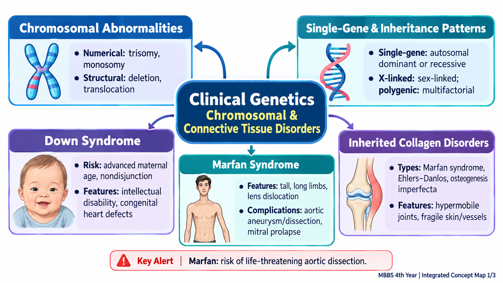
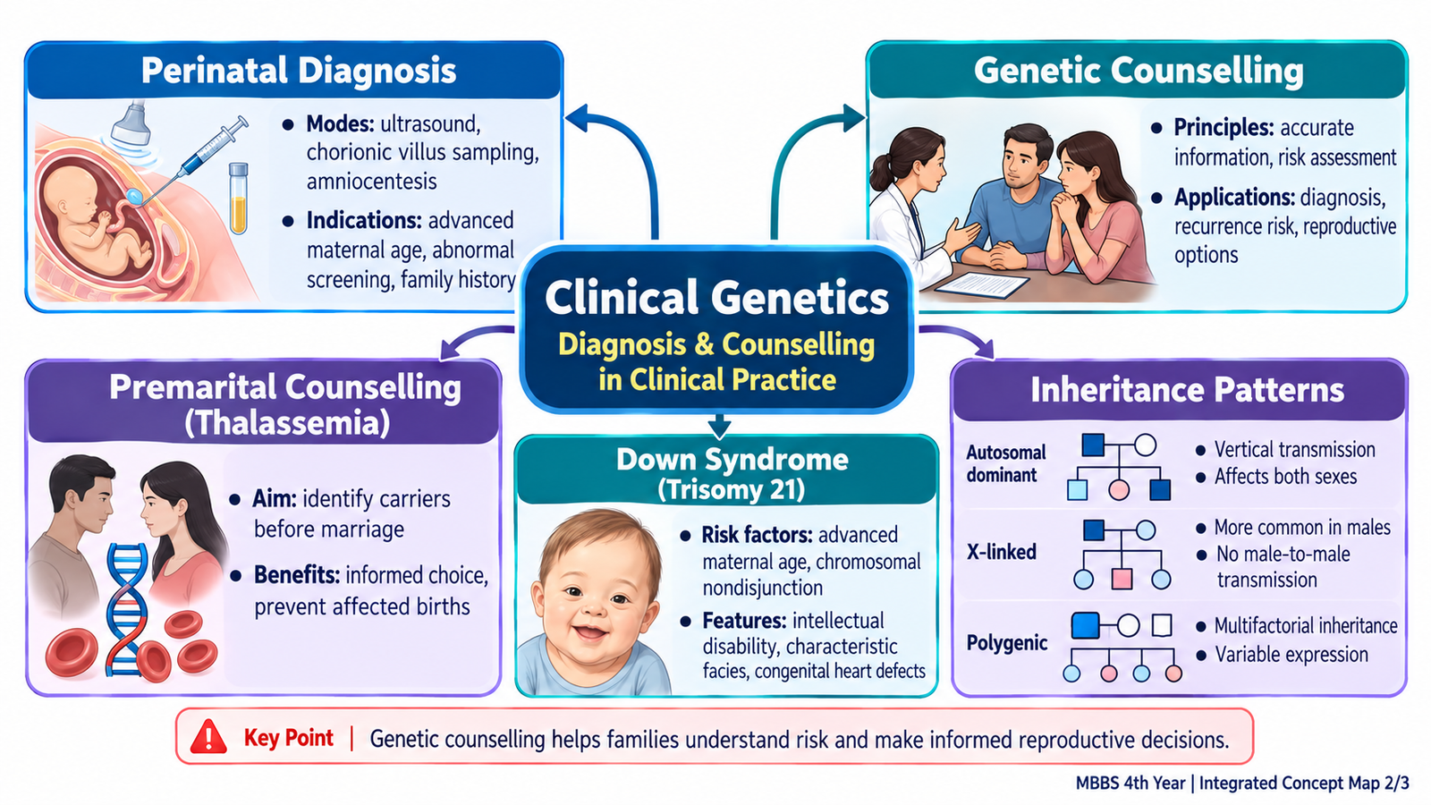
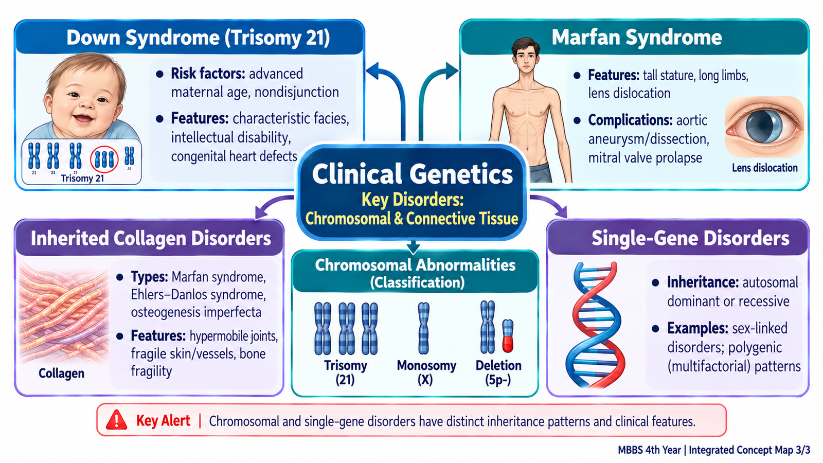
A. Principles of Genetic Inheritance and Chromosomal Abnormalities
Human genetic information is organized into DNA, which forms genes and chromosomes. Genes contain instructions for making functional proteins, while chromosomes carry large numbers of genes.
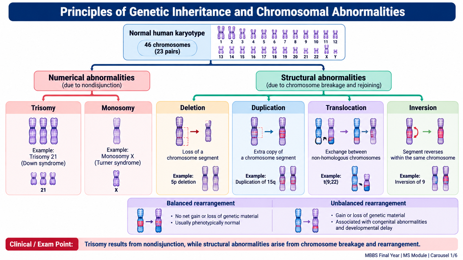
Normal human somatic cells usually contain 46 chromosomes arranged in 23 pairs. Twenty-two pairs are autosomes, while the remaining pair consists of sex chromosomes, usually XX or XY.
Genetic disorders can result from abnormalities in chromosome number, chromosome structure or individual genes. The type of abnormality helps predict how the disorder develops and whether it may recur within a family.
Classification of Chromosomal Abnormalities
Chromosomal abnormalities are broadly classified into numerical and structural abnormalities.
1. Numerical abnormalities
These occur when a cell contains an abnormal number of chromosomes. The most important mechanism is nondisjunction, in which chromosomes fail to separate correctly during cell division.
- Trisomy: Presence of an additional chromosome, producing three copies instead of two. Down syndrome commonly results from trisomy 21.
- Monosomy: Loss of one chromosome from a pair. Turner syndrome is an important example involving a missing or structurally abnormal X chromosome.
- Polyploidy: Presence of an additional complete chromosome set, such as triploidy. It is usually associated with severe developmental abnormalities and pregnancy loss.
2. Structural abnormalities
These occur when chromosomes break and rejoin abnormally, leading to loss, duplication or rearrangement of genetic material.
- Deletion: Loss of a chromosomal segment.
- Duplication: Presence of an additional copy of a chromosomal segment.
- Translocation: Transfer or exchange of chromosome segments between chromosomes.
- Inversion: A chromosome segment breaks and reinserts in the reverse orientation.
- Ring chromosome: A chromosome forms a ring after abnormal rearrangement of its ends.
A structural rearrangement may be balanced, with no substantial gain or loss of genetic material, or unbalanced, with extra or missing material. A healthy person carrying a balanced translocation may produce gametes with an unbalanced chromosome complement and therefore have an increased risk of miscarriage or an affected child.
Clinical Interpretation
Chromosomal abnormalities are particularly suspected when a child has multiple congenital anomalies, developmental delay, characteristic dysmorphic features or unexplained growth abnormalities. A chromosomal analysis may identify a numerical or major structural abnormality and guide recurrence-risk counselling.
For example, a child with characteristic features of Down syndrome may have free trisomy 21 or a translocation involving chromosome 21. Although both can produce the same syndrome, their recurrence implications differ.
🖼️ AIM VISUAL 01 — Chromosomal Abnormalities
B. Single-Gene, Sex-Linked and Polygenic Disorders
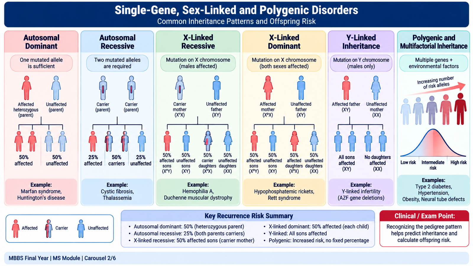
A single-gene disorder results from a pathogenic change in one gene. The pattern of inheritance depends on whether the gene lies on an autosome or sex chromosome and whether one or two altered copies are needed to produce disease.
Autosomal Dominant Inheritance
In autosomal dominant disorders, one pathogenic gene variant is usually sufficient to produce the condition. An affected heterozygous parent has a 50% chance of passing the variant to each child, regardless of sex.
Examples include Marfan syndrome and some forms of osteogenesis imperfecta.
- Males and females can be affected equally.
- The condition may appear in successive generations.
- Father-to-son transmission is possible.
- A new pathogenic variant can produce disease without a previous family history.
Clinical application: When a patient with Marfan syndrome plans to have children, genetic counselling should explain the possibility of transmission to offspring. Disease severity can vary even among affected members of the same family.
Autosomal Recessive Inheritance
Autosomal recessive disorders usually develop when both copies of a gene contain pathogenic variants. Parents carrying one altered copy are commonly asymptomatic carriers.
When both parents carry pathogenic variants in the same gene, each pregnancy has the following probabilities:
- 25% chance of an affected child.
- 50% chance of a carrier child.
- 25% chance of a child who has inherited neither familial pathogenic variant.
These probabilities apply independently to every pregnancy. They do not mean that exactly one out of four children must be affected.
Beta-thalassemia major is a clinically important example of autosomal recessive inheritance.
Consanguineous marriage increases the likelihood that both partners have inherited the same rare recessive variant from a shared ancestor. However, consanguinity does not by itself establish that a child will have a genetic disorder.
Sex-Linked Inheritance
Sex-linked disorders result from pathogenic variants located on the sex chromosomes. Most clinically important examples involve the X chromosome.
X-linked recessive disorders
Males generally have one X chromosome; therefore, a pathogenic recessive variant on that chromosome can produce disease. Females with one pathogenic variant are often carriers, although some may show clinical manifestations.
Examples include hemophilia A and Duchenne muscular dystrophy.
- An affected father does not transmit his X-linked variant to his sons.
- All daughters of an affected father inherit his affected X chromosome, assuming the usual chromosomal pattern.
- For a carrier mother, each son has a 50% chance of inheriting the variant, and each daughter has a 50% chance of inheriting it.
X-linked dominant disorders
One pathogenic variant on the X chromosome can produce disease. An affected father generally transmits the variant to all daughters and no sons, while a heterozygous affected mother has a 50% chance of transmission to each child.
Y-linked inheritance involves variants on the Y chromosome and follows father-to-son transmission. It is uncommon in routine clinical genetics.
Polygenic and Multifactorial Inheritance
Polygenic inheritance involves the combined effects of multiple genes. Multifactorial disorders arise through the interaction of genetic susceptibility with environmental or lifestyle influences.
Common examples include type 2 diabetes mellitus, hypertension and some congenital anomalies.
Unlike a typical single-gene disorder, these conditions do not usually follow a predictable Mendelian ratio. Family history may indicate increased susceptibility, but it does not determine the outcome with certainty.
The overall risk often becomes greater when multiple close relatives are affected, especially when disease is severe or begins early.
🖼️ AIM VISUAL 02 — Patterns of Genetic Inheritance
C. Down Syndrome — Genetics, Clinical Features and Complications
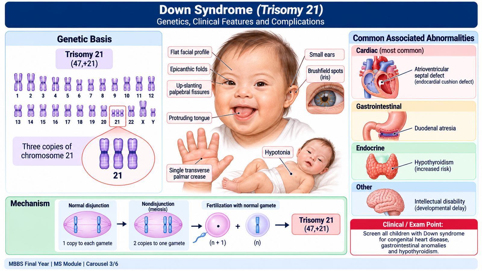
Down syndrome is a chromosomal disorder caused by the presence of additional genetic material from chromosome 21. It is associated with characteristic physical features, variable intellectual disability and increased risk of several congenital and acquired medical conditions.
Chromosomal Basis and Risk Factors
There are three principal cytogenetic mechanisms.
- Free trisomy 21: The commonest form, accounting for approximately 95% of cases. It usually results from meiotic nondisjunction.
- Robertsonian translocation: Additional chromosome 21 material is attached to another chromosome, commonly chromosome 14 or 21. Some cases are associated with a parental balanced translocation.
- Mosaic Down syndrome: Two or more cell populations exist, typically including both normal and trisomy 21 cells. Clinical features can vary.
Advanced maternal age is an important risk factor for meiotic nondisjunction and trisomy 21. However, Down syndrome can occur at any maternal age, and many affected babies are born to younger mothers because more pregnancies occur in this age group.
A previous pregnancy affected by a chromosomal abnormality and a parental balanced translocation may also influence recurrence risk.
Clinical Features
The physical features of Down syndrome arise from altered gene dosage, which affects growth and development across multiple organ systems.
Important features include:
- Generalized hypotonia during infancy.
- Flat facial profile and depressed nasal bridge.
- Upward-slanting palpebral fissures.
- Epicanthic folds.
- Small ears and relatively small oral cavity, sometimes producing the appearance of a protruding tongue.
- Single transverse palmar crease in some children.
- Short broad hands and fifth-finger clinodactyly.
- Wide gap between the first and second toes.
- Developmental delay and variable intellectual disability.
No single physical feature establishes the diagnosis. Clinical suspicion is based on the overall pattern, followed by appropriate chromosomal testing.
Important Complications
Congenital heart disease: Atrioventricular septal defect is particularly associated with Down syndrome. Ventricular septal defects and other congenital lesions may also occur. Because significant heart disease may initially be clinically silent, neonatal cardiac assessment and echocardiography are important.
Gastrointestinal abnormalities: Duodenal atresia and Hirschsprung disease are important associations. Persistent bilious vomiting, abdominal distension or delayed passage of meconium warrants urgent evaluation.
Endocrine disorders: Hypothyroidism is relatively common. It can worsen growth and developmental difficulties, so appropriate thyroid screening and follow-up are necessary.
Hematological complications: Children have increased susceptibility to certain leukemias, particularly acute megakaryoblastic leukemia and acute lymphoblastic leukemia. Neonates may develop transient abnormal myelopoiesis.
Other complications: Hearing impairment, visual disorders, obstructive sleep apnea, recurrent respiratory infections and cervical spine instability can affect long-term health and function.
Clinical Assessment and Diagnostic Confirmation
When Down syndrome is suspected, the clinician should assess the child’s growth, feeding, muscle tone, developmental milestones and associated congenital abnormalities.
Karyotyping confirms the chromosomal diagnosis and distinguishes free trisomy from translocation and detectable mosaicism. This distinction is important for estimating recurrence risk in future pregnancies.
Early developmental support, appropriate medical screening, hearing and vision assessment and family-centred follow-up improve health and quality of life.
🖼️ AIM VISUAL 03 — Down Syndrome: Clinical Recognition
D. Marfan Syndrome and Inherited Collagen Disorders
Connective tissue provides structural support to blood vessels, bones, joints, skin and many internal organs. Important inherited connective tissue disorders result from abnormalities in proteins that provide strength or elasticity to these tissues.
Marfan syndrome is primarily a disorder of fibrillin-containing microfibrils, while classical inherited collagen disorders involve abnormalities of collagen structure or processing.
Marfan Syndrome
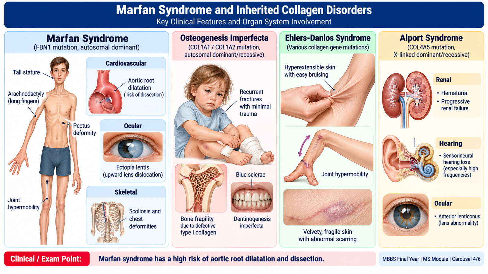
Marfan syndrome is an autosomal dominant connective tissue disorder, most commonly associated with pathogenic variants in the FBN1 gene, which encodes fibrillin-1.
Fibrillin-1 is an important component of extracellular microfibrils that support elastic connective tissue. Abnormal fibrillin compromises tissue integrity and alters growth-factor signalling, particularly involving transforming growth factor-beta.
The principal clinical consequences involve the skeletal, cardiovascular and ocular systems.
1. Skeletal features
- Tall, slender body habitus.
- Disproportionately long limbs.
- Arachnodactyly, meaning unusually long, slender fingers.
- Increased arm-span-to-height ratio.
- Pectus excavatum or pectus carinatum.
- Scoliosis or kyphoscoliosis.
- Joint hypermobility and characteristic hand signs.
2. Cardiovascular features and complications
The most clinically dangerous abnormality is progressive dilatation of the aortic root.
Weakness of the aortic wall increases susceptibility to aortic aneurysm and dissection. Aortic root dilatation may also produce aortic regurgitation because the valve leaflets can no longer close effectively.
Mitral valve prolapse is another important cardiovascular association.
Important Clinical Danger
Sudden severe chest or back pain in a patient with Marfan syndrome may indicate acute aortic dissection and requires emergency assessment.
3. Ocular features
Ectopia lentis, or displacement of the crystalline lens, is a characteristic manifestation. It commonly occurs in a superotemporal direction. Myopia and retinal complications may also occur.
The combination of long slender limbs, arachnodactyly, ectopia lentis and aortic root dilatation should strongly suggest Marfan syndrome.
Clinical Assessment and Complication Prevention
Assessment includes relevant family history, body proportions, skeletal abnormalities, eye examination and evaluation of the cardiovascular system.
Echocardiography is important for identifying aortic root enlargement and valvular abnormalities. Ophthalmological assessment helps detect lens displacement and other visual complications.
Patients require ongoing cardiovascular surveillance. Measures to reduce aortic stress may include beta-blockers or angiotensin receptor blockers under specialist supervision. Significant progressive aortic enlargement may require planned surgical intervention.
High-intensity competitive sports and activities involving marked cardiovascular strain are generally discouraged when aortic disease is present.
Classification of Inherited Collagen Disorders
Inherited collagen disorders can be understood according to the collagen type or processing pathway affected and the tissues most severely involved.
1. Osteogenesis imperfecta
Osteogenesis imperfecta is a genetically heterogeneous group of disorders, commonly involving abnormalities of type I collagen or its processing. The principal clinical feature is increased bone fragility.
Characteristic manifestations include:
- Recurrent fractures after minor trauma.
- Bone deformities in more severe forms.
- Blue sclerae in some patients.
- Dentinogenesis imperfecta.
- Hearing impairment in some forms.
The important clinical connection is that defective type I collagen reduces the strength of bone matrix, making fractures more likely.
2. Ehlers-Danlos syndromes
These are inherited connective tissue disorders involving abnormalities of collagen, collagen processing or related extracellular matrix proteins.
Common manifestations include joint hypermobility, skin hyperextensibility and tissue fragility. Some forms cause abnormal scarring and easy bruising.
Vascular Ehlers-Danlos syndrome is particularly important because it can cause spontaneous arterial or organ rupture.
3. Alport syndrome
Alport syndrome is caused by abnormalities of type IV collagen, an important component of basement membranes.
It is associated with persistent hematuria, progressive renal disease, sensorineural hearing loss and characteristic ocular abnormalities. Many cases follow an X-linked inheritance pattern, although autosomal forms also occur.
The relationship between the affected collagen and the clinical presentation helps distinguish these conditions:
- Type I collagen abnormalities → bone fragility.
- Collagen or related extracellular matrix abnormalities in Ehlers-Danlos syndromes → skin, joint and tissue fragility.
- Type IV collagen abnormalities → basement membrane dysfunction affecting kidneys, ears and eyes.
🖼️ AIM VISUAL 04 — Marfan Syndrome and Collagen Disorders
🩺 AIM Clinical Bridge
A 16-year-old boy is referred because of his unusually tall stature, long fingers and progressive chest-wall deformity. His father has similar physical features. Examination shows arachnodactyly and increased arm span, while echocardiography demonstrates enlargement of the aortic root.
Think clinically:
- The combination of skeletal features and aortic root enlargement strongly suggests Marfan syndrome.
- The family history supports autosomal dominant inheritance.
- The major concern is progression of aortic enlargement and the possibility of dissection.
The patient requires cardiovascular surveillance, ophthalmological assessment and appropriate counselling about family risk and activity-related precautions.
E. Prenatal and Perinatal Genetic Diagnosis
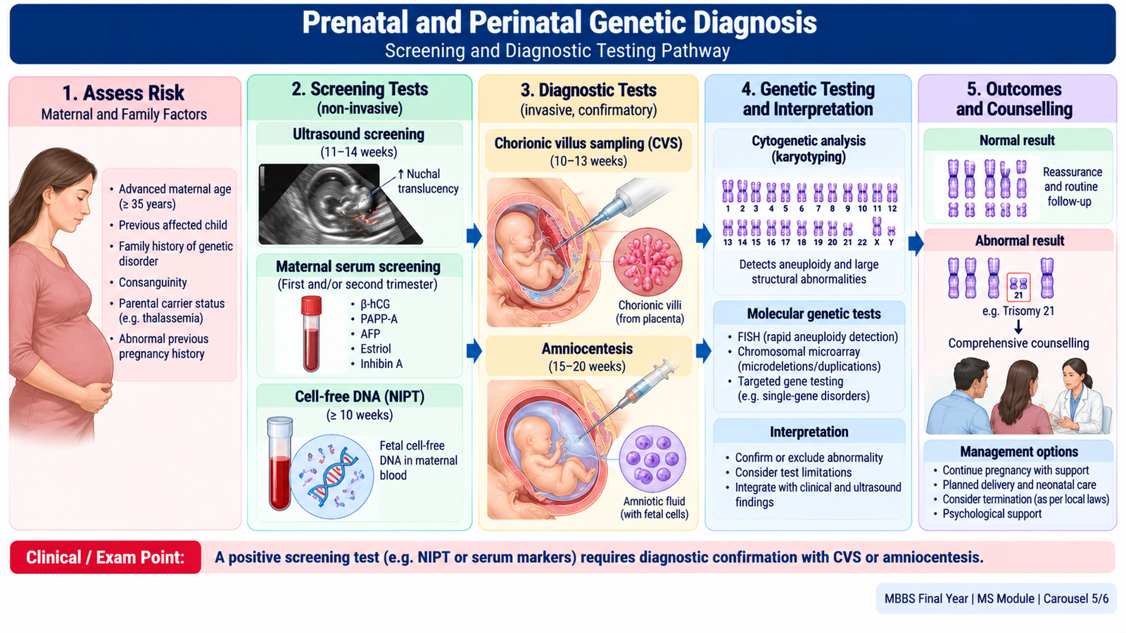
Perinatal genetic diagnosis involves identifying genetic or chromosomal disorders during pregnancy or around the time of birth. It is especially valuable when a pregnancy has an increased risk because of family history, previous affected children, parental carrier status or abnormal screening findings.
An important distinction is that screening estimates risk, whereas diagnostic testing can confirm or exclude a specific abnormality within the limits of the test.
Indications for Prenatal Genetic Assessment
Common indications include:
- Previous child or pregnancy affected by a chromosomal or inherited genetic disorder.
- Known parental carrier status for a serious inherited condition.
- Balanced chromosomal rearrangement in one parent.
- Family history suggesting a Mendelian genetic disorder.
- Abnormal fetal ultrasound findings.
- Positive or high-risk prenatal screening results.
- Advanced maternal age, particularly when assessing aneuploidy risk.
Non-Invasive Prenatal Screening
Ultrasound examination may identify structural abnormalities and markers associated with chromosomal disorders. First-trimester nuchal translucency measurement can contribute to aneuploidy screening.
Maternal serum screening uses biochemical markers, sometimes combined with maternal age and ultrasound measurements, to estimate the probability of certain fetal chromosomal abnormalities.
Cell-free DNA screening, also called non-invasive prenatal testing (NIPT), analyzes placental DNA fragments circulating in maternal blood. It is a highly sensitive screening test for common aneuploidies, particularly trisomy 21.
However, an abnormal NIPT result does not independently confirm fetal Down syndrome. Confirmatory diagnostic testing should be discussed before irreversible pregnancy-related decisions.
Invasive Prenatal Diagnostic Methods
Chorionic villus sampling (CVS)
CVS obtains placental tissue for genetic testing, commonly from approximately 10–13 weeks of pregnancy. It allows relatively early chromosomal or molecular diagnosis.
Because the sample is placental, confined placental mosaicism can occasionally produce results that differ from the fetal genetic constitution.
Amniocentesis
Amniocentesis obtains amniotic fluid containing fetal cells, generally from 15 weeks onward. These cells or extracted genetic material can be tested for relevant chromosomal or molecular abnormalities.
Cordocentesis
Cordocentesis involves sampling fetal blood from the umbilical cord under ultrasound guidance. It is reserved for selected clinical indications and is not a routine first-line genetic diagnostic procedure.
Invasive sampling carries procedure-related risks, including pregnancy loss. The magnitude of risk depends on the procedure, patient factors and operator experience, and should be discussed before consent.
Interpreting Genetic Tests
Karyotyping identifies chromosome number and many large structural abnormalities. It is especially useful when aneuploidy or a major chromosomal rearrangement is suspected.
Chromosomal microarray can detect smaller gains and losses of chromosomal material that may not be visible on routine karyotyping. However, it does not reliably detect balanced rearrangements.
Targeted molecular testing is used when a specific familial pathogenic variant is known, such as a variant associated with an inherited hemoglobin disorder.
Test selection depends on the clinical indication, family history, gestational age and the type of abnormality being investigated.
All results should be interpreted in the context of pre-test probability and the limitations of the method. A normal test for one condition does not exclude every genetic or structural abnormality.
🖼️ AIM VISUAL 05 — Prenatal Genetic Diagnosis
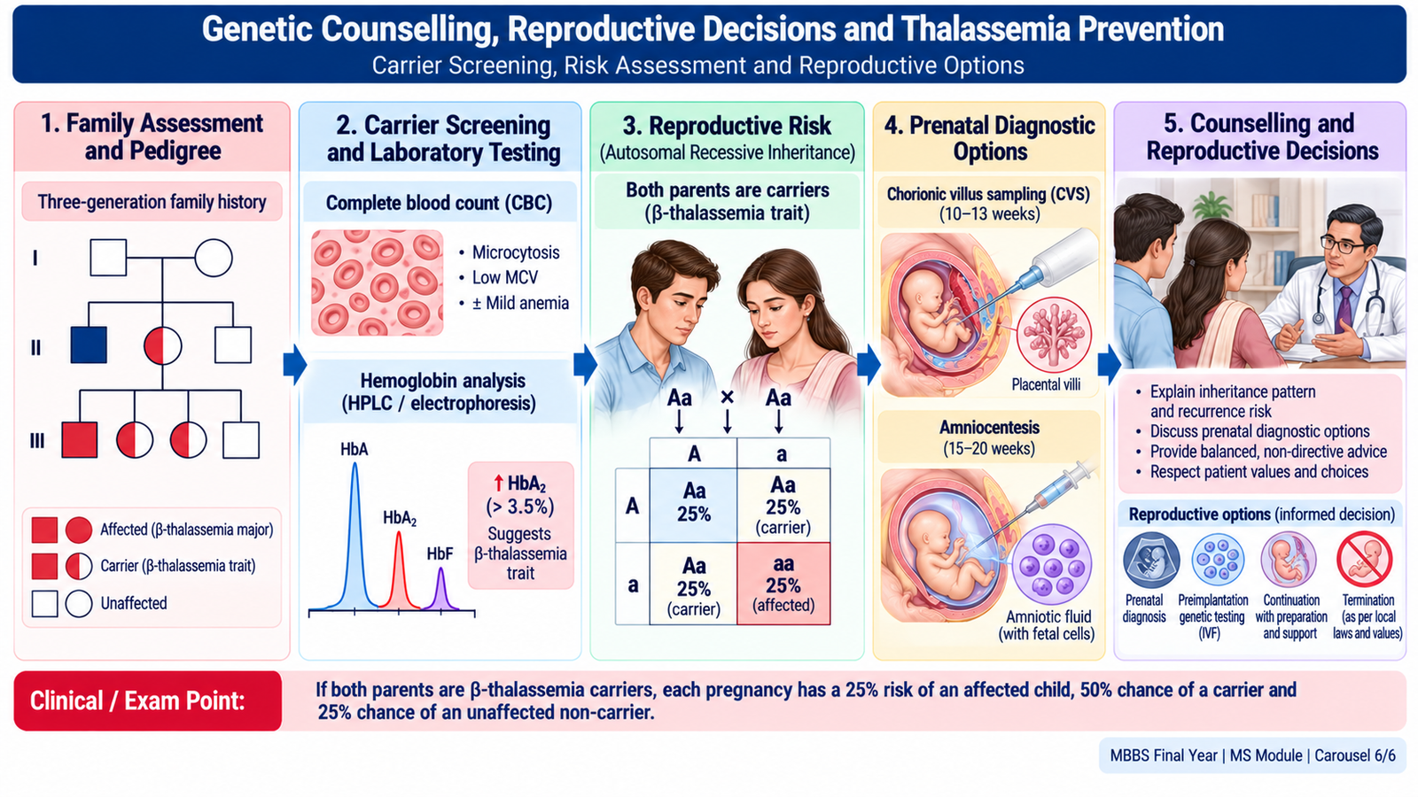
F. Genetic Counselling, Reproductive Decisions and Thalassemia Prevention
Genetic counselling is a communication process that helps individuals and families understand a genetic condition, its pattern of inheritance, the likelihood of recurrence and the available options for testing, prevention and reproductive decision-making.
The purpose is not to make decisions for the patient. It is to support informed, voluntary choices using accurate information presented with empathy and respect.
Principles of Genetic Counselling
Genetic counselling generally includes several connected activities.
1. Establishing the clinical and family history
A pedigree covering approximately three generations may identify repeated disease patterns, consanguinity, affected siblings, recurrent pregnancy losses or unexplained early deaths.
2. Identifying the likely inheritance pattern
Understanding whether the condition is autosomal dominant, autosomal recessive, X-linked or multifactorial helps estimate risk to relatives and future offspring.
3. Explaining the condition and available tests
Patients should understand what a test can detect, what it cannot detect and what a positive, negative or uncertain result could mean.
4. Communicating reproductive risk
Risk should be explained in understandable numerical terms whenever possible. For example, a 25% recurrence probability means one chance in four in each independent pregnancy.
5. Supporting informed decisions
The clinician should present available options without pressure or judgment. Counselling should respect religious, cultural and personal values while maintaining patient autonomy.
6. Maintaining confidentiality and arranging follow-up
Genetic information may have implications for other relatives. Its disclosure requires careful attention to confidentiality, consent and professional responsibilities.
Premarital Counselling in Families Affected by Thalassemia
Beta-thalassemia is an inherited disorder caused by reduced or absent production of beta-globin chains. Severe disease may develop when a child inherits clinically significant pathogenic variants from both parents.
Thalassemia carrier status is particularly important in communities where inherited hemoglobin disorders are prevalent and consanguineous marriage is common.
Identification of carrier status
Carrier evaluation may include:
- Family history of thalassemia or unexplained severe anemia.
- Complete blood count and red-cell indices.
- Peripheral blood film when appropriate.
- Hemoglobin analysis by HPLC or electrophoresis.
- Targeted molecular testing when indicated.
Beta-thalassemia trait commonly produces microcytosis with a relatively preserved or increased red-cell count. An elevated HbA2 level supports the diagnosis in many carriers.
Iron deficiency can also cause microcytosis and should be considered during interpretation. A carrier should not automatically be prescribed iron unless iron deficiency is established.
Risk When Both Partners Are Carriers
If both prospective parents carry a pathogenic variant causing autosomal recessive beta-thalassemia, the typical probability for each pregnancy is:
- 25%: Child affected by the disorder.
- 50%: Child who is a carrier.
- 25%: Child who has inherited neither familial pathogenic variant.
These probabilities must be individualized when the partners carry different variants, including variants associated with other hemoglobin disorders. The severity of disease depends on the specific genotypes inherited.
Reproductive Options and Counselling
When a couple has an increased risk of an affected child, counselling should explain the available options and their implications.
Options may include prenatal diagnosis using CVS or amniocentesis with appropriate molecular testing, and preimplantation genetic testing where available and acceptable.
Couples may also consider other reproductive choices according to their preferences, values and applicable legal and healthcare context.
A couple should not be pressured to marry, avoid marriage, conceive or avoid pregnancy on the basis of carrier status. The clinician’s role is to provide accurate risk information and support an informed decision.
Premarital counselling can reduce the occurrence of severe inherited disease through awareness, informed partner testing and reproductive planning while protecting individuals from discrimination and stigma.
🖼️ AIM VISUAL 06 — Genetic Counselling and Thalassemia
Important Comparison — Major Genetic Inheritance Patterns
| Feature | Autosomal Dominant | Autosomal Recessive | X-Linked Recessive |
|---|---|---|---|
| Typical family pattern | Successive generations | Affected siblings; parents often unaffected | Predominantly affected males |
| Father-to-son transmission | Possible | Possible | Absent |
| Typical risk | 50% transmission from affected heterozygous parent | 25% affected if both parents are carriers | 50% of sons affected if mother is a carrier |
| Example | Marfan syndrome | Beta-thalassemia | Hemophilia A |
⭐ AIM High-Yield Review
- Chromosomal numerical abnormalities commonly result from nondisjunction. Trisomy 21 is the classic clinical example.
- Balanced translocation carriers may be clinically healthy but can have an increased risk of pregnancy loss or offspring with unbalanced chromosomal abnormalities.
- Autosomal dominant inheritance: An affected heterozygous parent generally has a 50% chance of transmitting the variant in each pregnancy.
- Autosomal recessive inheritance: When both parents carry pathogenic variants in the same gene, the typical risk of an affected child is 25% per pregnancy.
- X-linked recessive disorders: Predominantly affect males; father-to-son transmission does not occur.
- Polygenic and multifactorial inheritance: Multiple genes and environmental influences contribute to disease susceptibility.
- ⭐ Down syndrome: Free trisomy 21 is the commonest mechanism. Advanced maternal age increases the risk of meiotic nondisjunction.
- Down syndrome complications: Congenital heart disease, gastrointestinal anomalies, hypothyroidism, hearing or visual impairment and increased leukemia risk.
- ⭐ Marfan syndrome: An FBN1-related connective tissue disorder characterized by skeletal abnormalities, aortic root disease and ectopia lentis.
- Marfan syndrome emergency: Sudden severe chest or back pain may indicate aortic dissection.
- Inherited collagen disorders: Osteogenesis imperfecta produces bone fragility; Ehlers-Danlos syndromes produce variable skin, joint and tissue fragility; Alport syndrome affects basement membranes.
- ⭐ Prenatal screening versus diagnosis: Ultrasound, serum screening and NIPT estimate risk; CVS or amniocentesis may provide diagnostic genetic material.
- Genetic counselling: Must be informed, confidential, patient-centred and non-directive.
- ⭐ Thalassemia prevention: Carrier identification, partner testing and informed reproductive counselling are important in at-risk families.
- Recurrence probabilities apply independently to each pregnancy and must be interpreted using the actual parental genotype.
🎥 AIM Video Learning
Topic 6 — Clinical Genetics, Chromosomal & Connective Tissue Disorders
Final Year MBBS | KMU Curriculum
Video 1. Genetic Inheritance & Pedigree Analysis
Understand autosomal dominant, autosomal recessive, sex-linked inheritance and pedigree interpretation.
Video 2. Down Syndrome — Trisomy 21
Learn the chromosomal basis, characteristic clinical features and important complications of Down syndrome.
Video 3. Marfan Syndrome
Understand the genetic basis and important skeletal, cardiovascular and ocular manifestations of Marfan syndrome.