
Station 5 — Microscopic Identification of Carcinoma of the Breast
🎯 Candidate Task
Examine the provided breast histopathology slide or image. Identify the lesion and state the important microscopic features supporting your diagnosis.
⚡ Model Station Answer
Identification: This histopathology slide shows carcinoma of the breast.
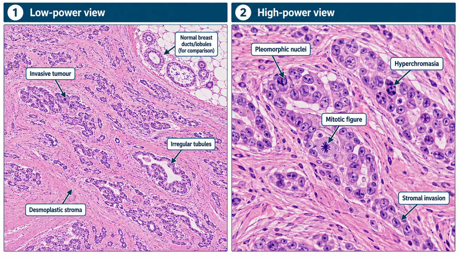
Key findings: Malignant epithelial cells are arranged in irregular nests, cords and poorly formed glandular or tubular structures infiltrating the surrounding breast stroma.
The tumour cells show nuclear pleomorphism, hyperchromasia and increased mitotic activity.
A surrounding desmoplastic fibrous stromal reaction may be prominent.
🔎 How to Perform / Approach
- Step 1 — Identify the tissue: Recognize breast tissue by ducts, lobules and surrounding fibrofatty stroma where preserved.
- Step 2 — Assess architecture: Look for loss of normal breast architecture and irregular infiltration by abnormal epithelial cells.
- Step 3 — Examine cell arrangement: Look for malignant cells forming irregular nests, cords, sheets or poorly formed tubules.
- Step 4 — Assess cytology: Identify nuclear enlargement, pleomorphism, hyperchromasia and mitotic figures.
- Step 5 — Examine the stroma: Look for a desmoplastic fibrous reaction surrounding infiltrating tumour cells.
- Step 6 — State the diagnosis: Combine invasive malignant epithelial architecture and cytological atypia to identify carcinoma of the breast.
🖼️ Visual Learning
Histopathology of Breast Carcinoma
📝 Important Viva Questions
1. What is the key microscopic evidence of invasion in breast carcinoma?
Answer: Malignant epithelial cells infiltrate beyond normal ductal or lobular boundaries into the surrounding breast stroma.
2. What cytological features suggest malignancy?
Answer: Nuclear pleomorphism, hyperchromasia, an increased nuclear-to-cytoplasmic ratio and increased mitotic activity are important malignant features.
3. What is meant by a desmoplastic reaction?
Answer: It is a dense fibrous stromal response produced around infiltrating malignant tumour cells.
4. How may malignant epithelial cells be arranged microscopically?
Answer: They may form irregular nests, cords, sheets or poorly formed glandular and tubular structures.
5. What feature distinguishes invasive carcinoma from carcinoma in situ?
Answer: In invasive carcinoma, malignant cells breach their normal ductal or lobular boundaries and infiltrate the surrounding stroma; in situ disease remains confined within them.