Course Content
🩺 OSCE Station 1 — Prescription Writing in Graves’ Disease
KMU clinical activity: Graves’ disease — formulation of an appropriate prescription. Learning Objectives The student should be able to: Formulate a prescription for a patient with Graves’ disease.
0/1
🩸 OSCE Station 2 — Diabetes Mellitus: Glucose Testing and Prescription Writing
KMU clinical activities: Diabetes mellitus prescription + glucose estimation in urine and blood. Learning Objectives The student should be able to: Check glucose in urine. Check blood glucose in a given sample. Formulate an appropriate prescription for a patient with Type 1 diabetes mellitus. Formulate an appropriate prescription for a patient with Type 2 diabetes mellitus.
0/1
🔬 OSCE Station 3 — Microscopic Identification of Multinodular Goitre
KMU clinical activity: Goitre — microscopic identification. Learning Objectives The student should be able to: Identify the important microscopic features of multinodular goitre. Recognize multinodular goitre when presented as a histopathology slide or image at the station.
0/1
🔬 OSCE Station 4 — Gynecological Pathology: Pap Smear and Uterine Fibroid
KMU clinical activities: Pap smear interpretation + microscopic identification of uterine fibroids. Learning Objectives The student should be able to: Interpret a Pap smear slide. Identify the important microscopic features of uterine fibroids. Differentiate the two when presented as cytology/histopathology specimens or images at the station.
0/1
🎗️ OSCE Station 5 — Microscopic Identification of Carcinoma of the Breast
KMU clinical activity: Carcinoma breast — microscopic identification. Learning Objectives The student should be able to: Identify the important microscopic features of carcinoma of the breast. Recognize carcinoma of the breast when presented as a histopathology slide or image at the station.
0/1
⚖️ OSCE Station 6 — Forensic Examination in Sexual Offences: Examination, Sampling and Preservation of Evidence
KMU clinical activities: Examination of a rape victim and assailant + examination of a sodomy victim and assailant. Learning Objectives The student should be able to: Take appropriate consent from a rape victim. Demonstrate or describe the examination of a rape victim/manikin. Describe the procedure for taking samples from the victim/manikin/item. Describe the correct sealing and preservation of specimens. Describe the procedure for examining a victim of sodomy. Describe the procedure for taking appropriate samples in such a case. Describe the correct sealing and preservation of specimens obtained during the examination.
0/1
💊 OSCE Station 7 — Contraceptive Identification and Counselling
KMU clinical activity: Contraception. Learning Objectives The student should be able to: Identify a given contraceptive device or drug. Explain its merits and demerits. Discuss the appropriate method of administration/use of the given contraceptive device or drug.
0/1
🤰 OSCE Station 8 — Antenatal Care: Maternal Health Chart and WHO Visit Schedule
KMU clinical activity: Mother and child health. Learning Objectives The student should be able to: Identify the relevant mother-and-child-health chart presented at the station. Devise an appropriate schedule for antenatal visits according to WHO criteria.
0/1
💉 OSCE Station 9 — EPI Schedule, Vaccine Identification and Immunization Counselling
KMU clinical activities: EPI schedule + vaccination and immunization. Learning Objectives The student should be able to: Counsel a mother regarding the EPI schedule. Identify a given vaccine. Explain the uses of the vaccine. Discuss its schedule of administration. Interpret the VVM (vaccine vial monitor). Discuss the use of VVM in epidemics.
0/1
👶 OSCE Station 10 — Child Growth Assessment: Anthropometry, Shakir’s Tape and Growth Chart
KMU clinical activities: IMCI anthropometric measures/Shakir’s tape + growth chart. Learning Objectives The student should be able to: Identify the relevant anthropometric model/instrument. Measure the mid-upper arm circumference using Shakir’s tape. Calculate or determine the weight and height of the child as required by the station. Interpret the anthropometric results. Identify a growth chart. Plot data on the growth chart using a given clinical scenario. Interpret the different parts and findings of the growth chart.
0/1
📊 OSCE Station 11 — Interpretation of Demographic Indicators
KMU clinical activity: Demographic indicators. Learning Objectives The student should be able to interpret a given demographic indicator, including: Population pyramid Human Development Index (HDI) Physical Quality of Life Index (PQLI) Population growth rate Dependency ratio
0/1
AIM OSPE/OSCE Lab — Endocrine & Reproduction-II Module | 4th Year MBBS
AIM OSCE / OSPE • MBBS

Station 3 — Microscopic Identification of Multinodular Goitre

4th Year • GIT / Hepatobiliary • Rapid Revision • Practical Skills • Viva

🎯 Candidate Task

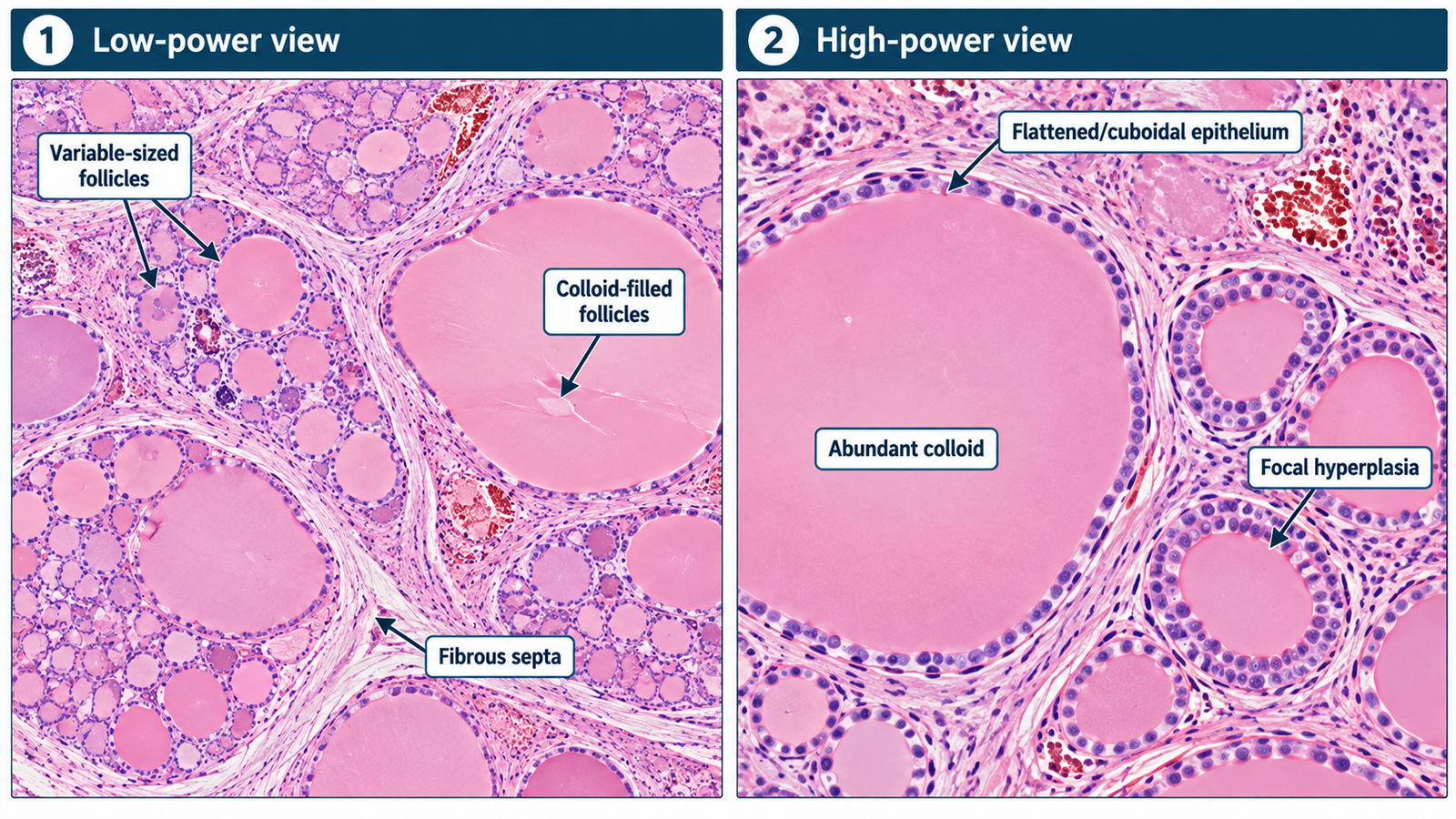
Examine the provided thyroid histopathology slide or image. Identify the lesion and state the important microscopic features supporting your diagnosis.

⚡ Model Station Answer

Identification: This is a histopathology slide of multinodular goitre.

Key findings: There are thyroid follicles of markedly variable size, with many enlarged follicles distended by colloid and lined by flattened to cuboidal follicular epithelium.

Areas of follicular hyperplasia may alternate with inactive colloid-rich follicles.

Secondary changes such as fibrosis, hemorrhage, cystic degeneration or calcification may also be present.

Exam shortcut: Variable-sized follicles + abundant colloid + mixed hyperplastic and inactive areas strongly support multinodular goitre.

🔎 How to Perform / Approach

  1. Step 1 — Identify the tissue: Recognize thyroid tissue by follicles containing eosinophilic colloid.
  2. Step 2 — Assess the architecture: Look for marked variation in follicular size rather than a uniform follicular pattern.
  3. Step 3 — Examine the follicles: Identify enlarged colloid-filled follicles mixed with smaller or hyperplastic follicles.
  4. Step 4 — Examine the lining: Note flattened to cuboidal follicular epithelium, with focal areas of epithelial hyperplasia.
  5. Step 5 — Look for secondary changes: Search for fibrosis, hemorrhage, cystic change or calcification where visible.
  6. Step 6 — State the diagnosis: Combine the characteristic variable nodular follicular pattern and colloid accumulation to identify multinodular goitre.

🖼️ Visual Learning

VISUAL 1

Histopathology of Multinodular Goitre

.

📝 Important Viva Questions

1. What is the most characteristic microscopic pattern in multinodular goitre?

Answer: Marked variation in thyroid follicular size, with large colloid-filled follicles alternating with smaller or hyperplastic follicles.

2. What type of material is present within the thyroid follicles?

Answer: The follicles contain colloid, which appears as homogeneous eosinophilic material on H&E staining.

3. How is the epithelium commonly seen in enlarged inactive follicles?

Answer: It is usually flattened to low cuboidal because the follicles are distended with colloid.

4. Name two secondary microscopic changes that may occur in multinodular goitre.

Answer: Fibrosis and hemorrhage are common secondary changes; cystic degeneration and calcification may also occur.

5. Which feature helps distinguish multinodular goitre from the typical microscopic pattern of Graves’ disease?

Answer: Multinodular goitre shows a heterogeneous mixture of variably sized colloid-rich follicles, whereas Graves’ disease typically shows more diffuse follicular epithelial hyperplasia with tall crowded cells and scalloped colloid.

Scroll to Top
💬 WhatsApp Support