
Station 4 — Gynecological Pathology: Pap Smear and Uterine Fibroid
🎯 Candidate Task
Examine the provided cytology and histopathology slides or images. Interpret the Pap smear specimen, identify the second specimen and describe the important microscopic features supporting your identification.
⚡ Model Station Answer
- Pap smear: This is a cervical cytology specimen. I would assess specimen adequacy and examine the squamous cells for nuclear and cytoplasmic abnormalities before giving the cytological interpretation.
- Second specimen: This is a uterine leiomyoma (fibroid).
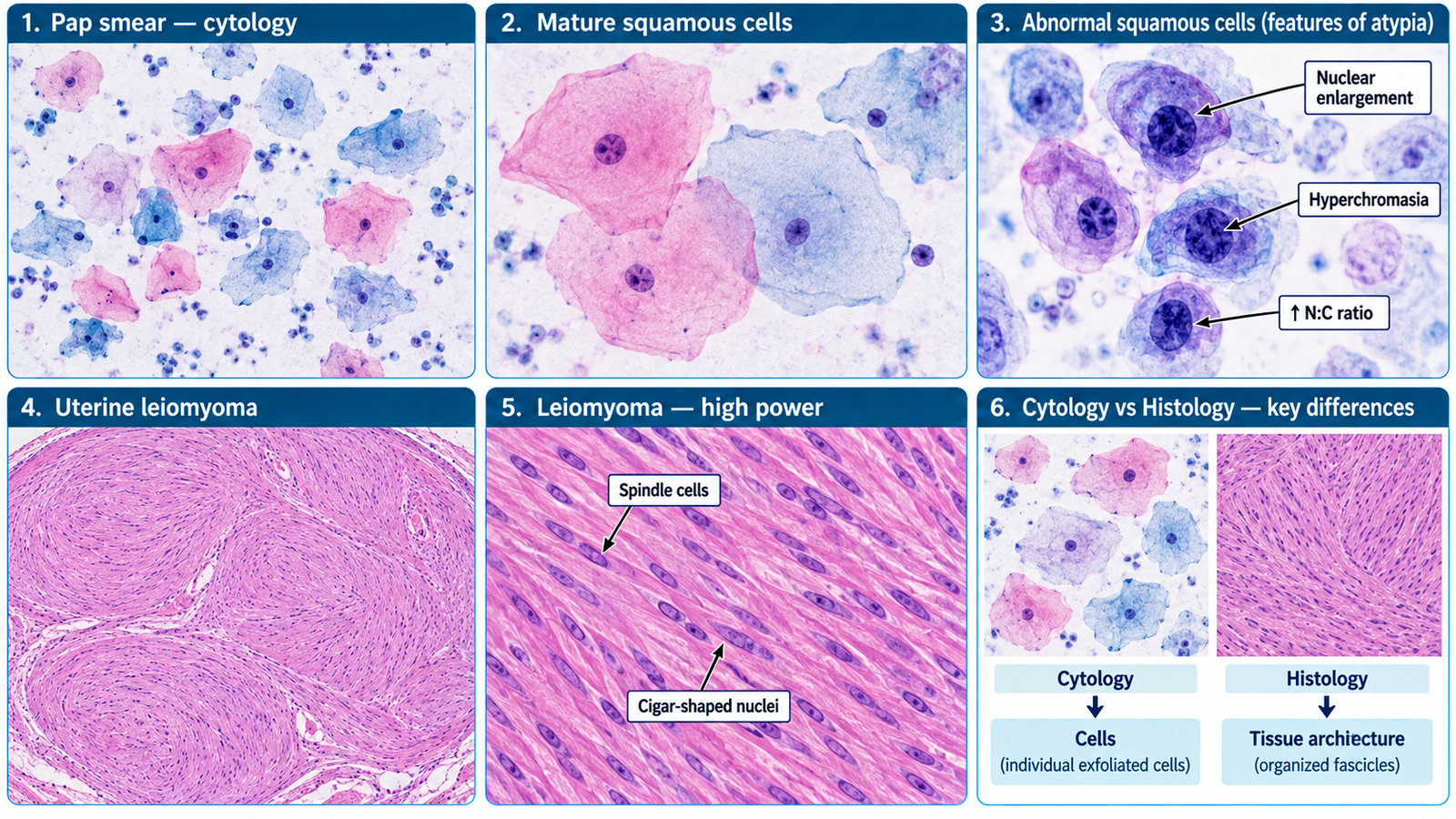
- Microscopy: It shows intersecting or whorled fascicles of uniform spindle-shaped smooth-muscle cells with elongated blunt-ended nuclei and eosinophilic cytoplasm.
- Key distinction: A Pap smear is a cytological preparation of exfoliated cervical cells, whereas a uterine fibroid is identified on tissue histology by organized smooth-muscle fascicles.
🔎 How to Perform / Approach
- Step 1 — Identify the preparation.
Decide whether the specimen is a cytology smear containing individual exfoliated cells or a histological tissue section showing tissue architecture. - Step 2 — Review the Pap smear systematically.
Assess cellularity and adequacy, then examine squamous cells for changes in cell size, nuclear size, chromatin, nuclear-to-cytoplasmic ratio and cellular arrangement. - Step 3 — Interpret the cervical cytology.
State whether the smear shows no obvious epithelial abnormality or whether abnormal squamous-cell features are present, according to the provided specimen. - Step 4 — Examine the tissue section.
Look for bundles of spindle-shaped smooth-muscle cells arranged in intersecting fascicles. - Step 5 — Identify the cellular features.
Note uniform elongated cells with eosinophilic cytoplasm and blunt-ended, cigar-shaped nuclei with minimal atypia in a typical fibroid. - Step 6 — State the diagnosis.
Identify the smooth-muscle lesion as uterine leiomyoma and mention the characteristic fascicular or whorled microscopic pattern. - Step 7 — Differentiate the specimens.
Pap smear = exfoliative cervical cytology; uterine fibroid = histological smooth-muscle tissue with preserved architecture.
🖼️ Visual Learning
Pap Smear Cytology and Uterine Leiomyoma Microscopy
📝 Important Viva Question
1. What type of specimen is examined in a Pap smear?
Answer: It is an exfoliative cytology specimen containing cells collected from the cervix, particularly the transformation-zone region.
2. Which cytological features suggest an abnormal squamous cell?
Answer: Important features include nuclear enlargement, hyperchromasia, irregular nuclear contours and an increased nuclear-to-cytoplasmic ratio.
3. What is the characteristic microscopic arrangement of a uterine leiomyoma?
Answer: It consists of intersecting or whorled fascicles of uniform smooth-muscle cells.
4. What do the nuclei of typical leiomyoma cells look like?
Answer: They are elongated, uniform and blunt-ended or cigar-shaped, usually without significant atypia.
5. How can you rapidly distinguish a Pap smear from a leiomyoma slide?
Answer: A Pap smear shows dispersed exfoliated epithelial cells without tissue architecture, whereas leiomyoma histology shows organized bundles of spindle-shaped smooth-muscle cells.