
AIM OSCE / OSPE • MBBS
Station 10 — Identification of Common Abnormalities on Chest X-Ray
Rapid Revision • Systematic Interpretation • Clinical Correlation • Viva
🎯 Task
CANDIDATE TASK
You are shown a chest radiograph together with a brief respiratory clinical scenario. Examine the radiograph systematically, identify and describe the major abnormality, and state the most likely interpretation in the clinical context.
You are shown a chest radiograph together with a brief respiratory clinical scenario. Examine the radiograph systematically, identify and describe the major abnormality, and state the most likely interpretation in the clinical context.
⚡ Model Station Answer
Film: “This is a chest radiograph. I would first confirm the patient details, projection and image quality.”
Findings: “On systematic ABCDE review, the major abnormality is [state abnormality and location], demonstrated by [2–3 key radiographic features].”
Interpretation: “In the supplied clinical context, these appearances are most consistent with [most likely diagnosis].”
Completion: “I would review the remainder of the film for associated abnormalities and compare with previous imaging if available.”
🔎 Stepwise Procedure / Approach
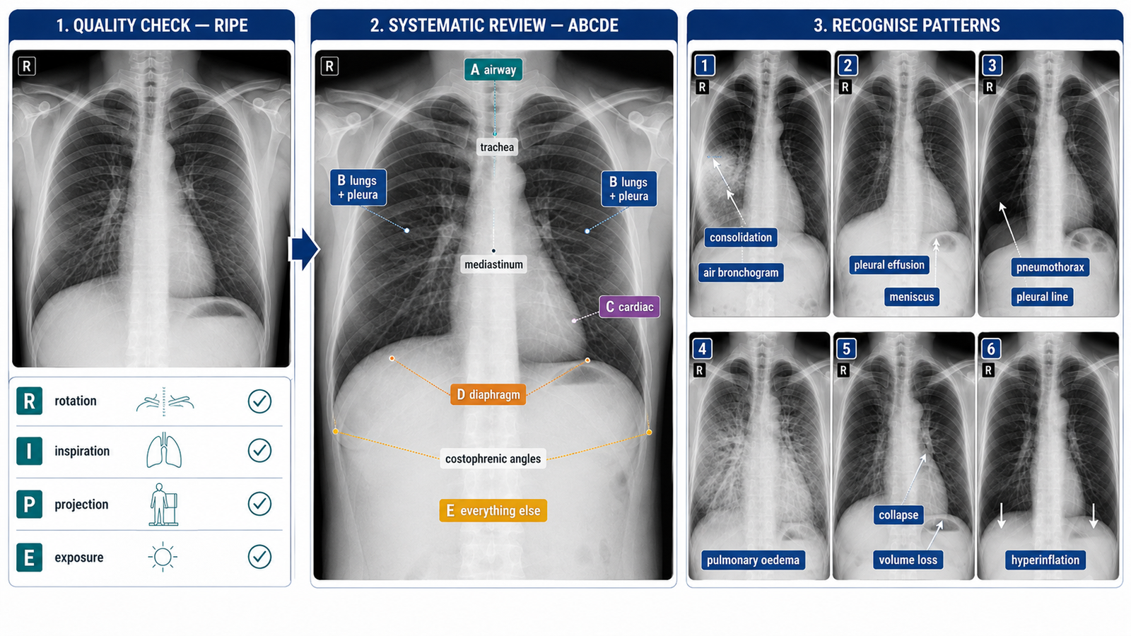
- Confirm the film. Check patient identity, date/time and whether the image is a chest radiograph.
- Assess image quality. Review rotation, inspiration, projection and exposure before interpreting pathology.
- A — Airway. Check the trachea for central position or deviation, then inspect the carina and hilar regions.
- B — Breathing. Compare both lung fields and pleura from apices to costophrenic angles for opacity, hyperlucency, volume loss or absent lung markings.
- C — Cardiac. Assess heart size and cardiac borders, remembering that apparent enlargement is less reliable on an AP film.
- D — Diaphragm. Inspect both hemidiaphragms and costophrenic angles for blunting, elevation or free subdiaphragmatic gas.
- E — Everything else. Review mediastinum, bones, soft tissues, hidden review areas and any tubes or devices.
- Describe the dominant abnormality. State its side, zone/lobe where possible, pattern, extent and important associated signs.
- Conclude and correlate. Give the most likely radiological interpretation and relate it directly to the supplied respiratory scenario.
Consolidation
Focal or lobar air-space opacity; air bronchograms may be visible.
Pleural effusion
Blunted costophrenic angle with basal homogeneous opacity and a meniscus pattern when sufficiently large.
Pneumothorax
Visible visceral pleural line with absent peripheral lung markings beyond it.
Pulmonary oedema
Bilateral predominantly perihilar/interstitial or air-space opacification, often with cardiomegaly or pleural effusions.
Lobar collapse
Increased opacity accompanied by signs of volume loss such as fissural, hilar or mediastinal displacement.
Hyperinflation
Increased lung volumes with flattened diaphragms and relatively hyperlucent lung fields.
Exam tip: Describe what you can actually see before naming the diagnosis. A structured description earns marks even when the final diagnosis is uncertain.
🖼️ Visual Learning
VISUAL 1
Systematic CXR Interpretation + Common Abnormal Patterns
🎥 Practical Video
🎥 Chest X-ray Interpretation — Structured OSCE Approach
Source: Geeky Medics
Demonstrates image-quality assessment, systematic ABCDE interpretation and practical recognition of important chest X-ray abnormalities using clinical examples.
📝 Important Viva Questions
Q1. Why should image quality be assessed before interpreting a chest X-ray?
Answer: Poor rotation, inspiration, projection or exposure can mimic pathology or obscure genuine abnormalities.
Q2. What systematic approach can you use to examine a chest radiograph?
Answer: After confirming details and image quality, use an ABCDE approach: Airway, Breathing, Cardiac, Diaphragm and Everything else.
Q3. What are typical radiographic features of lung consolidation?
Answer: Consolidation produces air-space opacity, often in a segmental or lobar distribution, and may contain visible air bronchograms.
Q4. What finding strongly suggests a pneumothorax?
Answer: A visible visceral pleural line with absence of lung markings peripheral to that line is characteristic.
Q5. What are common chest X-ray features of pleural effusion?
Answer: Blunting of the costophrenic angle and a basal homogeneous opacity are typical; a larger effusion may produce a meniscus-shaped upper border.
Q6. How do you distinguish collapse from simple consolidation?
Answer: Collapse is associated with volume loss, producing signs such as displacement of fissures, hilum, diaphragm or mediastinum toward the affected region.
Q7. Why must a chest X-ray finding be correlated with the clinical scenario?
Answer: Radiographic appearances are not interpreted in isolation; symptoms, examination findings and clinical context help determine the most likely diagnosis and significance.