
This chapter connects the supplied KMU outcomes through recognition, assessment, investigation and treatment of inflammatory arthritis. Understand the explanations first, then use the AIM High-Yield Review for revision.
📖 AIM Learning Material
TOPIC 1. INFLAMMATORY POLYARTHRITIS IN ADULTS AND CHILDREN
Musculoskeletal Module — Final Year MBBS
A connected approach to rheumatoid arthritis, systemic lupus erythematosus and juvenile idiopathic arthritis, from clinical assessment to preservation of function.
Topic Introduction
Inflammatory polyarthritis means inflammation affecting several joints. The clinical task is to recognize true arthritis, identify the pattern of involvement and determine whether the problem is mainly joint disease or part of a wider systemic disorder. Rheumatoid arthritis commonly produces persistent symmetrical inflammation of small peripheral joints. Systemic lupus erythematosus may produce a similar joint presentation, but associated skin, blood, kidney or other organ involvement changes assessment and treatment. In children, juvenile idiopathic arthritis requires an age-appropriate approach that also protects growth, vision and participation in school. These conditions share important principles: early recognition, carefully interpreted investigations, control of inflammation and preservation of function. The following sections connect these principles while explaining the differences that matter for patient care.
A. Recognizing and Classifying Inflammatory Arthritis
Arthritis is inflammation within a joint. It differs from arthralgia, which means joint pain without definite clinical evidence of inflammation. Inflammatory arthritis commonly produces soft swelling, warmth, tenderness and restricted movement. Stiffness is particularly troublesome after rest because inflamed tissues become less mobile during inactivity; gentle movement often helps.
Why inflammation produces joint symptoms
The synovium is the lining of a synovial joint. When it becomes inflamed, its blood supply increases and inflammatory fluid accumulates. This causes warmth and swelling. Pain and capsular distension restrict movement. Persistent inflammation may eventually damage cartilage, bone, tendons and ligaments, producing deformity and loss of function.
Synovial inflammation → swelling and pain → restricted movement → persistent tissue injury → deformity and disability.
Inflammatory pain commonly occurs at rest and is accompanied by prolonged morning stiffness. Mechanical pain usually worsens with use and has shorter stiffness after inactivity. These are useful patterns, but the diagnosis depends on the whole clinical assessment.
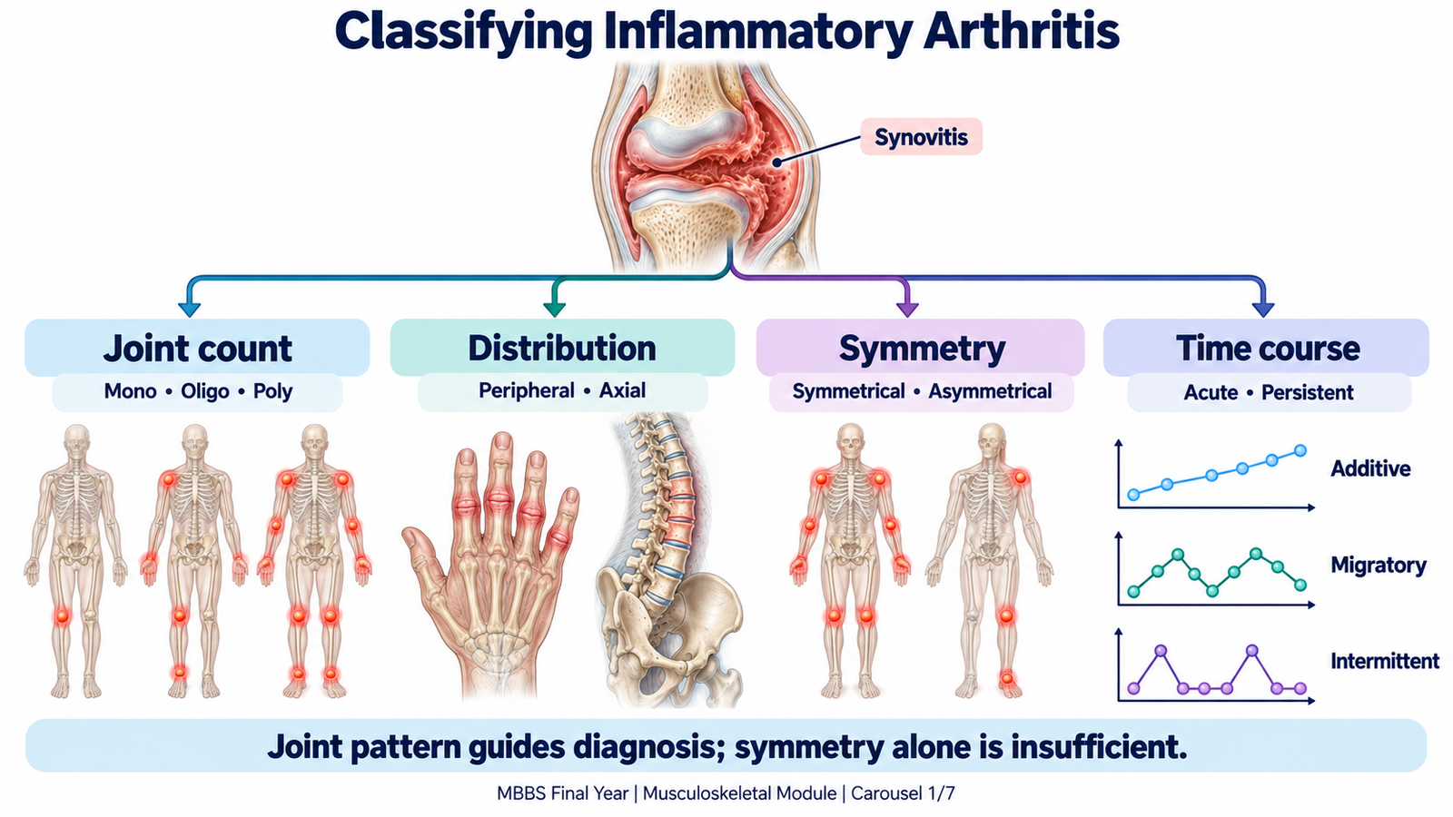
Classify the pattern before naming the disease
- Number of joints: monoarthritis affects one joint, oligoarthritis two to four, and polyarthritis five or more.
- Peripheral or axial: peripheral arthritis affects limb joints. Axial disease mainly involves the spine and sacroiliac joints. Rheumatoid arthritis is predominantly peripheral, although the cervical spine can be affected.
- Symmetrical or asymmetrical: matching joints on both sides suggest rheumatoid arthritis or lupus, but symmetry alone does not establish the diagnosis.
- Small or large joints: wrists, metacarpophalangeal joints and proximal interphalangeal joints are characteristic sites in rheumatoid arthritis. Childhood inflammatory disease may involve both small and large joints.
- Time course: sudden, short-lived arthritis suggests a different differential from persistent, gradually progressive synovitis.
- Evolution: additive arthritis involves new joints while previous joints remain inflamed. Migratory arthritis moves from one joint to another. Intermittent arthritis occurs in episodes with improvement between attacks.
Use associated findings to refine the classification
A symmetrical peripheral pattern supports rheumatoid arthritis when persistent small-joint synovitis is present. Photosensitivity, oral ulcers or renal abnormalities suggest lupus. Inflammatory back pain, enthesitis— inflammation where a tendon or ligament attaches to bone—psoriasis or a preceding gastrointestinal or genitourinary infection suggests a spondyloarthritis pattern. These clues guide further assessment; they do not justify diagnosing a disease from one feature.
B. Assessing the Adult or Child with Polyarthritis
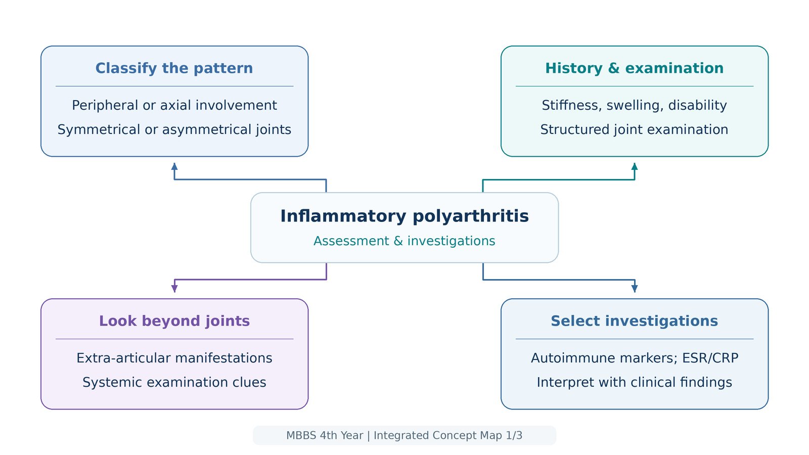
Assessment must establish whether joints are genuinely inflamed, define the pattern and search for systemic involvement. It must also determine how the disease affects daily life. A patient with mild-looking swelling may have substantial difficulty dressing, walking or working.
Focused history in an adult
Ask when symptoms began, which joints were affected first and whether involvement has spread. Clarify morning stiffness, swelling, pain at rest, fluctuation and response to movement. Persistent bilateral wrist and finger-joint symptoms are more informative than a general statement that “all joints hurt.”
- Function: difficulty opening containers, fastening buttons, gripping, climbing stairs or walking suggests clinically important limitation.
- Systemic clues: fatigue, fever, weight loss, rash, photosensitivity, oral ulcers, hair loss, dry eyes or mouth, chest symptoms and changes in urine help identify extra-articular disease.
- Alternative causes: recent infection, psoriasis, trauma and inflammatory bowel symptoms may redirect the differential.
- Risk and treatment context: previous autoimmune disease, family history, smoking, occupation, current medicines and previous treatment responses affect diagnosis and management.
- Patient priorities: establish the effects on work, caring responsibilities, sleep and mood. Discuss reproductive plans when treatment choices may affect pregnancy.
Examination findings and their meaning
Examine the distribution of tenderness and swelling, compare both sides and assess movement and function. Boggy swelling suggests thickened inflamed synovium; hard bony enlargement is more consistent with established structural change. Painful restriction of both active and passive movement supports a joint problem, whereas isolated pain on a particular resisted movement may suggest a tendon disorder.
In the hands, assess wrists, metacarpophalangeal and proximal interphalangeal joints, grip and tendon function. Distal interphalangeal involvement is less typical of rheumatoid arthritis. Examine the feet as well, because metatarsophalangeal disease may impair walking before the patient volunteers foot symptoms.
Look beyond the joints for pallor, fever, skin rash, nodules, oral ulcers, lymphadenopathy, edema and cardiopulmonary abnormalities. These findings may indicate systemic disease, a complication or an alternative diagnosis. Measure blood pressure when lupus is suspected because hypertension may accompany renal involvement.
Age-appropriate orthopedic assessment
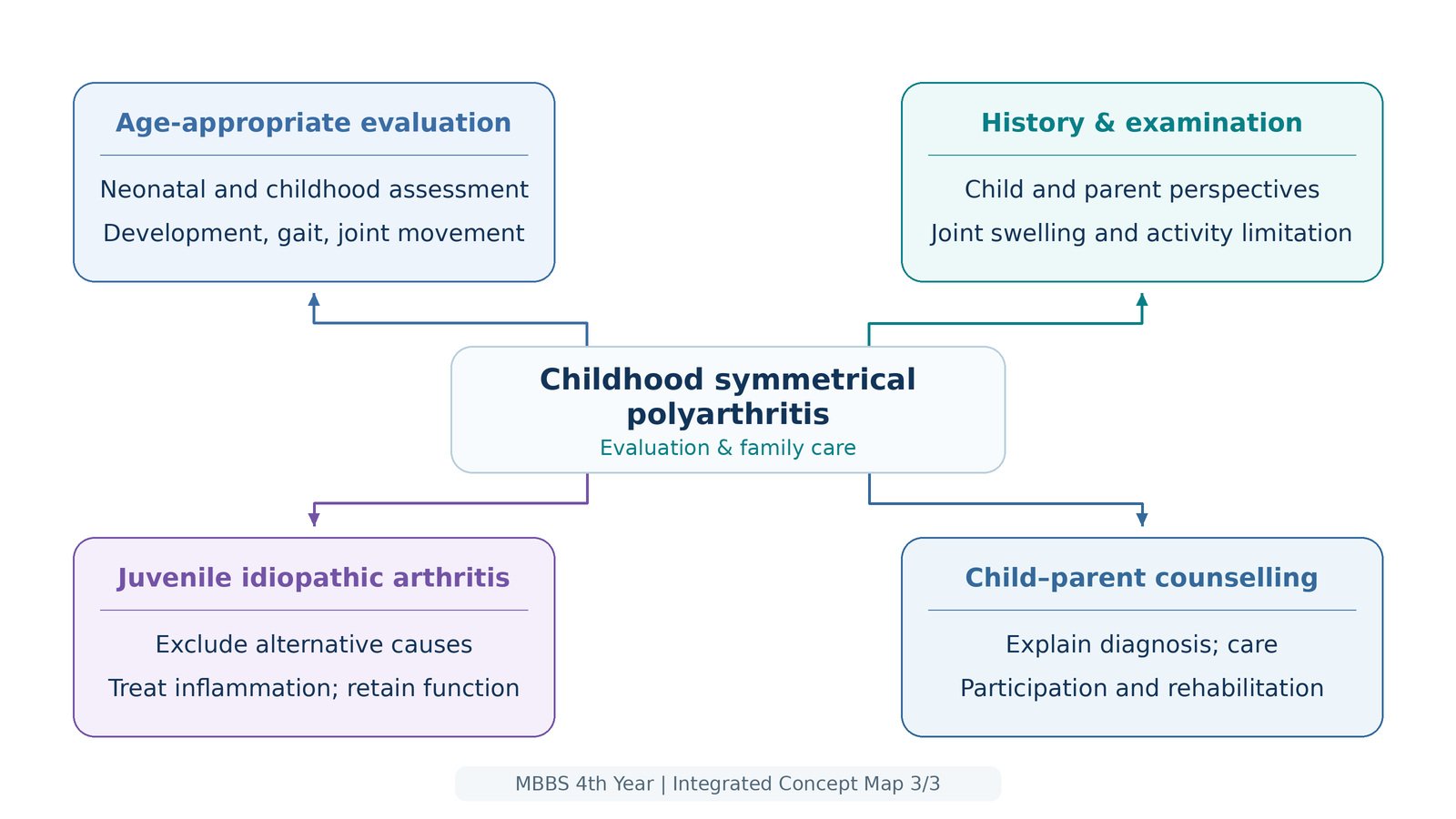
A child’s musculoskeletal assessment depends on developmental stage. Young children may show stiffness through behavior rather than describing it: refusing to walk, asking to be carried, avoiding play or struggling after waking. Obtain the parent’s observations and include the child’s account whenever possible.
- Neonate and infant: observe spontaneous movement, symmetry, limb posture and apparent pain with gentle handling. Assess hips, feet, spine and limb alignment appropriately for age. Reduced movement of a painful limb may represent pseudoparalysis from infection rather than a neurological disorder.
- Preschool child: observe walking, getting up, reaching and play. Compare joint movement gently; do not force a painful joint.
- School-age child: assess gait, joint swelling, range of movement, limb length, alignment and participation in physical activity.
- Adolescent: assess joint symptoms alongside growth, puberty, school attendance, sport, mood and increasing responsibility for treatment.
Record growth and look for muscle wasting, contractures and limb-length differences. Inflammation and reduced use can impair growth and muscle development, while local inflammation near a growth plate may alter growth of the affected limb. Hip disease may present as knee pain, so an unexplained painful knee requires assessment of the hip.
Important alternatives and urgent findings
In adults, osteoarthritis favors mechanical pain and bony enlargement; viral arthritis may follow a recent infection and resolve; psoriatic arthritis is supported by psoriasis, nail changes or a different joint pattern. In children, exclude infection, injury and malignancy before attributing persistent symptoms to juvenile idiopathic arthritis. Pallor, bruising, night pain, weight loss or an abnormal blood count should raise concern for leukemia or another serious disorder.


C. Selecting and Interpreting Investigations
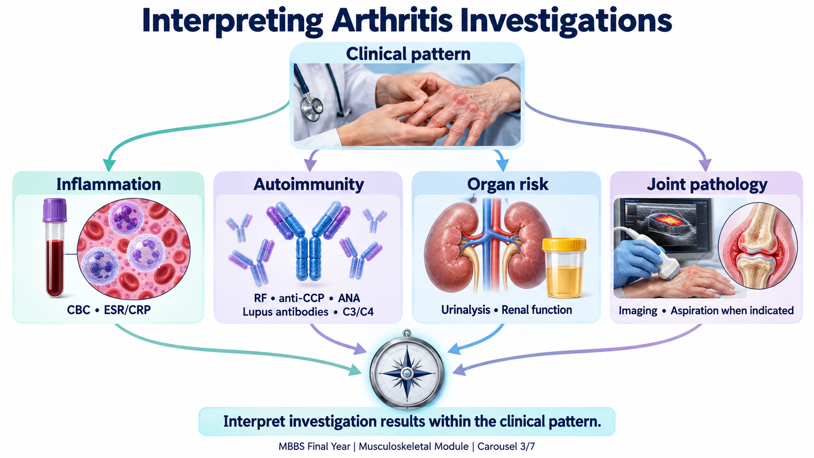
Investigations should answer a clinical question. They support the diagnosis, detect organ involvement, establish a baseline before treatment and monitor disease or drug toxicity. No single blood test replaces assessment of the patient.
Initial tests and clinical decisions
- Complete blood count: anemia may reflect chronic inflammation. Leukopenia or thrombocytopenia can support lupus but also require consideration of infection, marrow disease or medicines. An unexpected abnormal count in a child may redirect assessment away from JIA.
- ESR and CRP: raised values support inflammation but are nonspecific. Normal results do not exclude inflammatory arthritis. In lupus, ESR may rise while CRP is relatively modest; a marked CRP rise should prompt assessment for infection, although some inflammatory manifestations can also raise it.
- Renal function and urinalysis: proteinuria, hematuria or urinary casts may indicate lupus nephritis. Quantify protein loss and assess renal function; normal creatinine alone does not exclude early renal involvement.
- Liver function and albumin: help assess systemic illness and establish a baseline before medicines that may affect the liver.
Autoimmune markers: interpret them in context
Rheumatoid factor (RF) is an autoantibody commonly directed against part of immunoglobulin G. A positive result supports rheumatoid arthritis in a patient with compatible synovitis, but it can occur in other diseases and healthy people. A negative result does not exclude rheumatoid arthritis.
Anti-cyclic citrullinated peptide antibodies (anti-CCP) are more specific for rheumatoid arthritis than RF. In a compatible presentation, positivity strengthens the diagnosis and may indicate a greater risk of erosive disease. Some patients remain negative for both antibodies; this is termed seronegative rheumatoid arthritis.
Antinuclear antibodies (ANA) are sensitive for lupus but lack specificity. Request and interpret them when the clinical picture suggests connective tissue disease. An isolated positive ANA is insufficient to diagnose lupus. :chatgpt-content-reference{index=”0″}
Anti-double-stranded DNA antibodies support lupus and, in some patients, help assess activity, particularly renal disease. Anti-Smith antibodies are relatively specific but are not present in every patient. Low C3 and C4 complement may reflect consumption during immune-complex inflammation. Rising anti-dsDNA with falling complement and new urinary abnormalities should prompt evaluation for active lupus nephritis; antibody changes alone do not establish a flare.
In JIA, diagnosis is principally clinical after excluding other causes. ANA helps identify children who need particular attention to uveitis risk; it does not confirm JIA. RF helps characterize a subgroup of polyarticular disease rather than acting as a general screening test. HLA-B27 is reserved for a clinically compatible enthesitis or axial pattern, because positivity alone does not establish disease.
Imaging and joint fluid
Plain radiographs establish structural baseline findings. Early rheumatoid arthritis may show soft-tissue swelling with no erosions; later disease can show periarticular osteopenia, joint-space narrowing and marginal erosions. A normal early film does not exclude active disease.
Ultrasound can show synovial thickening, effusion and increased blood flow associated with active inflammation. It is useful when clinical swelling is uncertain. MRI is selected when deeper structures, early damage or a particular joint problem require further assessment; it is not necessary for every patient.
Synovial fluid aspiration is important when infection or crystal arthritis is suspected. Send fluid for appropriate cell assessment, microscopy and microbiological testing. An inflammatory cell pattern alone does not distinguish autoimmune arthritis from infection, and finding crystals does not automatically exclude coexisting infection.
Connect results to the patient
For the woman in the Clinical Bridge, persistent symmetrical small-joint synovitis plus anti-CCP positivity would strongly support rheumatoid arthritis. The same joint symptoms with photosensitivity, cytopenias, lupus-associated antibodies and proteinuria would require assessment for lupus. Referral for suspected persistent inflammatory arthritis should not wait for antibody positivity or radiographic damage.
D. Rheumatoid Arthritis: Disease, Complications and Treatment
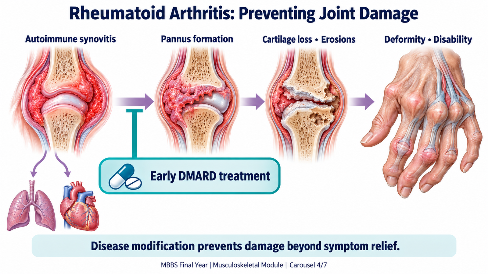
Rheumatoid arthritis is a chronic systemic autoimmune disease characterized by persistent synovitis. It commonly affects peripheral joints symmetrically and can produce irreversible structural damage. Genetic susceptibility and environmental factors, including smoking, contribute to loss of immune tolerance; the disease is not caused by ordinary joint use.
From synovitis to deformity
Activated immune cells release inflammatory mediators, including tumor necrosis factor and interleukin-6. The synovium thickens into invasive inflammatory tissue called pannus. This damages cartilage and promotes bone erosion. Tendon and ligament injury then destabilizes joints, explaining why persistent inflammation can produce deformity even after pain improves.
Typical disease involves wrists, metacarpophalangeal, proximal interphalangeal and metatarsophalangeal joints. Later findings may include ulnar deviation, swan-neck or boutonnière deformities, tendon rupture and painful forefoot deformity. These are manifestations of established damage; early disease need not show deformity.
Extra-articular disease and complications
Rheumatoid arthritis affects more than joints because the inflammatory process is systemic. Important manifestations include rheumatoid nodules, anemia of inflammation, dry eyes or mouth, episcleritis or scleritis, pleuritis and interstitial lung disease. Cardiovascular risk is increased by chronic inflammation and associated risk factors.
Local complications include carpal tunnel syndrome from swelling around the median nerve, tendon rupture and cervical instability, particularly at the atlantoaxial joint. Severe longstanding disease may cause vasculitis; Felty syndrome combines rheumatoid arthritis, splenomegaly and neutropenia. Treatment-related infection and osteoporosis also contribute to morbidity.
New limb weakness, gait disturbance or other neurological symptoms with neck pain require urgent evaluation for cervical disease. Suspected cervical instability is important before anesthesia or neck manipulation. A painful red eye, new breathlessness or evidence of infection also needs prompt assessment.
Treat inflammation early and preserve function
The treatment goal is remission or low disease activity, with prevention of damage and preservation of daily function. Prompt rheumatology assessment enables disease-modifying treatment before irreversible damage develops.
- Disease-modifying antirheumatic drugs (DMARDs): methotrexate is a common initial treatment. It modifies inflammatory activity and reduces progression of damage. Alternatives or additional conventional agents include sulfasalazine, leflunomide and hydroxychloroquine, selected according to disease and patient factors.
- NSAIDs: reduce prostaglandin-mediated pain and inflammation, but do not prevent structural damage. Consider gastrointestinal bleeding, renal impairment and cardiovascular risk.
- Glucocorticoids: may provide short-term control while slower-acting treatment takes effect or during a flare. Prolonged exposure increases infection, osteoporosis and metabolic complications.
- Escalation: persistent activity despite appropriate conventional treatment may require specialist-directed biologic or targeted therapy. These interrupt selected immune pathways and require infection-risk assessment.
- Rehabilitation: physiotherapy maintains movement and strength; occupational therapy, joint protection, splints and footwear adaptations reduce strain and support independence.
Monitor the disease and the treatment
Review joint swelling, stiffness, functional ability and inflammatory markers to assess response. Methotrexate requires blood-count, liver and renal monitoring because marrow suppression, hepatotoxicity and impaired clearance may occur. Folic acid reduces some adverse effects. Hydroxychloroquine requires appropriate retinal monitoring. New fever or respiratory symptoms during immunosuppression need assessment for infection or drug-related complications.
Medication safety: methotrexate for inflammatory arthritis is usually taken weekly, not daily. Confirm understanding of the prescribed schedule and discuss pregnancy planning before treatment.
E. Systemic Lupus Erythematosus: Arthritis within Multisystem Disease
Systemic lupus erythematosus is an autoimmune disease in which loss of tolerance to self-antigens leads to autoantibody production and immune-mediated tissue injury. Immune complexes contribute to inflammation in organs such as the skin and kidneys. Arthritis may resemble rheumatoid arthritis, but the wider clinical pattern determines diagnosis, severity and treatment.
Recognize the joint and systemic pattern
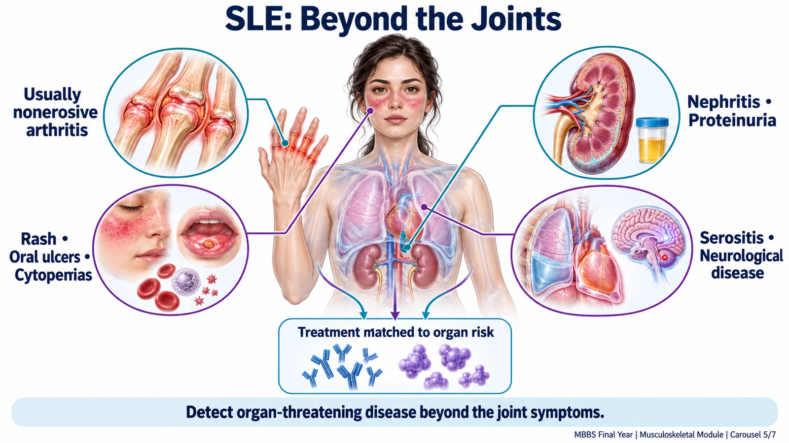
Lupus commonly causes symmetrical arthralgia or inflammatory arthritis involving small joints. The arthritis is usually nonerosive. Some patients develop deformity from tendon and ligament laxity, called Jaccoud arthropathy; the deformity may initially be reducible. Destructive erosions are less characteristic than in rheumatoid arthritis.
Associated manifestations include photosensitive rash, malar rash, oral or nasal ulcers, alopecia, fatigue and fever. Pleuritis or pericarditis causes inflammatory chest pain. Cytopenias may result from immune-mediated destruction or other disease-related processes. Renal involvement may cause proteinuria, hematuria, edema and hypertension, but can initially be clinically silent. Neurological disease can produce seizures, psychosis or other focal or diffuse abnormalities.
Antiphospholipid antibodies may be associated with thrombosis or pregnancy morbidity. Their presence must be interpreted alongside clinical history; a positive antibody result alone does not establish antiphospholipid syndrome.
Assess severity by organ involvement
Joint and skin disease can cause substantial distress, but kidney, central nervous system or severe hematological involvement changes treatment urgency. Assess urine, renal function, blood count and relevant organ symptoms even when joint pain is the presenting complaint.
Management follows disease severity
Management aims to suppress inflammation and prevent organ damage. Hydroxychloroquine is a foundation treatment for most patients unless contraindicated. Glucocorticoids provide faster control during active disease, while steroid-sparing treatment is selected according to the affected organs and severity. :chatgpt-content-reference{index=”1″}
NSAIDs may help selected patients with musculoskeletal symptoms, but renal disease or other contraindications may limit their use. Persistent arthritis may require a disease-modifying agent. Significant nephritis or other organ-threatening disease requires specialist-directed immunosuppression, such as mycophenolate or cyclophosphamide in appropriate settings; the undergraduate principle is that therapy must match the organ risk.
Sun protection reduces ultraviolet-triggered skin activity. Follow-up includes blood pressure, urine and renal assessment, blood counts, treatment toxicity and cardiovascular risk. Discuss adherence, reproductive planning and the distinction between disease activity and infection.
Prognosis
Lupus usually follows a relapsing and remitting course. Outcomes have improved with earlier recognition and treatment, and many patients maintain good function. Prognosis is less favorable with severe renal or neurological involvement, recurrent thrombosis, infection or accumulated organ damage. Sustained disease control and reduced treatment toxicity are both important for long-term health.
F. Juvenile Idiopathic Arthritis: Diagnosis, Growth and Treatment
Juvenile idiopathic arthritis is a group of conditions characterized by arthritis beginning before 16 years of age, persisting for at least six weeks and remaining unexplained after other causes are excluded. “Idiopathic” means that a specific cause has not been identified. JIA is not simply adult rheumatoid arthritis occurring in a smaller patient.
Recognize childhood polyarthritis
Children may present with joint swelling, morning stiffness, limping or reduced activity. Pain can be less prominent than swelling and loss of movement. A parent may notice difficulty dressing, a change in handwriting or reluctance to walk after rest.
Polyarticular JIA affects five or more joints during the first six months. It may be RF-positive or RF-negative. RF-positive disease often resembles adult rheumatoid arthritis and has greater potential for erosive damage. Oligoarticular disease affects fewer joints; systemic JIA can involve recurrent fever, an evanescent rash and systemic inflammation. This classification helps anticipate complications and choose treatment.
Diagnosis requires exclusion and interpretation
History and examination establish the arthritis pattern. Blood counts and inflammatory markers help assess inflammation and identify alternative explanations, but neither normal markers nor negative autoantibodies exclude JIA. Ultrasound may clarify synovitis; imaging is selected according to the affected joints and clinical question.
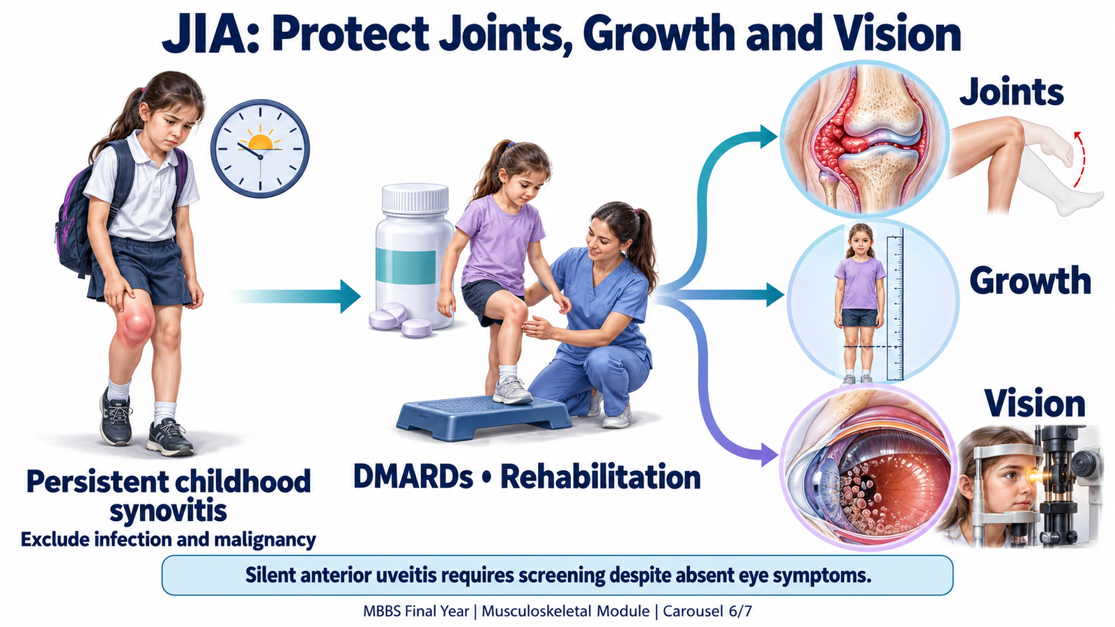
Do not wait for the six-week definition before referring a child with concerning persistent synovitis. Infection, malignancy and other serious causes must be assessed promptly. A toxic child with a very painful joint requires an infection-focused approach rather than an assumption of JIA.
Protect vision, growth and mobility
Chronic anterior uveitis may be silent, so the absence of a painful or red eye does not exclude eye inflammation. Ophthalmology screening with slit-lamp examination is therefore essential, with frequency determined by the child’s risk and specialist plan. Untreated uveitis can cause cataract, glaucoma and visual loss.
Persistent arthritis can cause contractures, muscle wasting, altered limb growth and reduced bone health. Systemic inflammation and prolonged glucocorticoid exposure may impair overall growth. Follow height, weight, joint movement, limb alignment and functional participation.
Systemic JIA can be complicated by macrophage activation syndrome, an uncontrolled inflammatory state. Persistent fever with rapid clinical deterioration, falling blood counts, liver dysfunction or coagulation abnormalities requires emergency specialist assessment.
Treatment must control disease and support development
Management is coordinated by pediatric rheumatology. The goals are to control inflammation, prevent damage and allow normal growth and participation. NSAIDs can relieve symptoms; selected joints may be treated with intra-articular glucocorticoids. Polyarticular disease commonly requires a DMARD such as methotrexate. Biologic therapy is considered when response is inadequate or the disease phenotype warrants it. Physiotherapy and occupational support preserve movement and everyday function. :chatgpt-content-reference{index=”2″}
Use systemic glucocorticoids selectively because growth, bone and infection risks matter particularly in children. Monitor treatment safety alongside clinical response. Encourage appropriate activity, with adaptations during flares rather than unnecessary prolonged inactivity.
Prognosis varies by subtype, disease activity and complications. Some children achieve sustained remission; others require continuing treatment into adulthood. Early control of inflammation and reliable eye screening improve the opportunity to preserve function and vision.
G. Counselling, Rehabilitation and Surgery for Persistent Disability
Inflammatory arthritis is a long-term condition whose treatment depends on partnership with the patient and family. Counselling should explain what the diagnosis means, why treatment is needed and how the person can remain active. Rehabilitation and, when necessary, surgery address limitations that medicines alone cannot fully reverse.
Counselling a patient with newly diagnosed rheumatoid arthritis
Explain that the immune system is driving inflammation and that the disease is not contagious or the patient’s fault. Discuss the potential for damage while emphasizing that early treatment can control inflammation and preserve function. Explore concerns about work, family responsibilities, pain, disability and medication risks before giving advice.
- Explain the treatment roles: pain relief acts quickly, while DMARDs control the underlying disease and may take longer to work. Improved pain alone does not prove that inflammation is controlled.
- Support safe adherence: clarify the prescribed schedule, monitoring appointments and what to do if adverse effects or infection occur. Reinforce weekly methotrexate administration when prescribed.
- Promote function: encourage suitable exercise, joint protection, rest during flares, smoking cessation and help with workplace or household adaptations.
- Discuss follow-up: assess symptoms, function and toxicity regularly. Review vaccination needs with the treating team before and during immunosuppression.
- Address pregnancy planning: some medicines require modification before conception. The patient should discuss plans early rather than stopping treatment without advice.
Seek prompt advice for fever, severe mouth ulcers, unusual bruising, new breathlessness, neurological symptoms or a painful red eye. Explain warning symptoms in language the patient understands and check understanding.
Counselling the child and parents after a JIA diagnosis
Speak directly to the child at an appropriate level and include parents without excluding the child’s voice. Explain that arthritis can affect children, is not contagious and is not usually caused by anything the family did. Discuss the uncertainty of the course honestly, alongside the realistic aim of maintaining normal activity and development.
Explain medication benefits, monitoring and the need for eye screening even when vision seems normal. Encourage school attendance and participation with practical adjustments, such as extra time for writing or temporary modification of sport. Address fears about growth, appearance, pain and future independence. Adolescents should gradually learn to manage appointments and medicines, with planned transition to adult services when needed.
Parents should report increasing limp or swelling, reduced movement, fever, visual symptoms or medicine-related concerns. A child who is acutely unwell or unable to bear weight requires urgent assessment.
When disabling rheumatoid arthritis needs surgical assessment
Medicines suppress inflammation, but established cartilage loss, tendon rupture or fixed deformity may leave pain and disability. Consider surgical referral for persistent pain, worsening function, progressive deformity or persistent localized synovitis despite appropriate nonsurgical treatment. Suspected tendon rupture, nerve compression or cervical instability may require earlier assessment.
- Rheumatic hand: tendon dysfunction, unstable joints and deformity may impair grip and dexterity. Selected procedures include synovectomy, tendon repair or reconstruction, joint replacement or fusion.
- Rheumatic foot: forefoot deformity, displaced toes and painful plantar pressure may impair walking and shoe wear. Footwear and orthoses are considered first where suitable; reconstructive surgery or fusion may help selected patients.
- Destroyed major joint: arthroplasty may relieve pain and restore useful movement. Arthrodesis stabilizes a joint by fusion when stability and pain relief are more valuable than preserving movement.
The operation is chosen according to pain, function, structural damage and the patient’s goals. Surgery does not cure systemic rheumatoid arthritis. Coordinate disease control, infection-risk assessment, perioperative medication planning and postoperative rehabilitation. Cervical disease must be considered before anesthesia when clinically indicated.

Important Comparison
The joint pattern must be interpreted alongside age, systemic findings and investigations. The following differences help organize the three principal diagnoses in this topic.
| Feature | Rheumatoid arthritis | Systemic lupus erythematosus | Polyarticular JIA |
|---|---|---|---|
| Typical context | Adult with persistent peripheral synovitis | Arthritis with possible multisystem disease | Child with persistent unexplained arthritis |
| Joint pattern | Commonly symmetrical small joints | Often symmetrical; usually nonerosive | Multiple small or large joints; symmetry varies |
| Helpful tests | RF and anti-CCP support the clinical diagnosis | ANA, anti-dsDNA, anti-Sm, complement and urine assessment | Clinical diagnosis; tests assess subtype, risk and alternatives |
| Major concerns | Erosions, tendon damage, disability and systemic complications | Renal, neurological, hematological or thrombotic disease | Growth disturbance, contractures and silent uveitis |
| Treatment principle | Early DMARD treatment and rehabilitation | Control inflammation according to organ severity | Control disease while protecting growth, vision and participation |
⭐ AIM High-Yield Review
- Arthritis requires evidence of inflammation; arthralgia means joint pain without definite inflammatory findings.
- Classify arthritis by joint number, peripheral or axial distribution, symmetry and time course.
- Persistent symmetrical wrist, MCP and PIP synovitis suggests rheumatoid arthritis; assess feet and daily function too.
- Pannus damages cartilage, bone and supporting tissues, linking persistent inflammation to deformity.
- RF and anti-CCP support rheumatoid arthritis, but negative tests do not exclude it.
- ANA positivity alone does not diagnose lupus; clinical features and targeted investigations determine its meaning.
- Proteinuria, hematuria or casts in suspected lupus require renal assessment, even if creatinine is normal.
- Lupus arthritis is usually nonerosive; organ involvement determines severity and treatment urgency.
- DMARDs modify disease progression. NSAIDs relieve symptoms without preventing structural damage.
- Methotrexate is usually prescribed weekly and requires laboratory monitoring and reproductive counselling.
- JIA begins before 16 years, persists for at least six weeks and requires exclusion of other causes.
- Children may present with limp or reduced activity; monitor growth, movement and school participation.
- JIA-associated uveitis can be silent, making planned ophthalmology screening essential.
- A hot painful joint with systemic illness requires urgent assessment for infection; known autoimmune disease does not exclude septic arthritis.
- Counselling and rehabilitation are integral to care. Surgery addresses selected painful or disabling structural problems but does not cure systemic rheumatoid arthritis.
🎥 AIM Video Learning — Rheumatoid Arthritis
Explore the causes, mechanism, clinical features, diagnosis and treatment of rheumatoid arthritis. Use this video alongside the Topic 1 learning material on adult and childhood inflammatory polyarthritis.
Source: Osmosis from Elsevier
Open video on YouTube