
This chapter connects the supplied KMU outcomes through recognition, investigation and treatment of a painful single joint. Understand the explanations first, especially how suspected infection changes urgency, then use the High-Yield Review for revision.
📖 AIM Learning Material
TOPIC 2. ACUTE MONOARTHRITIS AND INFECTIVE ARTHRITIS
Musculoskeletal Module — Final Year MBBS
Assess the painful single joint, recognize infection and connect diagnostic findings with medical treatment, drainage and recovery.
Topic Introduction
Monoarthritis means inflammation affecting one joint. A painful swollen knee or a child refusing to walk may result from infection, crystal deposition, trauma, bleeding or another inflammatory process. These conditions can look similar initially, but their consequences and treatment differ. Acute bacterial infection can rapidly damage cartilage and cause systemic illness, so it must be assessed promptly. Tuberculous arthritis generally develops more slowly and requires a different diagnostic and treatment approach. This chapter follows one clinical journey: establish whether the problem is within the joint, identify danger, interpret appropriate investigations and choose treatment. Adult and childhood differences are included where they change recognition, diagnosis or management.
A. Understanding Monoarthritis and Its Important Causes
A synovial joint contains cartilage-covered bone ends, a capsule and a synovial lining. Inflammation of the lining increases blood flow and fluid production. The resulting warmth, effusion and capsular distension cause pain and restricted movement. A deep joint such as the hip may be inflamed without visible external swelling. First distinguish a joint problem from pain arising in a tendon, bursa, muscle or adjacent bone. Intra-articular disease commonly restricts both active and passive movement. A periarticular problem may produce more localized tenderness or pain during a particular resisted movement.
A focused adult differential
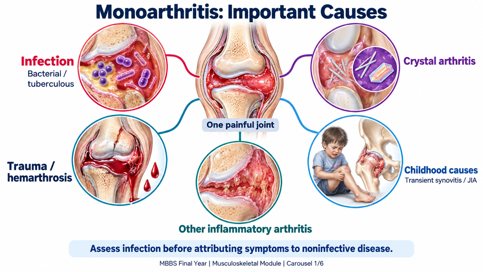
The time course, risk context and examination guide the differential. The important causes can be organized into five groups:
- Infection: acute bacterial septic arthritis causes a painful inflamed joint and may progress rapidly. Tuberculosis is an important cause of persistent, gradually progressive monoarthritis.
- Crystal arthritis: gout results from monosodium urate crystals; calcium pyrophosphate deposition can also cause an acute inflammatory attack. Either can closely resemble infection.
- Trauma or mechanical injury: a clear injury, bruising or mechanical locking suggests fracture, ligament injury or internal joint damage.
- Hemarthrosis: bleeding into a joint may follow trauma or occur with a bleeding disorder or anticoagulant treatment. Blood distends the capsule and causes painful swelling.
- Other inflammatory disease: reactive arthritis or an initial presentation of another inflammatory arthritis may begin in one joint. Persistent unexplained synovitis requires reassessment rather than repeated symptomatic treatment.
How the differential changes in children
In children, prioritize septic arthritis, adjacent osteomyelitis, trauma, transient synovitis and juvenile idiopathic arthritis. Hemarthrosis is important when there is a bleeding history. Osteomyelitis affects bone but may mimic arthritis or coexist with it. Transient synovitis is a self-limited inflammatory condition, particularly of the hip. A child may limp after a recent viral illness while remaining relatively well. This diagnosis should follow appropriate assessment for infection, not replace it. Juvenile idiopathic arthritis becomes a consideration when arthritis persists and other causes have been excluded; swelling or stiffness may be more prominent than pain.
Central distinction: an acutely inflamed joint needs an infection-focused assessment. Neither a history of gout nor an existing autoimmune diagnosis makes a new episode automatically noninfective.

B. Clinical Assessment and Recognition of Urgent Disease
Assessment must answer three questions: is the problem within the joint, how likely is infection, and is the patient systemically unstable? The severity of pain and loss of movement matter, but no single symptom or sign reliably excludes septic arthritis.
Focused history and risk context
Establish onset, speed of progression, previous episodes and the ability to use the limb. Sudden severe symptoms suggest bacterial infection, crystals, injury or bleeding. Progressive symptoms over weeks or months raise concern for tuberculosis or another chronic process.
- Infection clues: fever, rigors, recent skin infection, bacteremia, an open wound, joint injection or surgery increase concern.
- Host risk: diabetes, immunosuppression, pre-existing joint damage and injecting drug use can increase susceptibility to bacterial infection.
- Crystal clues: previous similar attacks, known gout or characteristic previous joint involvement support crystal disease without excluding infection.
- Bleeding or injury clues: trauma, anticoagulants, easy bruising or an established bleeding disorder redirect assessment.
- TB clues: previous tuberculosis, household exposure, immunosuppression, weight loss or night sweats support suspicion. Their absence does not exclude joint TB.
- Relevant exposures: obtain a sensitive sexual history when gonococcal infection is a realistic possibility.
What to examine and why
Assess vital signs, mental state, hydration and peripheral perfusion before concentrating on the joint. Fever, tachycardia, hypotension or altered consciousness may indicate systemic infection. Compare the affected and opposite joint for warmth, effusion, tenderness and movement. Pain through passive movement suggests intra-articular inflammation. Examine the surrounding skin for wounds or an infectious source and adjacent bone for focal tenderness. Avoid repeatedly forcing movement of an intensely painful joint. Knee disease may produce a visible effusion, warmth and flexed resting posture. Hip disease may produce groin or thigh pain, a limp and markedly painful rotation without obvious swelling. The hip may be held flexed, abducted and externally rotated because this position reduces capsular tension. Hip pain can be referred to the knee.
Childhood presentation
Young children may present with refusal to walk, reduced play or distress during dressing rather than a clear description of joint pain. Infants may stop moving a limb because movement hurts; this is pseudoparalysis. Observe spontaneous movement and gait when appropriate, then assess joints gently. A child with bacterial infection may be febrile and very unwell, but some infections in young children produce less dramatic fever and inflammatory findings. A relatively mild presentation therefore needs interpretation alongside examination and risk.

C. Investigations and Interpretation of Joint Findings
Investigations should identify the cause, assess severity and guide treatment. In suspected septic arthritis, joint sampling is central because examination and blood tests alone cannot reliably distinguish bacterial infection from other inflammation.
Blood tests: support and limitations
- Complete blood count: neutrophilia may support bacterial infection, but a normal count does not exclude it. Unexpected cytopenias, particularly with childhood bruising or night pain, require consideration of marrow disease.
- CRP and ESR: indicate inflammation and provide a baseline for follow-up. Raised values are nonspecific; normal values, especially early in illness, do not securely exclude infection.
- Blood cultures: may identify a bloodstream organism. Obtain them before antibiotics when feasible, particularly with fever or suspected systemic infection.
- Renal and liver function: assess systemic illness and help select and monitor medicines.
- Serum urate: supports assessment of gout in context, but a normal value during an attack does not exclude gout, and a raised value alone does not establish the cause of acute swelling.
- Coagulation studies: are selected when bleeding or anticoagulant-associated hemarthrosis is suspected.
Joint aspiration: what the sample tells you
Send synovial fluid for microbiological testing, leukocyte assessment and crystal analysis. The clinical question determines additional tests. A deep hip aspiration generally requires image guidance and an appropriate specialist setting.
- Appearance: cloudy or purulent fluid raises concern for infection; bloody fluid suggests bleeding but must be interpreted with the aspiration circumstances.
- Leukocytes: a high count with neutrophil predominance supports substantial inflammation. There is overlap between bacterial and crystal arthritis, so no cell-count threshold should be used alone to exclude infection.
- Gram stain: a positive finding can guide early antibiotic selection. A negative stain does not exclude infection.
- Culture and susceptibility: identify the organism and help narrow treatment. Prior antibiotics can reduce yield; negative culture does not automatically exclude infection.
- Polarized microscopy: needle-shaped, negatively birefringent monosodium urate crystals support gout. Rhomboid-shaped, weakly positively birefringent calcium pyrophosphate crystals support CPP crystal arthritis.
Interpretation trap: crystals and infection can coexist. Continue appropriate microbiological assessment when the presentation remains concerning.
Choosing imaging
Plain radiographs assess fracture, baseline structural disease and later destructive changes. An early normal film does not exclude septic arthritis. Ultrasound is particularly useful for detecting a hip effusion and guiding aspiration. It shows fluid but does not determine whether that fluid is bacterial, transient or otherwise inflammatory. MRI is selected when adjacent osteomyelitis, a deep abscess or the extent of infection needs clarification. It should not delay treatment in an unstable patient. For the child in the Clinical Bridge, ultrasound can establish whether the hip contains an effusion. Clinical assessment, blood investigations and specialist-directed sampling are then needed to determine its cause.
Sampling and antibiotic timing
In a clinically stable patient, obtain joint fluid promptly before antibiotics when this can be done safely. In sepsis or rapid deterioration, collect blood cultures if feasible and start treatment without waiting for delayed aspiration; obtain joint samples as soon as possible. :chatgpt-content-reference{index=”0″}

D. Septic Arthritis: Mechanism, Medical Treatment and Drainage
Septic arthritis is infection within a joint, most commonly caused by bacteria. Organisms may arrive through the bloodstream, enter through a wound or procedure, or spread from nearby infected tissue. The synovium is highly vascular, allowing bloodborne organisms to reach the joint.
Why the joint can deteriorate rapidly
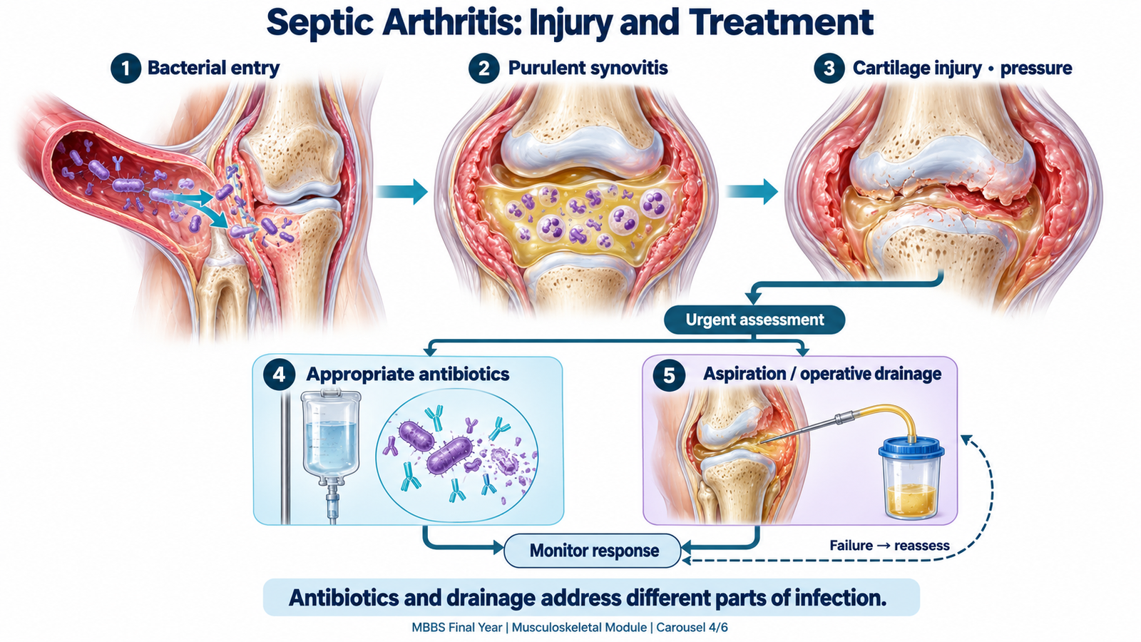
Bacterial growth triggers intense neutrophilic inflammation. Bacterial products and inflammatory enzymes damage cartilage, while pus and increased pressure cause pain and impair movement. In the hip, increased pressure can also threaten blood supply, particularly in children.
Bacterial entry → acute synovitis and pus → cartilage injury and increased pressure → pain, movement loss and possible permanent damage.
Staphylococcus aureus is a major pathogen across age groups. Streptococci and selected Gram-negative organisms are also important according to exposure and host risk. Gonococcal infection is considered in an appropriate sexual context. In young children, Kingella kingae is an important pathogen and may produce relatively subtle illness; antimicrobial selection must take age into account.
Immediate medical priorities
Arrange hospital assessment and urgent orthopedic input. Provide analgesia and temporarily protect the painful joint. If systemic instability is present, assess airway, breathing and circulation, provide appropriate oxygen and fluids, and escalate care while treating the infection. Empirical antibiotics should cover likely organisms, particularly staphylococci, with resistance patterns, age, allergies and renal function guiding selection. An antistaphylococcal beta-lactam may be appropriate when susceptible organisms are expected; MRSA risk or Gram-negative risk requires different or additional coverage. Examples of empirical IV antibiotics in adults include cloxacillin or cefazolin for staphylococcal coverage, with ceftriaxone added when Gram-negative coverage is indicated. Vancomycin may be required when MRSA is suspected. Adjust according to local resistance patterns and culture results. Narrow treatment when microbiological results become available. Initial intravenous treatment is appropriate for serious bacterial joint infection. Transition to oral therapy and total duration depend on clinical response, organism, drainage and whether adjacent bone is involved. Pediatric prescribing requires weight-based selection by the treating team; a single adult regimen should not be applied to every child.
Why drainage is part of treatment
Antibiotics treat the organism, but an infected collection also needs source control: removal of pus and, where necessary, infected or devitalized tissue. This reduces pressure and inflammatory burden.
- Aspiration: provides diagnostic material and may contribute to drainage in selected joints.
- Arthroscopic washout: permits lavage and assessment of the joint through small access points.
- Open drainage: may be needed according to the joint, disease extent, associated abscess or inadequate response to less invasive treatment.
The hip is a deep joint requiring prompt specialist-directed drainage planning; repeated bedside aspiration is not a universal substitute. The knee may be accessible to aspiration, but persistent infection or inadequate clearance can require operative washout.
Assessing response and treatment failure
Improvement should include less pain, reduced fever, better movement or weight-bearing and a falling inflammatory trend. Persistent fever, worsening pain, continued effusion or failure of markers to improve warrants reassessment for inadequate drainage, an abscess, adjacent osteomyelitis or ineffective antimicrobial coverage. Complications include cartilage destruction, secondary degenerative change, stiffness, osteomyelitis and systemic sepsis. Children may develop growth disturbance or damage to the femoral head after hip infection. Early source control and effective treatment aim to prevent these outcomes.
E. Tuberculous Arthritis of the Hip and Knee
Tuberculous arthritis is joint infection caused by Mycobacterium tuberculosis. It commonly results from hematogenous spread, with infection involving synovium or adjacent bone. Unlike typical acute pyogenic infection, it generally produces slowly progressive disease.
Mechanism and clinical presentation
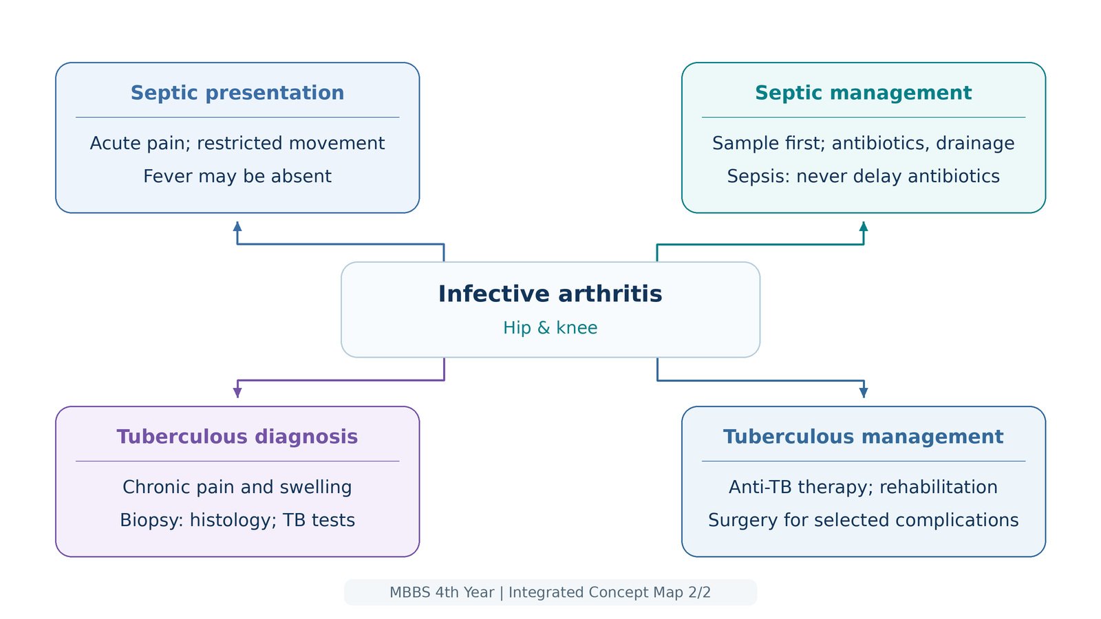
The host response forms granulomatous inflammation, often with caseous necrosis. Persistent synovial and bone disease gradually damages the joint. Pain, stiffness and restricted function increase over time rather than necessarily presenting as an intensely hot joint. Patients may have chronic swelling, muscle wasting, a limp or progressive contracture. Weight loss, night sweats or fever may occur, but constitutional symptoms and pulmonary symptoms may be absent. An abscess can develop with relatively little overlying warmth, traditionally termed a cold abscess; a draining sinus may appear in advanced disease.
- Hip TB: groin or referred knee pain, reduced rotation, limp and later deformity or shortening may occur. External swelling can be difficult to detect.
- Knee TB: persistent swelling, effusion, restricted movement and quadriceps wasting are important clues. Later disease may produce flexion deformity.
Consider TB when monoarthritis is prolonged, exposure or host risk is relevant, or an apparently routine inflammatory presentation does not improve as expected.
Diagnostic approach
Radiographs may show juxta-articular osteopenia, peripheral erosions and gradual joint-space narrowing, known as Phemister’s triad. These findings suggest a chronic destructive process but are not independently diagnostic. Ultrasound can identify an effusion; MRI can define synovial disease, bone involvement or an abscess. Assess for TB elsewhere when clinically indicated. Confirmation relies on appropriate joint fluid or tissue investigation. Synovial biopsy may provide material for histology, mycobacterial culture and molecular testing. Granulomas with caseation support TB, while organism identification strengthens confirmation. Joint-fluid acid-fast smears have limited sensitivity; a negative smear cannot exclude the disease. :chatgpt-content-reference{index=”2″} A tuberculin skin test or interferon-gamma release assay may indicate TB infection but does not prove that TB is causing the joint disease. Interpret these tests with the presentation and specimen findings.
Medical treatment
Combination antituberculosis treatment is the foundation of care. For drug-susceptible disease, treatment commonly begins with isoniazid, rifampicin, pyrazinamide and ethambutol, followed by a continuation phase selected by the treating TB team. A standard drug-susceptible TB regimen is 2 months of HRZE followed by 4 months of HR (2HRZE/4HR). Osteoarticular tuberculosis may require extended treatment of 9–12 months according to clinical response and specialist recommendations. Using several active drugs improves organism clearance and reduces selection of resistance. Isoniazid interferes with mycolic-acid synthesis; rifampicin inhibits bacterial RNA synthesis; ethambutol disrupts cell-wall formation. The prolonged course must be completed even if pain improves early. Regimen and duration depend on susceptibility, bone or joint involvement and the applicable TB treatment pathway. Monitor adherence and toxicity, including liver injury, isoniazid-associated neuropathy and ethambutol-associated visual problems. Pyridoxine is used when indicated to reduce neuropathy risk.
The role of surgery
Not every patient needs an operation. Procedures may be required to obtain diagnostic tissue, drain a significant abscess, remove devitalized tissue or address inadequate response despite appropriate medical treatment. Advanced joint destruction may require later reconstructive or salvage surgery, such as joint replacement or fusion. The choice and timing depend on infection control, structural damage and functional goals. Surgery complements antituberculous treatment; it does not replace it. Follow pain, swelling, movement, weight-bearing and inflammatory trends. Rehabilitation protects function, while persistent or worsening symptoms prompt assessment of adherence, resistance, an alternative diagnosis or a complication.

F. Noninfective Management, Childhood Decisions and Recovery
Once the cause is established, management should target that cause rather than treat every swollen joint identically. Relief of pain is important, but response to an analgesic does not prove that infection has been excluded.
Adult noninfective monoarthritis
Crystal arthritis: anti-inflammatory treatment may use an NSAID, colchicine or a glucocorticoid according to renal function, gastrointestinal risk, other medicines and diagnostic certainty. NSAIDs reduce prostaglandin-mediated symptoms; colchicine disrupts microtubule-dependent inflammatory-cell activity. Suspected infection must be assessed before using joint-directed steroid treatment. Acute symptom control and long-term prevention are separate decisions. After the attack, assess recurrent disease and contributing factors; selected patients with gout need a urate-lowering strategy. Do not interpret pain relief as removal of the underlying crystal burden. Traumatic disease: protect the limb, provide analgesia and investigate suspected fracture or significant internal injury. Hemarthrosis: address the bleeding cause, such as a factor deficiency or anticoagulant-associated problem, with appropriate specialist input. Joint procedures require particular care when hemostasis is impaired. Persistent inflammatory monoarthritis requires reassessment and, where appropriate, rheumatology referral for disease-directed treatment rather than repeated empirical anti-inflammatory courses.
Childhood decisions
A child with suspected bacterial arthritis needs coordinated pediatric and orthopedic care. Investigate for adjacent osteomyelitis when bone tenderness, persistent symptoms or imaging findings suggest it. Young children may need molecular testing to identify organisms that are difficult to recover on routine culture. A well child with a compatible presentation may be managed for transient synovitis with rest from painful activity, suitable analgesia and planned review after infection has been appropriately assessed. Worsening pain, fever, inability to walk or failure to improve requires reassessment. Persistent swelling or morning stiffness raises concern for JIA, but the duration should not become a reason to postpone evaluation. Bruising, pallor, night pain or unexpected cytopenias require assessment for a serious alternative such as leukemia.
Recovery and follow-up
Brief protection of a painful joint can help initially, but prolonged unnecessary immobilization promotes stiffness and muscle wasting. Introduce movement and progressive function when infection is controlled and the treating team considers it safe. Review symptoms, joint movement, weight-bearing and treatment tolerance. Children need attention to growth and limb development; hip infection may require longer orthopedic surveillance because growth or vascular injury can become apparent later. Explain the diagnosis, medication plan and review arrangements to the patient or parents. New fever, recurrent swelling, worsening pain or reduced function should prompt reassessment. For TB, emphasize completion of the full course and reporting toxicity or failure to improve.

Important Comparison
Acute pyogenic and tuberculous arthritis both threaten joint function, but their time course and diagnostic pathways differ.
| Feature | Acute pyogenic arthritis | Tuberculous arthritis |
|---|---|---|
| Course | Rapid onset and progression | Gradual, persistent progression |
| Presentation | Severe pain, warmth and movement restriction | Chronic swelling, stiffness and muscle wasting |
| Imaging clue | Early films may be normal | Osteopenia, peripheral erosions, gradual narrowing |
| Diagnostic material | Joint fluid for bacterial testing | Fluid or tissue for mycobacterial tests and histology |
| Treatment | Prompt antibiotics and appropriate drainage | Combination TB therapy; surgery for selected indications |
⭐ AIM High-Yield Review
- Monoarthritis is a presentation; establish its cause before choosing definitive treatment.
- Septic arthritis can rapidly damage cartilage and requires prompt assessment and source control.
- Absence of fever or normal inflammatory markers does not securely exclude infection.
- Hip disease may cause knee pain without visible hip swelling.
- An infant avoiding limb movement may have pain-related pseudoparalysis.
- Ultrasound detects an effusion; it does not establish whether the fluid is infected.
- Joint aspiration supports microbiological diagnosis and crystal identification.
- Negative Gram stain or culture does not independently exclude septic arthritis.
- Crystals can coexist with infection; serum urate alone cannot diagnose an acute attack.
- Obtain samples promptly before antibiotics when safe; do not delay treatment in sepsis.
- Persistent illness after treatment suggests inadequate source control, an unsuitable antibiotic or adjacent infection.
- TB should be considered in chronic unexplained hip or knee monoarthritis, even without constitutional symptoms.
- Phemister’s triad suggests joint TB but requires clinical and specimen-based interpretation.
- Combination antituberculous treatment is central; selected surgery addresses diagnosis, collections or structural complications.
- Childhood follow-up must protect movement, growth and long-term joint function.
Topic 2: Acute Monoarthritis and Infective Arthritis
Video focus: Septic arthritis—causes, pathophysiology and treatment. Use alongside the topic learning material for the complete differential diagnosis, childhood monoarthritis and tuberculous arthritis.
Source: Armando Hasudungan
Watch on YouTube if the embedded player is unavailable