
This chapter follows the KMU outcomes for oligoarticular and axial inflammatory arthritis. First understand the pattern of joint involvement, then connect that pattern with the clinical clues, investigations and management; use the High-Yield Review at the end for revision.
Topic 3. Oligoarticular and Axial Inflammatory Arthritis
Musculoskeletal Module • A clinical approach to adult and childhood oligoarthritis, ankylosing spondylitis, reactive arthritis and psoriatic arthritis.
Topic Introduction
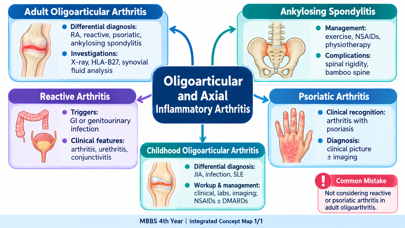
Oligoarthritis means inflammation affecting a small number of joints, while axial inflammatory arthritis predominantly involves the sacroiliac joints and spine. The pattern itself is an important diagnostic clue: an adult with asymmetric lower-limb arthritis, inflammatory back pain, psoriasis, or a recent gastrointestinal or genitourinary infection requires a different approach from a patient with symmetrical polyarthritis. In children, persistent oligoarthritis raises the possibility of juvenile idiopathic arthritis but infection and other important causes must first be considered. This chapter builds a practical approach from recognition of the joint pattern to focused investigations and management, then examines ankylosing spondylitis, reactive arthritis and psoriatic arthritis in the clinical contexts in which students are likely to encounter them.
A. Recognizing and Approaching Oligoarticular Arthritis
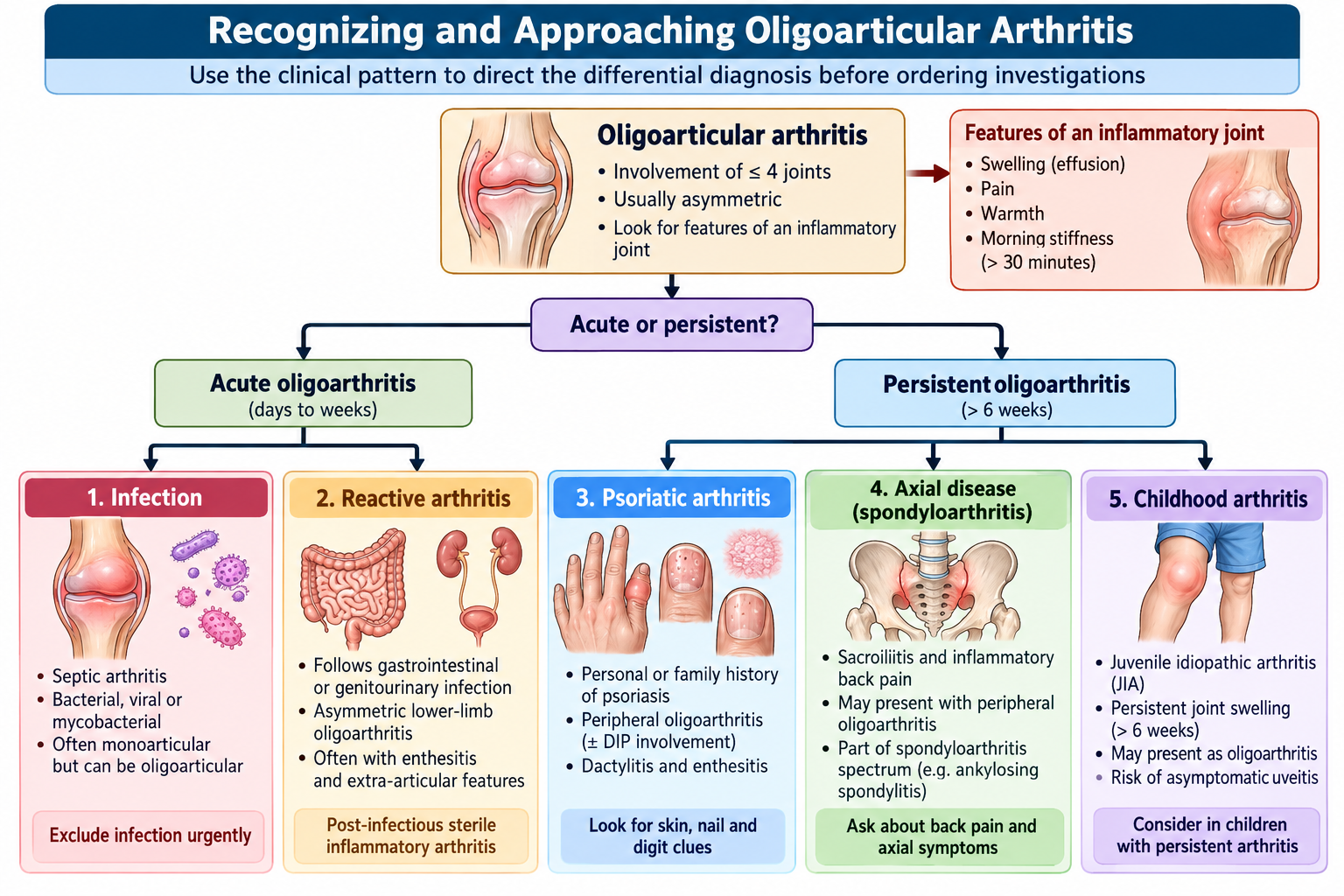
Oligoarticular arthritis is best approached as a clinical pattern rather than a single disease. The first task is to confirm that the symptoms arise from an inflamed joint and then use the distribution, duration and associated features to narrow the differential diagnosis. A swollen knee and ankle after an infection, for example, carries different diagnostic implications from persistent small-joint disease or predominantly spinal symptoms.
What makes a joint inflammatory?
An inflammatory joint commonly produces pain at rest, prolonged morning stiffness, swelling, warmth and restriction of movement. Symptoms may improve somewhat with movement rather than becoming progressively worse with use. In contrast, mechanical disorders tend to produce activity-related pain and shorter periods of stiffness.
Build the differential from the pattern
In an adult with oligoarthritis, several clues should be considered together rather than relying on a single laboratory test:
- Acute, very painful swollen joint with systemic illness: septic arthritis must be excluded urgently.
- Sudden episodic inflammation: crystal arthritis becomes an important possibility, particularly in an appropriate adult risk context.
- Recent gastrointestinal or genitourinary illness: consider reactive arthritis.
- Psoriasis, nail changes or characteristic digit involvement: consider psoriatic arthritis.
- Inflammatory back pain or sacroiliac symptoms: consider axial spondyloarthritis, including ankylosing spondylitis.
- Persistent arthritis in a child: consider juvenile idiopathic arthritis after excluding alternative causes.
Focused history and examination
The history should establish the number and distribution of joints, onset, duration, morning stiffness and previous episodes. Ask specifically about back pain, psoriasis, nail disease, eye symptoms, recent diarrhea or genitourinary symptoms, fever and systemic illness. These associated features can transform an apparently isolated joint problem into a recognizable inflammatory syndrome.
Examination should confirm true synovitis and document which joints are involved. Look beyond the painful joint for skin and nail psoriasis, enthesitis, dactylitis, limitation of spinal movement, sacroiliac features and evidence of extra-articular disease.
B. Investigation and Management of Adult Oligoarticular Arthritis
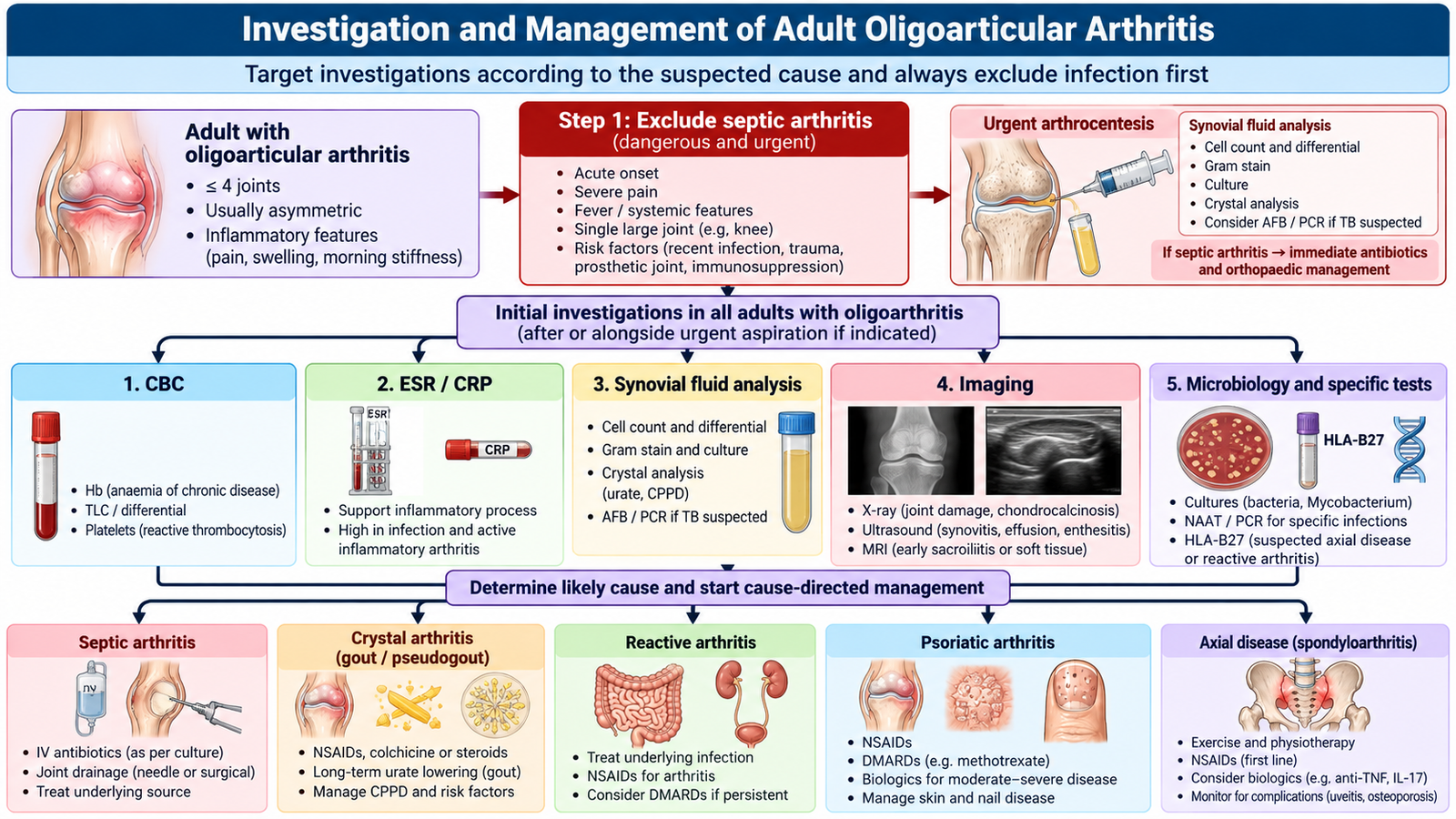
Investigations should answer a clinical question. They are not ordered as a fixed “arthritis panel.” The most important question in an acutely inflamed joint is whether infection or crystal disease must be excluded; in persistent inflammatory disease, testing is directed by the suspected rheumatological pattern.
Investigations and their interpretation
- CBC: leukocytosis may support infection or systemic inflammation, but is not diagnostic by itself.
- ESR and CRP: indicate inflammatory activity but do not identify the specific cause.
- Synovial fluid examination: becomes particularly important when septic or crystal arthritis is suspected; the sample can be assessed for inflammatory characteristics, organisms and crystals as clinically appropriate.
- Plain radiographs: help assess structural damage and may provide baseline information in persistent disease.
- Ultrasound: can demonstrate synovitis or effusion and may help when a clinically accessible fluid collection requires assessment.
- Targeted microbiological testing: is selected when the history suggests an infectious trigger or direct joint infection.
- HLA-B27: may support the assessment of a suspected spondyloarthritis phenotype but does not independently establish the diagnosis.
A positive HLA-B27 result is a supporting clue in the correct clinical setting. It should not replace clinical assessment or imaging when axial inflammatory disease is suspected.
Management follows the cause
There is no single treatment for “oligoarthritis.” Management starts by identifying conditions that require urgent treatment and then treating the underlying inflammatory disorder.
- Possible septic arthritis: requires urgent diagnostic assessment and appropriate antimicrobial and joint-management principles rather than routine anti-inflammatory treatment alone. Suspected septic arthritis requires urgent joint aspiration for microscopy, culture and sensitivity, preferably before antibiotics if the patient is stable. Start prompt empirical intravenous antibiotics and arrange joint drainage or washout when indicated. Do not delay antibiotics in sepsis or an unstable patient.
- Inflammatory spondyloarthritis pattern: treatment combines appropriate anti-inflammatory therapy with maintenance of mobility and function.
- Reactive arthritis: treatment is directed toward symptomatic arthritis and any clinically relevant active infection; the triggering infection and the sterile joint inflammation are conceptually different.
- Psoriatic arthritis: persistent inflammatory disease requires rheumatological assessment and disease-modifying treatment according to disease pattern and severity.
- Persistent or diagnostically uncertain disease: reassess the pattern and consider specialist evaluation rather than repeatedly treating undifferentiated joint pain.
C. Ankylosing Spondylitis: Axial Disease, Management and Complications
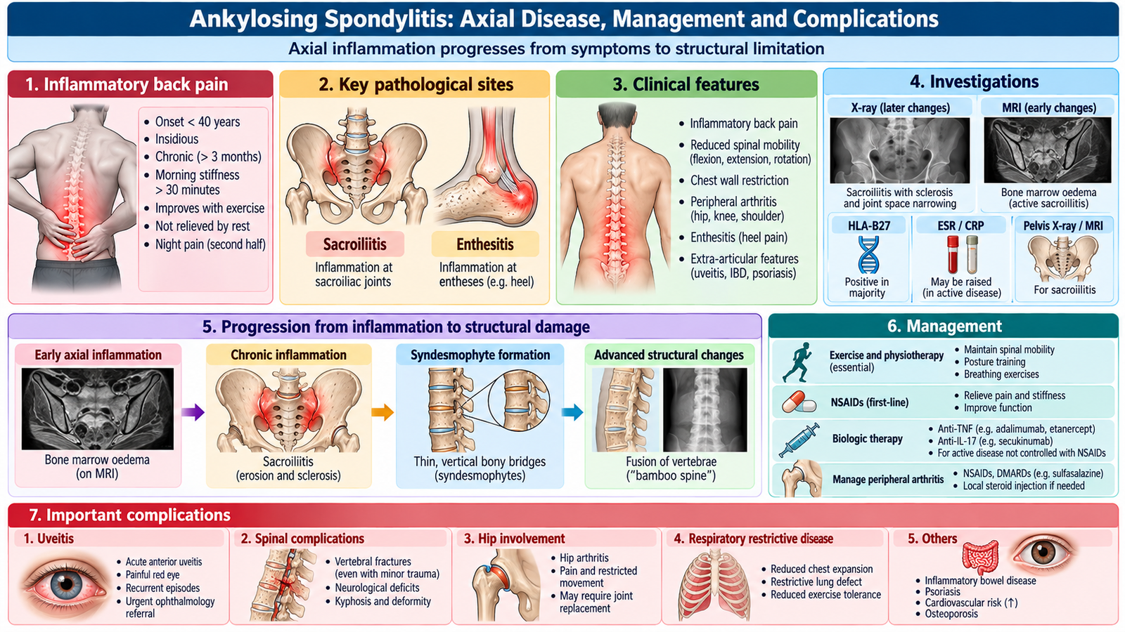
Ankylosing spondylitis is a chronic inflammatory disease within the axial spondyloarthritis spectrum. The principal sites are the sacroiliac joints and spine. Inflammation at entheses and axial joints produces pain and stiffness; persistent inflammation can eventually lead to structural change and reduced spinal mobility.
Recognizing the axial inflammatory pattern
The characteristic clinical problem is inflammatory back pain. It tends to begin in a younger patient, develops gradually, is associated with morning stiffness, improves with activity and is not simply relieved by prolonged rest. Sacroiliac involvement may produce buttock-region pain. Peripheral arthritis or enthesitis may coexist.
Examination focuses on spinal mobility, posture, chest expansion and peripheral manifestations where present. The important educational principle is to connect limitation of movement with the underlying axial inflammatory process rather than viewing back pain as a purely mechanical complaint.
Diagnosis
The diagnosis is based on the clinical pattern together with appropriate imaging and supportive investigations. Inflammatory markers may reflect disease activity but can be normal. Imaging of the sacroiliac joints is central to identifying sacroiliitis and structural disease. HLA-B27 supports the diagnosis in the correct clinical setting but is neither necessary nor sufficient on its own.
Management
Management aims to control inflammation while preserving posture, spinal mobility and function. Because immobility contributes to disability, exercise and physical therapy are central rather than optional additions.
- Education and regular exercise: encourage spinal mobility, posture and long-term physical function.
- Physiotherapy: supports posture, flexibility and chest movement.
- NSAIDs: are an important pharmacological option for symptomatic axial pain and stiffness when appropriate.
- Persistent active disease: specialist assessment may lead to biologic disease-modifying therapy when standard measures are inadequate.
- Monitoring: includes symptoms, function, mobility, treatment effects and important extra-articular manifestations.
Important complications
Chronic axial disease can cause marked stiffness, deformity and functional limitation. A rigid ankylosed spine is also more vulnerable to serious spinal injury. Important extra-articular complications include acute anterior uveitis. Cardiovascular and pulmonary involvement can occur in established disease, while reduced mobility and chronic inflammation may contribute to loss of bone strength.
A patient with an ankylosed, rigid spine who develops significant new spinal pain after trauma requires careful assessment because spinal fractures can occur and may be unstable.
🩺 AIM Clinical Bridge
A 25-year-old man reports several months of lower-back and alternating buttock pain. His stiffness is worst after waking and improves when he becomes active. He has no history of major trauma, and examination suggests reduced spinal mobility.
- Does this sound inflammatory or mechanical?
- Which anatomical region is particularly important to assess?
- Which complications and extra-articular features should you ask about?
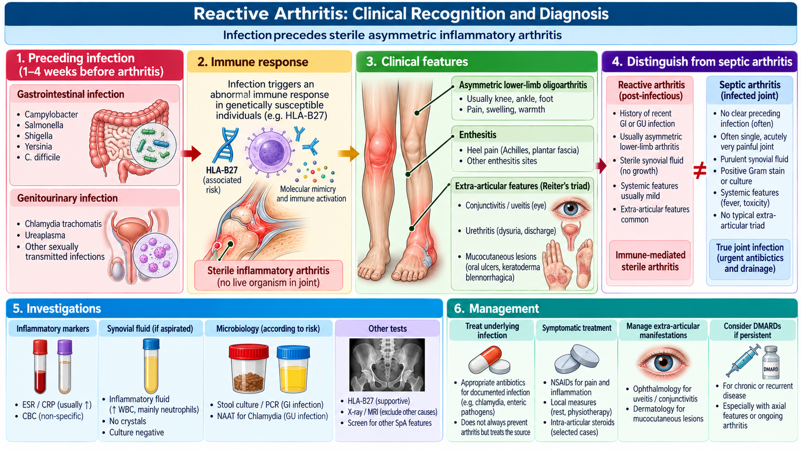
D. Reactive Arthritis: Clinical Recognition and Diagnosis
Reactive arthritis is an inflammatory arthritis occurring after an infection elsewhere in the body. The joint itself is typically not infected by the triggering organism. This distinction is fundamental: reactive arthritis is a post-infectious inflammatory syndrome, whereas septic arthritis represents direct infection within the joint.
How does the patient present?
The typical pattern is an acute asymmetric oligoarthritis predominantly involving the lower limbs following a gastrointestinal or genitourinary infection. Enthesitis may occur, particularly around the heel, and some patients develop axial symptoms.
Associated manifestations may provide the diagnostic clue. These include preceding diarrhea or genitourinary symptoms, eye inflammation, mucocutaneous manifestations and urethral symptoms. Not every patient develops the complete traditional combination, so diagnosis should be based on the overall clinical pattern rather than waiting for a classic triad.
Diagnostic approach
The diagnosis is primarily clinical: establish the characteristic arthritis pattern and seek evidence of a relevant preceding infection. Inflammatory markers may support the presence of inflammation. Tests for a suspected triggering infection should be guided by the history. HLA-B27 can be associated with the spondyloarthritis spectrum but does not diagnose reactive arthritis.
Reactive arthritis: infection precedes the sterile inflammatory arthritis. Septic arthritis: the joint itself is infected and requires urgent management.
E. Psoriatic Arthritis: Recognizing the Pattern
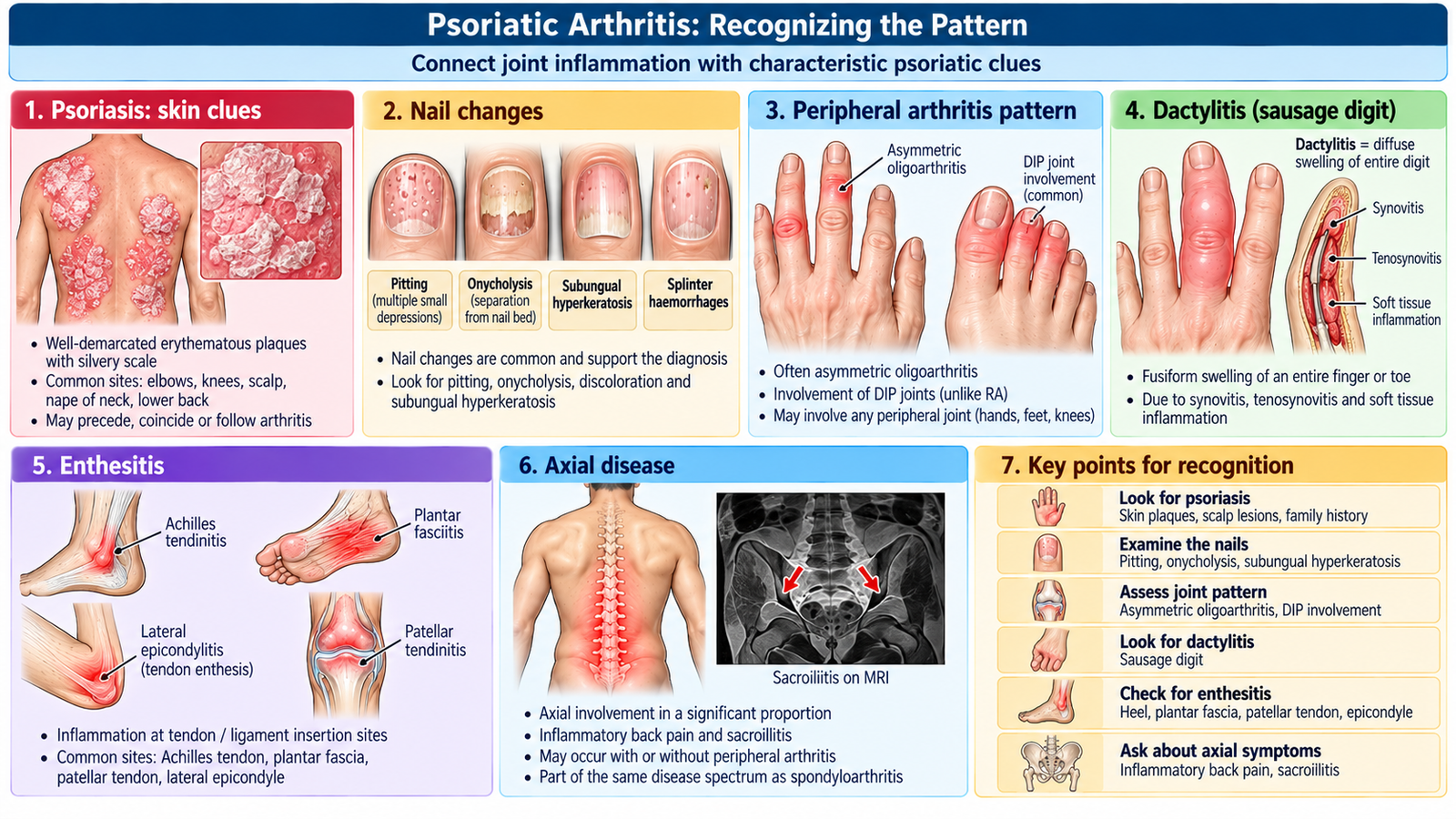
Psoriatic arthritis is an inflammatory musculoskeletal disease associated with psoriasis. Its presentation is variable: the key to recognition is to connect the joint pattern with skin, nail, digit, entheseal or axial clues. Arthritis may occasionally draw clinical attention before obvious psoriasis is recognized, so a careful skin and nail examination matters.
Clinical clues
- Peripheral inflammatory arthritis: may be oligoarticular and asymmetric.
- Dactylitis: diffuse inflammation of an entire digit produces the characteristic “sausage digit” appearance.
- Enthesitis: inflammation where tendons or ligaments insert into bone is an important spondyloarthritis feature.
- Distal interphalangeal joint involvement: particularly suggestive when associated with psoriatic nail disease.
- Nail changes: pitting and onycholysis provide useful diagnostic support.
- Axial disease: sacroiliac or spinal involvement may occur.
- Skin psoriasis: examine beyond obvious exposed sites when clinical suspicion is high.
Diagnosis
Psoriatic arthritis is diagnosed clinically by identifying inflammatory musculoskeletal disease in a patient with appropriate psoriatic features while excluding important alternatives. There is no single blood test that confirms it. Inflammatory markers may support active inflammation, while imaging can demonstrate inflammatory or structural changes according to the site and duration of disease.
Rheumatoid factor and anti-CCP antibodies are more strongly associated with rheumatoid arthritis and may assist when the clinical distinction is difficult, but the overall pattern remains central to diagnosis.
Inflammatory arthritis + psoriasis or nail disease + dactylitis/enthesitis strongly redirects the diagnostic reasoning toward psoriatic arthritis.
F. Childhood Oligoarticular Arthritis: Diagnostic Workup and Management
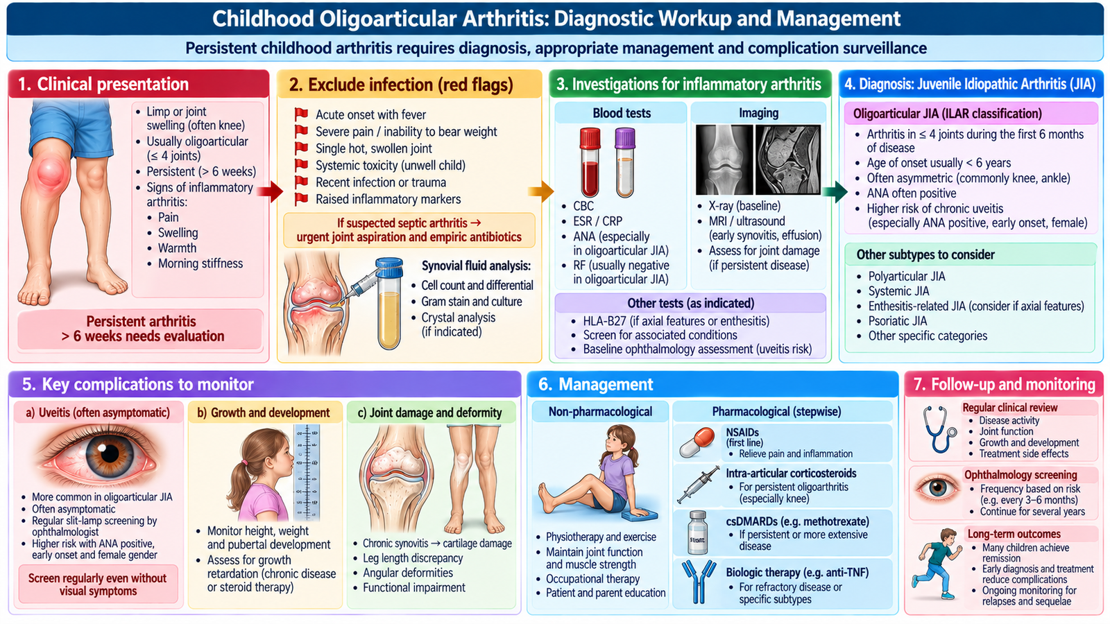
Persistent oligoarthritis in a child requires an age-appropriate approach. Juvenile idiopathic arthritis is an important cause, but it is a diagnosis reached after considering other explanations for childhood arthritis. The clinical priority is therefore to confirm arthritis, determine its pattern and duration, identify systemic or infectious warning features, and look for complications that may not initially produce symptoms.
Important differential diagnosis
The differential should remain focused. Important alternatives include infection, trauma or orthopedic disease, reactive/post-infectious arthritis and other inflammatory disorders. Severe acute pain, fever, systemic illness or marked restriction of movement should raise particular concern for infection.
Clinical assessment
History should establish onset, duration, number of joints, morning stiffness, functional change and systemic symptoms. In younger children, arthritis may present indirectly as limping, reluctance to walk or reduced use of a limb rather than a clear complaint of joint pain.
Examination identifies swelling, warmth, restriction of movement and functional limitation while also assessing growth, gait and other joints. The child should be assessed as a whole rather than examining only the visibly swollen joint.
Diagnostic workup
- CBC and inflammatory markers: help identify systemic inflammation and may reveal clues suggesting an alternative diagnosis.
- Joint aspiration: is important when septic arthritis is clinically suspected.
- Imaging: is selected according to the joint and clinical problem; ultrasound can help demonstrate synovitis or effusion.
- Autoimmune testing: should be interpreted in the clinical context rather than used as a screening substitute for history and examination.
- Eye assessment: is important in children with JIA because anterior uveitis can be clinically silent.
Management principles
Management depends on the identified cause. Infection requires urgent treatment. When juvenile idiopathic arthritis is established, the goals are to suppress inflammation, preserve joint movement and function, support normal activity and growth, and prevent complications.
Treatment may include anti-inflammatory therapy and, when disease persists or requires greater control, disease-modifying therapy under pediatric rheumatology care. Physiotherapy and maintenance of function are important. Follow-up must consider not only the joints but also growth, treatment effects and ocular complications. In oligoarticular JIA, initial treatment includes a short trial of an NSAID such as naproxen and/or intra-articular corticosteroid injection, preferably triamcinolone hexacetonide. Persistent active arthritis despite initial treatment generally requires methotrexate under pediatric rheumatology supervision. Routine systemic corticosteroids are avoided as initial treatment.
A child with an acutely painful swollen joint, fever or systemic illness must be assessed urgently for septic arthritis. Chronic inflammatory arthritis should not be assumed until dangerous alternatives have been considered.
Integrated Mechanism Flow
Important Comparison: Recognizing the Major Patterns
| Feature | Ankylosing spondylitis | Reactive arthritis | Psoriatic arthritis | Childhood oligoarthritis / JIA pattern |
|---|---|---|---|---|
| Dominant clue | Inflammatory axial symptoms | Recent infection | Psoriasis / nail disease | Persistent childhood arthritis |
| Typical pattern | Sacroiliac and spinal | Asymmetric lower-limb oligoarthritis | Peripheral and/or axial | Few persistently inflamed joints |
| Useful associated clue | Uveitis / enthesitis | Enthesitis / ocular or mucocutaneous features | Dactylitis / nail pitting | Possible silent uveitis |
| Key diagnostic principle | Clinical pattern + imaging | Arthritis pattern + preceding infection | Inflammatory arthritis + psoriatic features | Clinical assessment + exclusion of alternatives |
AIM High-Yield Review
- ⭐ Oligoarthritis is a clinical pattern; distribution and associated features guide the diagnosis.
- Acute severe joint inflammation with fever or systemic illness requires exclusion of septic arthritis.
- ⭐ Inflammatory back pain improves with activity and is associated with morning stiffness rather than behaving like simple mechanical pain.
- Ankylosing spondylitis primarily affects the sacroiliac joints and axial skeleton.
- HLA-B27 is supportive in an appropriate clinical context; it is not a stand-alone diagnostic test.
- ⭐ Exercise and physiotherapy are central to maintaining mobility and function in ankylosing spondylitis.
- Important ankylosing spondylitis complications include restricted spinal mobility, deformity, uveitis and vulnerability of a rigid spine to fracture.
- ⭐ Reactive arthritis classically produces asymmetric lower-limb oligoarthritis after a gastrointestinal or genitourinary infection.
- Reactive arthritis is post-infectious inflammatory arthritis; septic arthritis is direct infection of the joint.
- ⭐ Psoriasis, nail pitting, dactylitis and enthesitis are powerful clues to psoriatic arthritis.
- No single blood test confirms psoriatic arthritis; diagnosis depends mainly on the clinical inflammatory pattern and associated psoriatic features.
- In children, limping or reluctance to use a limb may be the presenting manifestation of arthritis.
- ⭐ Children with JIA require attention to ocular disease because anterior uveitis may be asymptomatic.
- Management of oligoarthritis is cause-directed: first identify urgent infection, then treat the specific inflammatory disorder and preserve function.
This video reviews the major seronegative spondyloarthropathies relevant to this topic, including ankylosing spondylitis, reactive arthritis and psoriatic arthritis, with emphasis on their clinical features, diagnosis and management principles.