
Topic 8 — Muscle Weakness, Myopathies and Childhood Muscular Dystrophies
A clinically organized approach to proximal muscle weakness, inflammatory myopathies, major muscular dystrophies, examination of an affected child, parent counselling and the orthopedic consequences of poliomyelitis.
Topic Introduction
Muscle weakness may result from disease of muscle itself, the nervous system, the neuromuscular junction or systemic illness. A useful first step is therefore to recognize the pattern of weakness. Myopathies typically produce symmetrical weakness that is more marked in proximal muscles, while sensation is usually preserved. This topic focuses on the diagnostic approach to proximal muscle weakness, polymyositis and dermatomyositis, and the major childhood muscular dystrophies. Particular attention is given to Duchenne muscular dystrophy, myotonic dystrophy and the distinguishing features of Becker, limb-girdle and facioscapulohumeral dystrophies. The chapter also covers examination and counselling of a child with muscular dystrophy and the major orthopedic complications of poliomyelitis.
A. Proximal Muscle Weakness: Etiology and Diagnostic Approach
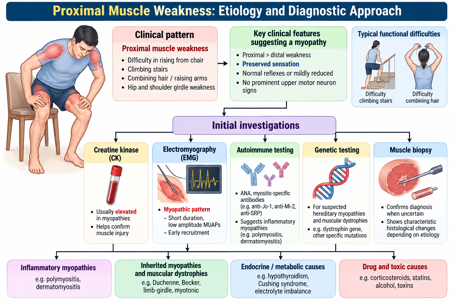
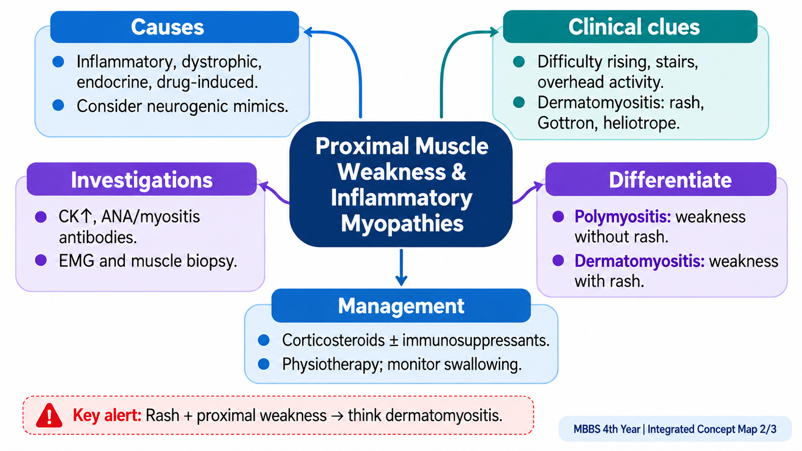
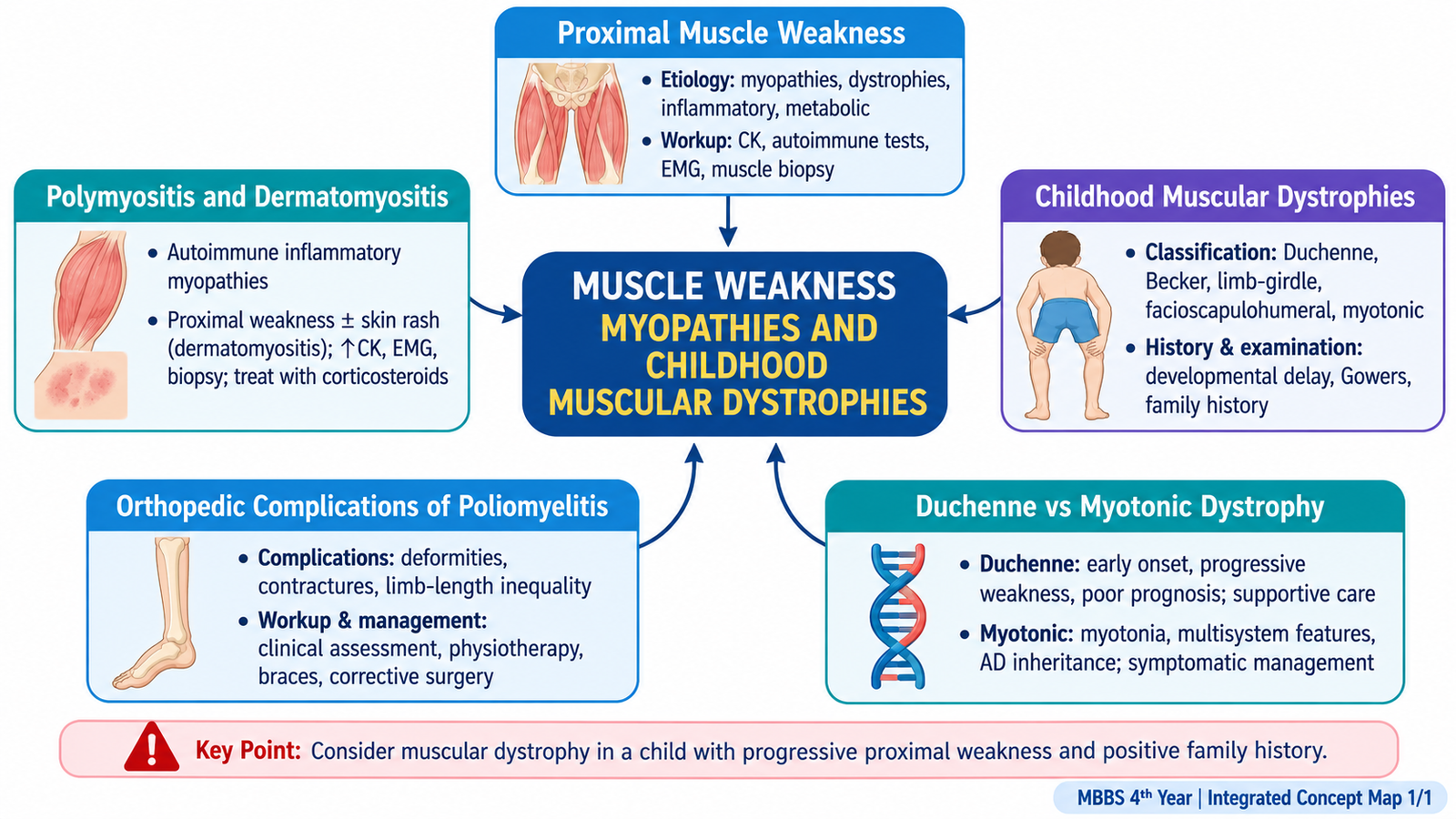
Proximal muscle weakness means reduced power predominantly in muscles close to the trunk, especially the shoulder and pelvic girdles. Patients may complain that they cannot rise easily from a chair, climb stairs, lift objects above shoulder level or comb their hair. This pattern immediately suggests a disorder affecting skeletal muscle, although neurological, endocrine, metabolic and drug-related causes must also be considered.
Recognizing a Myopathic Pattern
Primary muscle disease typically causes symmetrical proximal weakness. Distal muscles may become involved later in some disorders. Sensory symptoms are usually absent because the sensory nerves are not the primary site of disease. Deep tendon reflexes are often preserved until weakness becomes severe.
Important Causes
The cause can often be narrowed by age, tempo of illness, associated symptoms and family history.
- Inflammatory: polymyositis and dermatomyositis.
- Inherited: Duchenne, Becker, limb-girdle, facioscapulohumeral and myotonic dystrophies.
- Endocrine or metabolic: particularly thyroid-related and other metabolic muscle disorders.
- Drug or toxin related: suggested by an appropriate exposure history.
- Neurological mimics: anterior horn cell, peripheral nerve or neuromuscular-junction disorders must be considered when the pattern is atypical.
History
History should establish whether the weakness is truly muscular and whether it is acute, subacute or chronic.
- Onset and progression of weakness.
- Difficulty rising, climbing stairs, running or lifting the arms.
- Muscle pain or tenderness.
- Skin rash suggesting dermatomyositis.
- Dysphagia or respiratory symptoms indicating more extensive muscle involvement.
- Drug or toxin exposure.
- Family history of childhood weakness, delayed milestones or muscular dystrophy.
Examination
- Assess proximal and distal muscle power systematically.
- Look for muscle wasting or apparent enlargement such as calf pseudohypertrophy.
- Observe gait and the method used to rise from the floor or a chair.
- Assess tone and reflexes.
- Check sensation to identify neuropathic disorders.
- Inspect the skin for characteristic dermatomyositis changes.
Diagnostic Workup
Investigations should proceed from simple evidence of muscle injury toward tests that identify the specific cause.
| Investigation | Purpose |
|---|---|
| Serum creatine kinase | Supports active muscle-fibre injury when elevated. |
| Routine blood tests and selected metabolic/endocrine tests | Identify systemic or metabolic causes when clinically suspected. |
| Electromyography | Helps distinguish a myopathic pattern from neurogenic weakness. |
| Autoimmune investigations | Support suspected inflammatory myopathy. |
| Muscle imaging | May demonstrate muscle inflammation, edema, fatty replacement or help select a biopsy site. |
| Genetic testing | Important when an inherited muscular dystrophy is suspected. |
| Muscle biopsy | Useful when tissue morphology is required to establish or differentiate a myopathy. |
Diagnostic logic: pattern of weakness → evidence of muscle injury → distinguish inflammatory, inherited and other causes → use targeted autoimmune, genetic or tissue investigations.
B. Polymyositis and Dermatomyositis
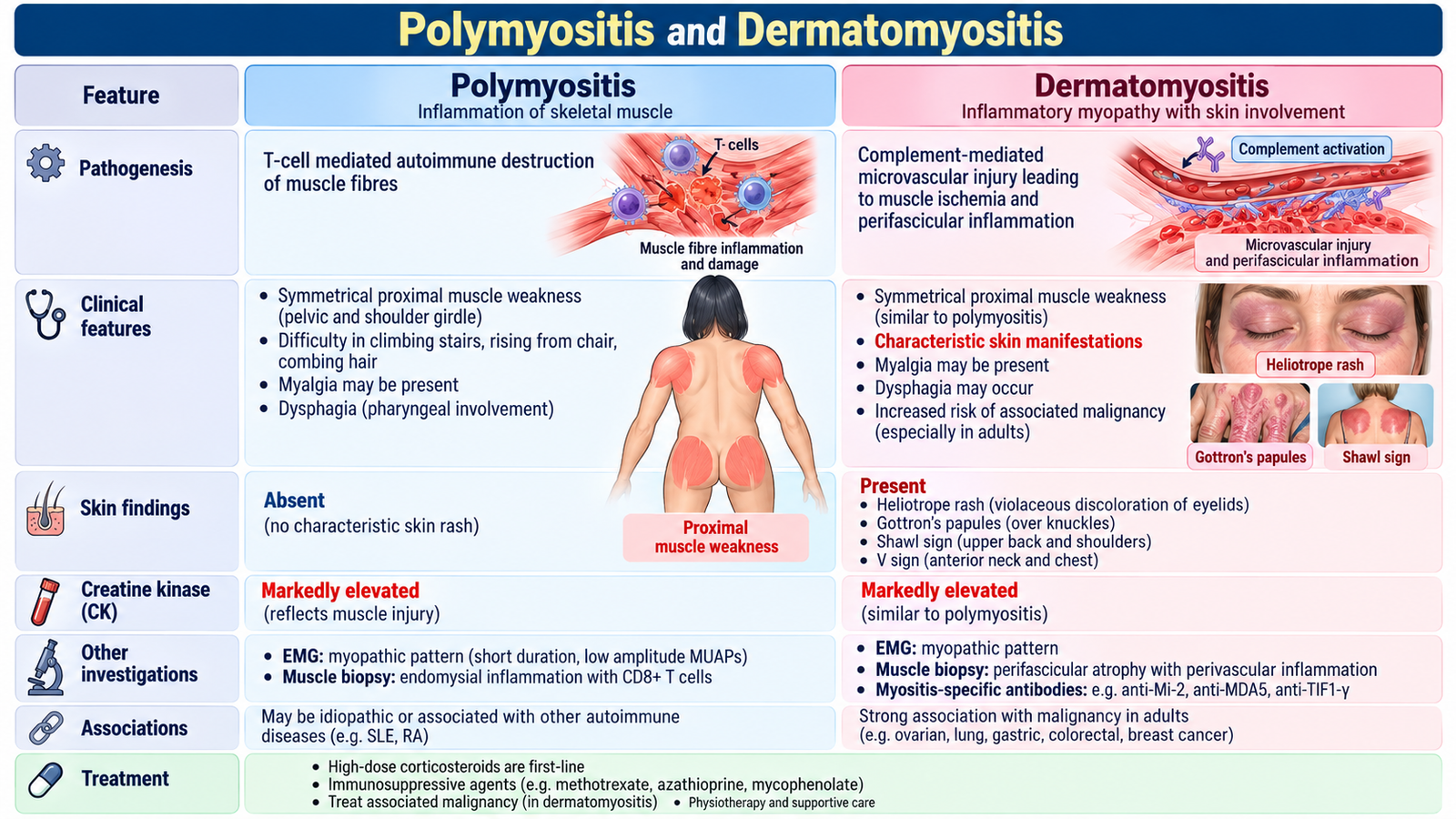
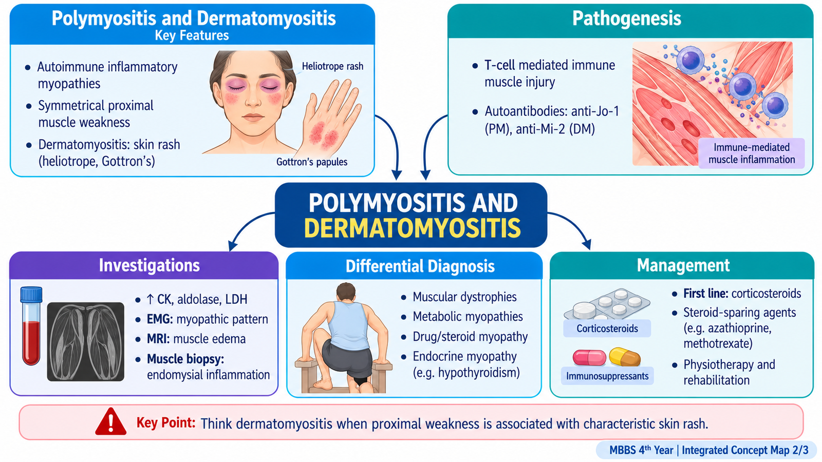
Polymyositis and dermatomyositis are inflammatory disorders in which immune-mediated injury damages skeletal muscle and produces progressive weakness. Both may cause symmetrical proximal muscle weakness, but dermatomyositis is distinguished by characteristic skin disease and has a different pattern of immune injury.
Pathogenesis
In polymyositis, the main process is cell-mediated injury directed against muscle fibres. Inflammatory cells infiltrate the muscle and contribute to fibre damage and weakness.
Immune-mediated muscle-fibre injury → muscle-fibre dysfunction and damage → loss of contractile strength → symmetrical proximal weakness.
In dermatomyositis, immune-mediated vascular injury affects small vessels supplying muscle and skin. This contributes to muscle injury and the characteristic cutaneous manifestations.
Immune-mediated microvascular injury → reduced perfusion and perifascicular muscle injury → proximal weakness + characteristic skin manifestations.
Clinical Features
The muscular presentation is usually a progressive, symmetrical weakness involving the pelvic and shoulder girdles.
- Difficulty climbing stairs.
- Difficulty rising from a low chair.
- Difficulty lifting the arms above the head.
- Neck flexor weakness may occur.
- Dysphagia may occur when pharyngeal muscles are involved.
- Respiratory muscle involvement is an important severe manifestation.
Dermatomyositis additionally produces characteristic skin changes. Important recognizable findings include a violaceous discoloration around the eyelids and inflammatory papules over the extensor surfaces of finger joints. These skin findings are useful because they direct the clinician toward dermatomyositis rather than an isolated muscle disorder.
Investigations
- Creatine kinase: commonly rises because damaged muscle fibres release intracellular enzymes.
- Electromyography: may demonstrate a myopathic pattern.
- Autoimmune testing: supports the diagnosis in appropriate clinical settings.
- Muscle imaging: may identify areas of active inflammation.
- Muscle biopsy: can demonstrate inflammatory muscle injury and help distinguish the inflammatory myopathies.
Differential Diagnosis
The main task is to separate inflammatory muscle disease from inherited dystrophy, endocrine or metabolic myopathy, drug-induced myopathy, neuropathy and neuromuscular-junction disease. Age, family history, skin findings, sensory examination, reflexes and laboratory evidence of muscle injury help make this distinction.
| Feature | Polymyositis | Dermatomyositis |
|---|---|---|
| Main presentation | Proximal muscle weakness | Proximal weakness with characteristic skin disease |
| Dominant immune pattern | Cell-mediated muscle-fibre injury | Immune-mediated microvascular injury |
| Skin manifestations | Absent | Characteristic cutaneous findings present |
Management
Management aims to suppress active inflammation, preserve muscle function and identify important complications. Systemic glucocorticoids are commonly used as initial immunosuppressive treatment. Additional immunosuppressive treatment may be required when disease is persistent or inadequately controlled. Physiotherapy and graded rehabilitation help preserve mobility and reduce disability.
Oral prednisolone is commonly used initially to control active myositis. Methotrexate or azathioprine may be added as steroid-sparing treatment. Severe swallowing difficulty, respiratory muscle weakness or interstitial lung disease requires urgent specialist assessment.
- Treat active inflammatory disease.
- Monitor muscle strength and functional recovery.
- Assess swallowing or respiratory involvement when symptoms suggest these complications.
- Use rehabilitation to maintain movement and function.
- In dermatomyositis, appropriate evaluation for associated systemic disease is important.
C. Muscular Dystrophies: Classification and General Principles
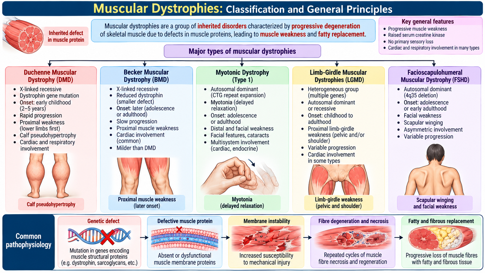
Muscular dystrophies are inherited disorders characterized by progressive dysfunction and degeneration of skeletal muscle. They differ in inheritance, age of onset, distribution of weakness and rate of progression. Recognizing these patterns is more useful clinically than memorizing isolated disease names.
Clinically Important Classification
- Duchenne muscular dystrophy — severe X-linked dystrophinopathy presenting in childhood.
- Becker muscular dystrophy — a milder dystrophinopathy with later onset and slower progression.
- Myotonic dystrophy — inherited multisystem disorder characterized particularly by myotonia and muscle weakness.
- Limb-girdle muscular dystrophies — heterogeneous inherited disorders predominantly affecting shoulder and pelvic girdle muscles.
- Facioscapulohumeral muscular dystrophy — predominantly affects facial and shoulder-girdle muscles.
How Muscular Dystrophy Produces Weakness
Although the exact genetic abnormality differs between dystrophies, the general principle is failure of normal muscle structure or function. Repeated muscle-fibre damage eventually exceeds the capacity for regeneration. Functional muscle tissue is progressively lost and replaced by fat and connective tissue.
Inherited protein abnormality → muscle-fibre instability or dysfunction → repeated fibre injury → degeneration and incomplete regeneration → fat/fibrous replacement → progressive weakness.
This process explains why many muscular dystrophies are progressive and why enlarged muscles do not necessarily indicate increased strength. In some conditions, apparent enlargement represents replacement of functional muscle by fat and connective tissue.
D. Duchenne Muscular Dystrophy
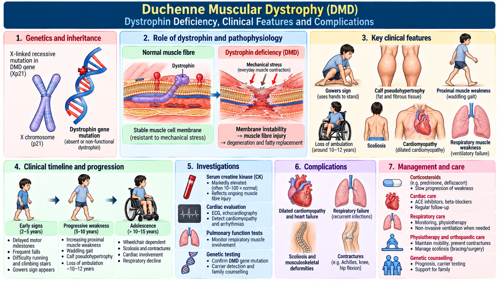
Duchenne muscular dystrophy is a severe, progressive childhood muscular dystrophy caused by deficiency of functional dystrophin. Because the dystrophin gene is located on the X chromosome, the disorder predominantly affects boys and typically follows an X-linked recessive pattern of inheritance.
Pathogenesis
Dystrophin helps connect the internal contractile apparatus of the muscle fibre to the cell membrane and surrounding supporting structures. When dystrophin is absent, contraction repeatedly stresses and damages the muscle-fibre membrane. Damaged fibres undergo degeneration, and over time muscle is replaced by fat and fibrous tissue.
Dystrophin gene abnormality → absent functional dystrophin → unstable muscle-fibre membrane → repeated contraction-related injury → fibre degeneration → fatty and fibrous replacement → progressive weakness.
Clinical Features
Weakness begins in early childhood and is most obvious in the pelvic-girdle muscles. The child may initially appear clumsy, fall frequently or have difficulty keeping up with other children.
- Delayed or impaired motor performance.
- Progressive proximal weakness, particularly around the pelvic girdle.
- Frequent falls.
- Difficulty running and climbing stairs.
- Waddling gait.
- Lumbar lordosis may become prominent as the child compensates for pelvic weakness.
- Gowers sign: the child uses the hands to push on the thighs and “climb up” the body when rising from the floor.
- Calf pseudohypertrophy: calves look enlarged despite reduced muscle function.
As the disorder progresses, contractures and skeletal deformity can develop. Cardiac and respiratory muscle involvement are clinically important because muscular dystrophy is not limited to limb movement.
Diagnosis
The combination of a typical childhood presentation, markedly increased muscle enzymes and a compatible family history raises strong suspicion. Genetic testing is central for confirming a dystrophin gene abnormality. Additional investigations assess disease effects and complications.
- Serum creatine kinase is typically markedly increased because muscle fibres continually leak intracellular enzymes.
- Genetic testing confirms the underlying dystrophin gene abnormality in most patients.
- Cardiac assessment is required because dystrophin deficiency may affect myocardium.
- Respiratory assessment becomes increasingly important as weakness progresses.
Differential Diagnosis
Duchenne muscular dystrophy must be distinguished from Becker muscular dystrophy, limb-girdle dystrophy, congenital myopathies, inflammatory myopathies, spinal muscular atrophy and other causes of childhood weakness. The age of onset, inheritance pattern, distribution of weakness, calf pseudohypertrophy and genetic findings are particularly helpful.
Management
Management is multidisciplinary and aims to preserve function, delay complications and support the child and family. Treatment does not depend on a single intervention.
- Physiotherapy and stretching to maintain mobility and reduce contractures.
- Appropriate physical activity while avoiding unnecessary muscle injury.
- Glucocorticoid therapy may slow functional decline in suitable patients.
- Prednisolone/prednisone or deflazacort may be used under specialist supervision to preserve muscle strength and delay functional deterioration. Long-term treatment requires monitoring of growth, weight, blood pressure and bone health. Corticosteroids should not be stopped abruptly.
- Orthopedic support for contractures and deformities.
- Cardiac surveillance and treatment of cardiac complications.
- Respiratory surveillance and respiratory support when required.
- Genetic counselling for the family.
- Psychological, educational and social support.
Prognosis
Duchenne muscular dystrophy is progressive. Muscle power gradually declines and mobility becomes increasingly limited. Long-term outcome is strongly influenced by cardiac and respiratory involvement, which is why anticipatory multidisciplinary care is essential.
E. Myotonic, Becker, Limb-Girdle and Facioscapulohumeral Dystrophies
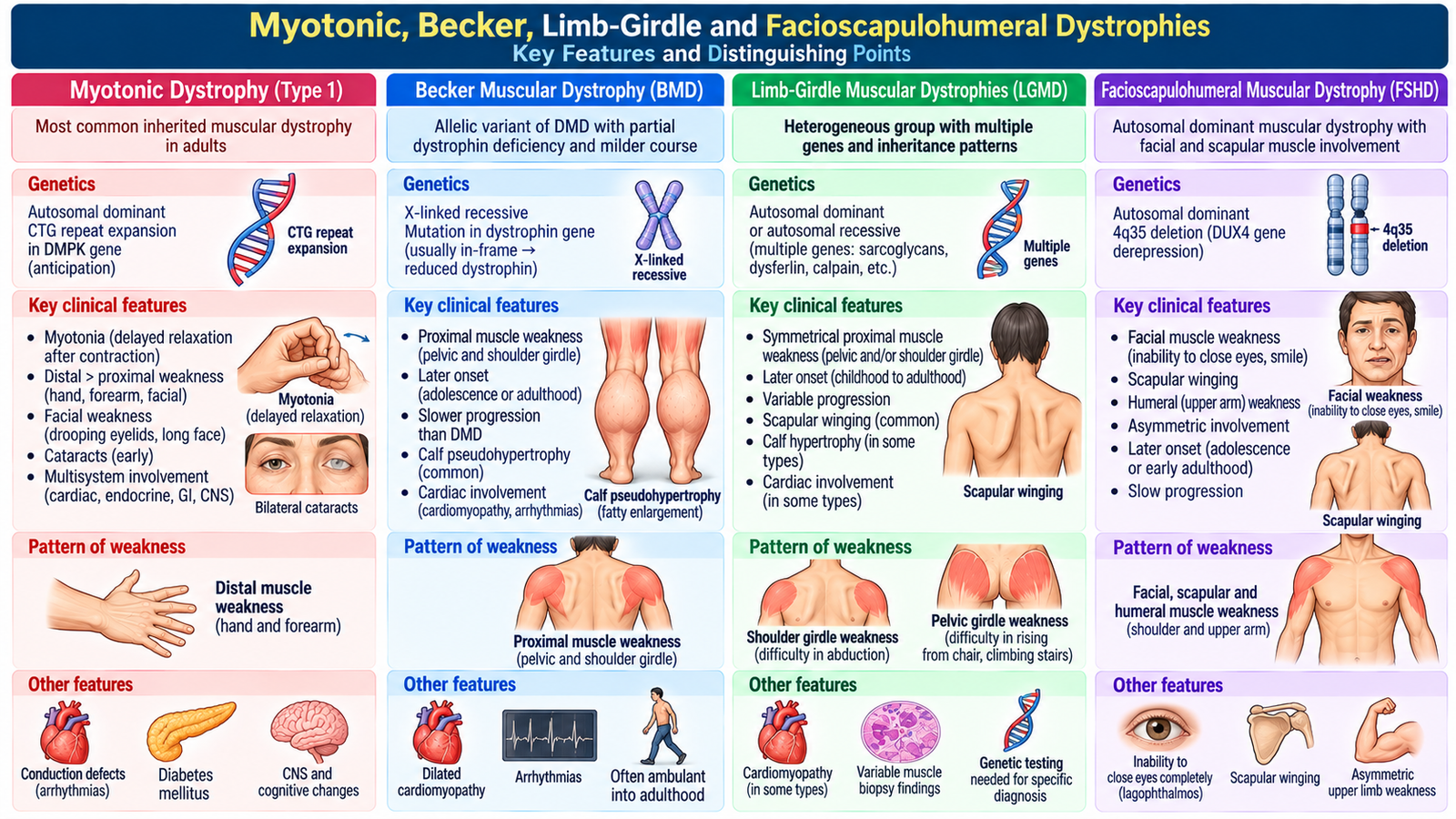
These dystrophies differ from Duchenne muscular dystrophy in age of presentation, inheritance and pattern of muscle involvement. Their clinical distribution provides useful diagnostic clues. The student should be able to recognize myotonic dystrophy and compare the major features of Becker, limb-girdle and facioscapulohumeral dystrophies.
Myotonic Dystrophy
Myotonic dystrophy is an inherited multisystem disorder in which muscle weakness occurs together with myotonia, meaning delayed relaxation after a muscle has contracted. A patient may therefore have difficulty releasing a handshake or relaxing the hand after gripping an object.
Unlike Duchenne muscular dystrophy, myotonic dystrophy is not simply a childhood dystrophin deficiency disorder. It commonly follows an autosomal dominant inheritance pattern and may involve other organ systems in addition to skeletal muscle.
- Muscle weakness and wasting.
- Myotonia with delayed relaxation.
- Facial and distal muscle involvement may be prominent.
- Systemic manifestations may accompany the muscular disease.
Diagnosis is based on the clinical pattern and confirmation by genetic testing. Management is mainly supportive and directed toward maintaining function and identifying clinically important systemic complications. Myotonic dystrophy requires regular cardiac assessment, including ECG screening, because conduction abnormalities may cause serious arrhythmias. Respiratory weakness, sleep-related breathing problems and cataracts should also be assessed. Symptomatic cardiac conduction disease requires cardiology referral and may necessitate pacemaker implantation.
Important Comparison
| Disorder | Typical Pattern | Useful Distinguishing Feature | General Course |
|---|---|---|---|
| Duchenne | Early childhood, pelvic-girdle weakness | Gowers sign, calf pseudohypertrophy, severe dystrophin deficiency | Rapidly progressive compared with Becker |
| Becker | Similar distribution to Duchenne but later onset | Some functional dystrophin is present | Usually slower progression |
| Limb-girdle | Shoulder and pelvic girdles | Prominent girdle weakness without the classic Duchenne pattern | Variable according to subtype |
| Facioscapulohumeral | Face and shoulder girdle predominantly | Facial weakness and scapular winging | Often slower and variable |
| Myotonic | Facial/distal or generalized involvement | Myotonia: delayed relaxation after contraction | Progressive multisystem disorder |
Becker muscular dystrophy is particularly important because it results from abnormalities involving the same dystrophin system as Duchenne, but dystrophin function is partly retained. Therefore onset is generally later and progression is less severe.
Limb-girdle muscular dystrophies form a heterogeneous group. Their common clinical feature is weakness centered on shoulder and pelvic girdle muscles. Exact inheritance and severity vary between forms.
Facioscapulohumeral muscular dystrophy is recognized by its distribution: facial weakness together with scapular and upper-arm involvement. Scapular winging may be an obvious physical finding.
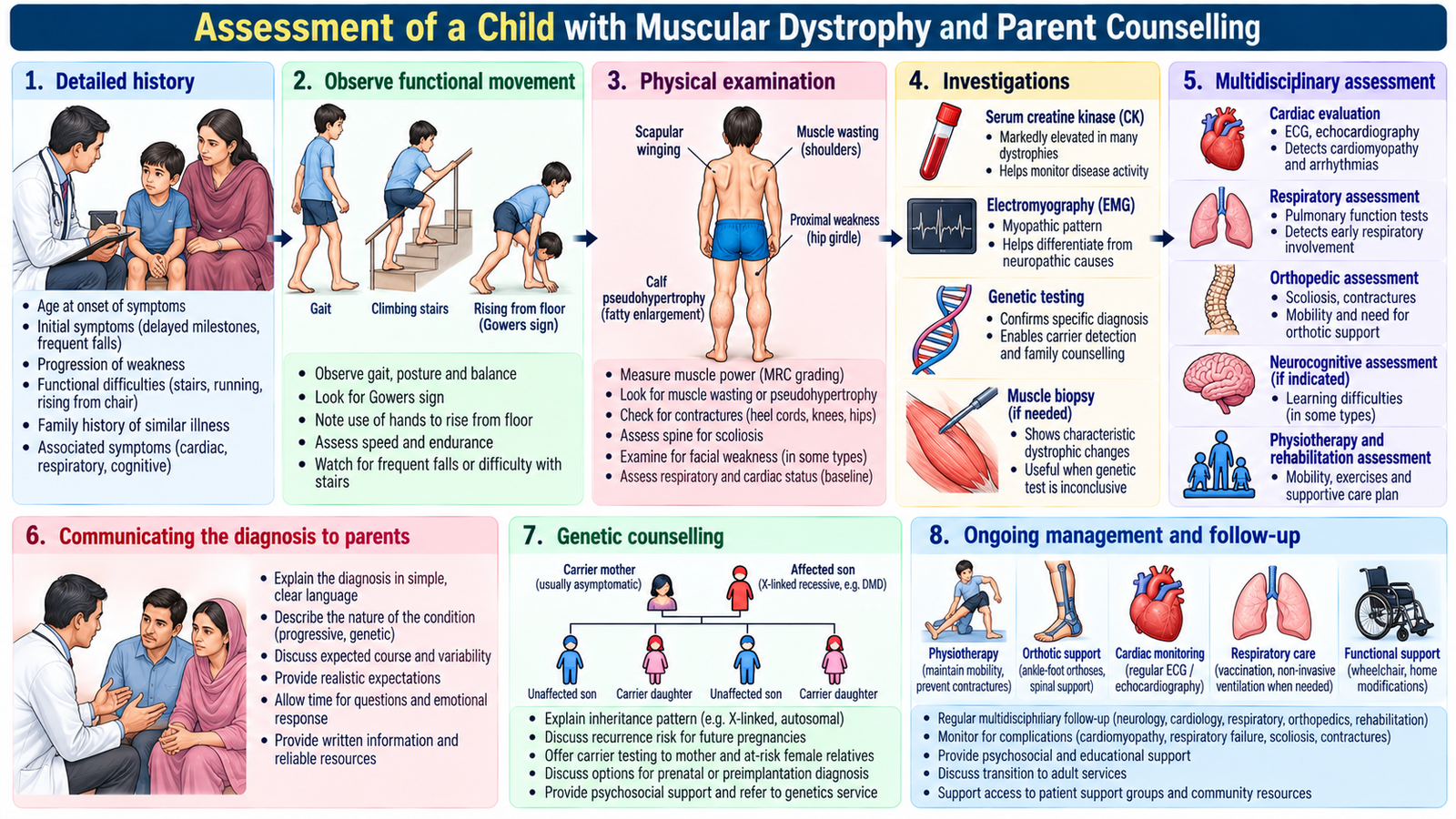
F. Assessment of a Child with Muscular Dystrophy and Parent Counselling
Clinical assessment of a child with suspected muscular dystrophy should establish the pattern and progression of weakness, identify important complications and determine whether the family history supports an inherited disorder. The examination should be systematic but child-friendly.
History
- Age at which weakness or motor difficulty was first noticed.
- Motor milestones and any loss of previously acquired abilities.
- Frequent falls or difficulty running, jumping or climbing stairs.
- Difficulty rising from the floor.
- Fatigue and limitation of everyday activities.
- Muscle cramps or stiffness when relevant.
- Swallowing or respiratory symptoms.
- Family history of male relatives or other family members with weakness, walking difficulty or diagnosed muscular dystrophy.
- Effect on schooling, mobility and family life.
Physical Examination
Begin by observing the child before formal testing. Gait and functional movements often reveal weakness more clearly than isolated power testing.
- Observe posture and gait.
- Ask the child to rise from the floor and look for Gowers sign.
- Inspect calves for pseudohypertrophy.
- Look for muscle wasting or asymmetry.
- Assess proximal and distal muscle power.
- Examine tone and tendon reflexes.
- Check for contractures or skeletal deformity.
- Assess respiratory effort when clinically relevant.
Counselling Parents
Counselling should be clear, empathetic and realistic. Parents should understand that muscular dystrophy is an inherited muscle disorder and that progression differs according to the exact diagnosis. Avoid overwhelming the family with technical genetic language during the first explanation.
Key counselling points include:
- Explain the diagnosis and the reason for the child’s weakness in understandable language.
- Explain that the disorder may be progressive.
- Discuss the purpose of genetic testing and genetic counselling.
- Explain the importance of regular assessment of mobility and complications.
- Encourage physiotherapy and preservation of useful function.
- Discuss school participation, independence and psychosocial support.
- Explain that cardiac or respiratory assessment may be required in particular dystrophies.
- Allow parents to ask questions and correct misconceptions about activity, inheritance and prognosis.
For an X-linked disorder such as Duchenne muscular dystrophy, genetic counselling is particularly important because the diagnosis may have implications for other family members and future pregnancies.
G. Orthopedic Complications of Poliomyelitis
Poliomyelitis may damage motor neurons and leave selected muscle groups permanently weak or paralyzed. The orthopedic problem develops because muscles acting across a joint no longer produce balanced forces. Stronger muscles may pull without effective opposition from weakened muscles, gradually producing deformity, contracture and abnormal loading during growth.
Motor-neuron injury → persistent muscle weakness/paralysis → muscle imbalance → abnormal joint position and loading → contracture/deformity → impaired gait and function.
Major Orthopedic Complications
- Muscle contractures: shortened soft tissues restrict normal joint movement.
- Joint deformity: persistent muscle imbalance gradually fixes the limb in an abnormal position.
- Foot and ankle deformities: may alter stance and gait.
- Limb-length discrepancy: may result when an affected limb grows less effectively.
- Joint instability: weak muscles provide inadequate dynamic support.
- Spinal deformity: trunk-muscle imbalance may contribute to scoliosis.
- Abnormal gait: develops from weakness, deformity and compensatory movement.
Diagnostic Workup
The purpose of assessment is to determine which muscles are weak, whether deformity is flexible or fixed, and how much the problem affects function.
- History of previous poliomyelitis and functional limitation.
- Detailed muscle-power examination.
- Assessment of joint range of movement.
- Observation of gait.
- Measurement for limb-length discrepancy when suspected.
- Assessment of spinal alignment.
- Appropriate radiographs when structural deformity requires evaluation.
Management Principles
Management is directed at maximizing useful function, preventing worsening deformity and correcting established problems where appropriate. The treatment plan depends on residual muscle power, age, the degree of deformity and functional need.
- Physiotherapy to maintain movement and strengthen functioning muscles appropriately.
- Stretching to reduce the development or progression of contractures.
- Orthoses or supportive devices to improve alignment and walking.
- Mobility aids when required.
- Corrective orthopedic procedures may be considered for significant fixed deformity, imbalance or functional impairment.
- Long-term rehabilitation should focus on independence and efficient mobility.
⭐ AIM High-Yield Review
- ⭐ A primary myopathy typically produces symmetrical proximal weakness with preserved sensation.
- ⭐ Difficulty rising from a chair, climbing stairs and lifting the arms suggests proximal muscle weakness.
- ⭐ Polymyositis and dermatomyositis both cause inflammatory proximal myopathy, but characteristic skin disease points toward dermatomyositis.
- ⭐ Serum creatine kinase rises when injured muscle fibres release intracellular enzymes.
- ⭐ Duchenne muscular dystrophy is a severe X-linked dystrophinopathy predominantly affecting boys.
- ⭐ Loss of dystrophin destabilizes the muscle-fibre membrane, causing repeated fibre injury and progressive weakness.
- ⭐ Gowers sign reflects proximal pelvic-girdle weakness.
- ⭐ Calf enlargement in Duchenne muscular dystrophy is pseudohypertrophy, not increased functional muscle.
- ⭐ Becker muscular dystrophy generally has later onset and slower progression than Duchenne because some functional dystrophin remains.
- ⭐ Myotonia means delayed relaxation after muscle contraction.
- ⭐ Limb-girdle dystrophies predominantly affect shoulder and pelvic girdle muscles.
- ⭐ Facial weakness with scapular involvement suggests facioscapulohumeral dystrophy.
- ⭐ Examination of a child with suspected muscular dystrophy should include gait, rising from the floor, calf appearance, power, reflexes and contractures.
- ⭐ Parent counselling should include progression, multidisciplinary care and the importance of genetic counselling.
- ⭐ Post-polio deformity develops mainly through persistent weakness and muscle imbalance, leading to contracture and abnormal joint loading.