
This chapter follows the supplied KMU outcomes for osteomyelitis and spinal tuberculosis. First understand how infection reaches bone or spine and produces the clinical picture; then connect those findings with investigations and management. Use the High-Yield Review at the end for revision.
Topic 7. Osteomyelitis and Spinal Tuberculosis
Musculoskeletal Module • Orthopedics
Understand the causes and presentation of bone and spinal infection, select and interpret appropriate investigations, and connect the diagnosis with medical and surgical management.
Topic Introduction
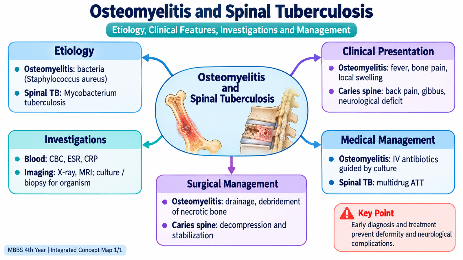
Osteomyelitis is infection and inflammation of bone and bone marrow, while spinal tuberculosis is tuberculosis involving the vertebral column, traditionally called Pott disease or caries spine. Both conditions can progressively damage bone if diagnosis is delayed. Their clinical behaviour, however, may differ: acute pyogenic osteomyelitis can present with prominent pain, fever and systemic illness, whereas spinal tuberculosis commonly develops more slowly with persistent back pain and progressive vertebral destruction. The central clinical task is to recognize the pattern of infection, determine its extent using appropriate laboratory tests and imaging, identify the causative organism where possible, and institute medical treatment while recognizing situations in which surgical intervention becomes necessary.
A. How Bone Infection Develops: Etiology and Routes of Osteomyelitis
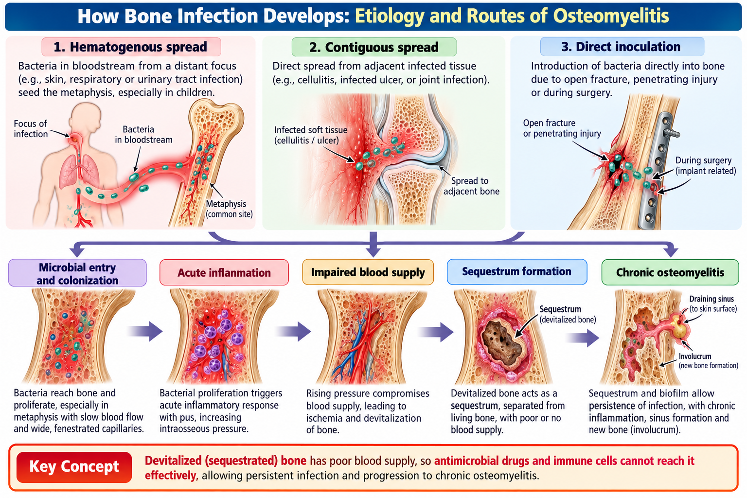
Osteomyelitis develops when microorganisms gain access to bone and establish infection. Understanding how organisms reach bone helps explain which patients are affected and which bones are likely to be involved.
Routes of infection
There are three clinically important routes.
- Hematogenous spread: organisms reach bone through the bloodstream from another site of infection. This route is particularly important in children.
- Contiguous spread: infection extends directly from nearby infected soft tissue or an adjacent focus.
- Direct inoculation: microorganisms enter bone following an open fracture, penetrating injury or orthopedic procedure.
Staphylococcus aureus is the most important general cause of pyogenic osteomyelitis. The likely organism may vary with the patient’s age, route of infection and clinical context, so microbiological confirmation is important whenever possible.
Why chronic osteomyelitis persists
When infection compromises the blood supply, a fragment of devitalized bone may separate from viable bone. This dead infected bone is called a sequestrum. Because its blood supply is lost, systemic antibiotics penetrate it poorly. Reactive new bone may form around the infected area, producing an involucrum. Persistent drainage through a sinus can occur in chronic disease.
B. Recognizing Osteomyelitis Clinically
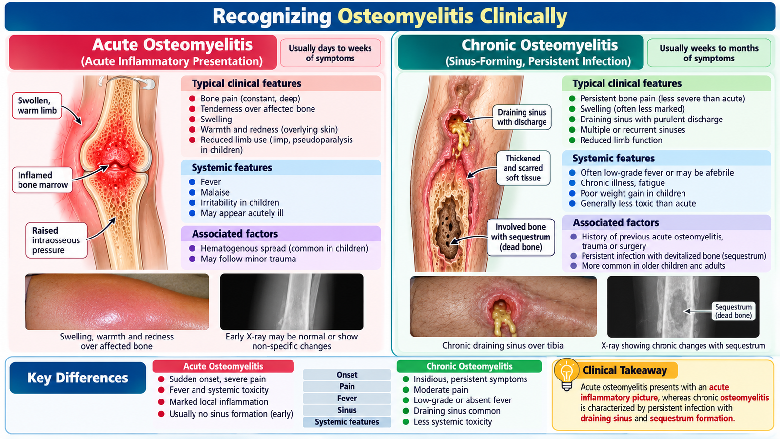
The clinical picture depends on whether infection is acute or chronic, the patient’s age and the site involved. The key principle is to suspect bone infection when localized bone pain and tenderness occur in an appropriate infective context.
Acute osteomyelitis
Acute infection commonly produces relatively rapid onset of symptoms. Local inflammation causes pain and tenderness, while systemic inflammatory response may produce fever and malaise.
- Localized deep bone pain
- Marked tenderness over the affected bone
- Swelling, warmth or reduced use of the affected limb
- Fever and systemic illness may be present
- Children may refuse to move or bear weight on the affected limb
In children, hematogenous infection commonly involves the highly vascular metaphyseal region of long bones. A child who becomes febrile and suddenly refuses to use a limb therefore requires careful assessment for musculoskeletal infection.
Chronic osteomyelitis
Chronic infection is more indolent. Previous trauma, surgery, an open fracture or inadequately controlled infection may be important historical clues.
- Persistent or recurrent localized pain
- Swelling or induration
- Recurrent discharge from a sinus
- Episodes of exacerbation separated by quieter periods
- Underlying sequestrum or chronically infected bone
Acute osteomyelitis tends to produce a more inflammatory presentation, whereas chronic osteomyelitis should be considered when symptoms persist or recur, particularly when a draining sinus or previous bone injury is present.
C. Spinal Tuberculosis: From Infection to Caries Spine
Spinal tuberculosis is infection of the vertebral column by Mycobacterium tuberculosis. The organisms usually reach the spine by hematogenous spread from another tuberculous focus. Unlike acute pyogenic infection, the process is commonly gradual and may initially produce few systemic features.
How vertebral disease develops
Tuberculous infection causes granulomatous inflammation and progressive destruction of vertebral bone. Adjacent vertebral bodies may become involved and the intervertebral disc can subsequently be affected. Progressive structural destruction may lead to vertebral collapse and angular deformity.
Tuberculous inflammatory material may extend into surrounding tissues and form a cold abscess. It is termed “cold” because prominent acute inflammatory features such as marked warmth and erythema may be absent.
Clinical presentation
Persistent localized back pain is an important presentation. Because progression is often slow, symptoms may be present for weeks or months before diagnosis.
- Persistent localized back pain
- Spinal tenderness and stiffness
- Reduced spinal movements
- Constitutional symptoms such as fever, night sweats, reduced appetite or weight loss may occur
- Progressive kyphotic or angular deformity may develop with vertebral collapse
- Neurological symptoms may appear when the spinal cord or nerve structures are compromised
Weakness, sensory disturbance, sphincter dysfunction or other evidence of neurological compromise in a patient with suspected spinal infection requires urgent assessment because neural structures may be threatened.

D. Clinical Assessment of Suspected Bone or Spinal Infection
Assessment should establish whether infection is likely, identify its probable route and duration, determine whether the disease is localized or complicated, and look specifically for neurological involvement when the spine is affected.
Focused history
- Onset, duration and progression of pain
- Fever or other systemic symptoms
- Previous wound, trauma, open fracture or surgery
- Previous episodes of bone infection or recurrent discharge
- Reduced limb use or difficulty walking
- For spinal disease: chronic back pain, constitutional symptoms and progression of deformity
- For suspected neurological involvement: weakness, altered sensation and bladder or bowel symptoms
Important examination findings
Examination is directed by the suspected site. In peripheral bone infection, localized tenderness, swelling, warmth and impaired function are important. In suspected spinal tuberculosis, assess spinal tenderness, deformity and movement and determine whether neurological dysfunction is present.
The combination of persistent localized bone or spinal pain, appropriate infective or exposure context, inflammatory findings and compatible imaging should raise suspicion of infection; microbiological or tissue confirmation should be pursued when appropriate.

A young adult presents with several months of progressively worsening back pain. He has lost weight and examination reveals localized spinal tenderness with a developing angular deformity. He now reports increasing weakness of both legs.
Think clinically: The chronic course and vertebral deformity suggest a destructive spinal process such as spinal tuberculosis. The new weakness is particularly important because it suggests neurological involvement and makes assessment of the extent of disease urgent.
E. Investigating Suspected Bone and Spinal Infection
Investigations have four main purposes: to support the presence of infection, define its anatomical extent, identify the causative organism, and detect complications. No single investigation should be interpreted in isolation.
Blood investigations
A complete blood count and inflammatory markers help assess inflammatory activity. Leukocytosis may support acute bacterial infection, while inflammatory markers such as ESR and CRP may be elevated. These tests are useful for supporting the diagnosis and following response to treatment, but they do not identify the anatomical site or organism by themselves.
Blood culture
Blood cultures should be considered in suspected hematogenous pyogenic osteomyelitis, particularly when systemic infection is present. A positive culture can identify the organism and guide antimicrobial therapy.
Plain radiography
Plain X-rays are useful as an initial structural assessment, but early osteomyelitis may not immediately produce visible bony changes. Later disease may show bone destruction, periosteal reaction or features of chronic infection. In spinal tuberculosis, radiographs may demonstrate vertebral destruction, narrowing of the involved disc space in established disease, collapse and deformity.
MRI
MRI is particularly valuable when early osteomyelitis or spinal infection is suspected. It demonstrates bone marrow and surrounding soft-tissue abnormalities and helps define the extent of disease. In spinal infection it is especially useful for assessing vertebral involvement, paravertebral collections and neural compression.
Microbiological and tissue diagnosis
When feasible, infected material or bone/tissue should be obtained for microbiological examination. In pyogenic infection, culture guides targeted antibiotics. In suspected spinal tuberculosis, appropriate tissue or aspirated material can provide microbiological and histopathological evidence supporting tuberculosis.

F. Medical and Surgical Management
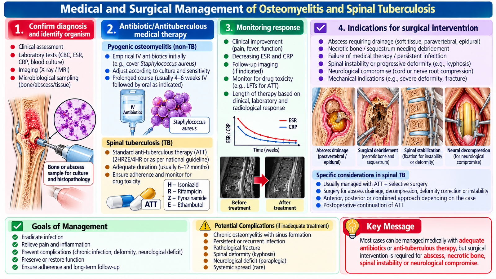
Management depends on the organism, duration of infection, extent of bone destruction and presence of complications. The overall goals are to eradicate infection, preserve viable bone and function, treat collections or necrotic tissue when necessary, and prevent structural or neurological complications.
Pyogenic osteomyelitis
Antibiotic treatment should cover the likely organism initially and then be adjusted according to microbiological results whenever available. Appropriate sampling before antibiotics is desirable when the clinical situation permits, but treatment should not be unnecessarily delayed in a seriously ill patient. Supportive measures include pain control and appropriate protection of the affected limb. Clinical condition and inflammatory markers can help assess response.
When surgery becomes important
Antibiotics alone may be insufficient when infected collections or devitalized tissue remain. Surgical treatment is therefore considered when there is:
- An abscess or collection requiring drainage
- Necrotic or devitalized bone requiring debridement
- Chronic infection associated with sequestrum
- Persistent infection despite appropriate medical treatment
- Need for operative management of associated structural problems
In chronic osteomyelitis, removal of necrotic infected tissue is particularly important because poorly vascularized dead bone can act as a continuing focus of infection.
Management of spinal tuberculosis
The foundation of treatment is appropriate anti-tuberculous therapy. Clinical symptoms, neurological status and structural disease must be followed during treatment. Surgical involvement becomes important when complications cannot be adequately managed medically, particularly where there is significant neurological compromise, major mechanical instability or deformity, a problematic collection, diagnostic uncertainty requiring tissue diagnosis, or failure of appropriate conservative treatment.
Neurological deterioration in spinal tuberculosis is not simply another symptom to observe. It indicates threatened neural function and requires prompt assessment and specialist management planning.
Integrated Mechanism Flow
Important Comparison: Pyogenic Osteomyelitis vs Spinal Tuberculosis
| Feature | Pyogenic Osteomyelitis | Spinal Tuberculosis |
|---|---|---|
| Typical course | May be acute with prominent inflammation | Usually gradual and chronic |
| Important organism | Staphylococcus aureus commonly | Mycobacterium tuberculosis |
| Major presentation | Bone pain, tenderness, fever ± swelling | Persistent back pain, stiffness ± constitutional symptoms |
| Important structural problem | Necrotic bone and sequestrum in chronic disease | Vertebral destruction, collapse and deformity |
| Major complication clue | Persistent abscess or chronic draining sinus | Neurological deficit from spinal involvement |
| Treatment principle | Antibiotics ± drainage/debridement | Anti-tuberculous therapy ± surgery when indicated |
AIM High-Yield Review
- Staphylococcus aureus is the major general cause of pyogenic osteomyelitis.
- Hematogenous spread is an important route of osteomyelitis, especially in children.
- Bone infection may also follow direct inoculation or contiguous spread from adjacent infection.
- ⭐ A sequestrum is devitalized bone separated from viable bone and can maintain chronic infection.
- A chronic draining sinus strongly suggests longstanding osteomyelitis in the appropriate clinical setting.
- A normal early plain X-ray does not reliably exclude osteomyelitis.
- ⭐ MRI is particularly useful for detecting and defining bone and spinal infection and associated soft-tissue or neural involvement.
- Culture or tissue sampling is important because identification of the organism allows specific antimicrobial treatment.
- Spinal tuberculosis commonly presents with a gradual course of persistent back pain and may cause vertebral destruction and collapse.
- A tuberculous collection may appear as a relatively non-inflammatory cold abscess.
- ⭐ Progressive vertebral destruction may produce kyphotic/angular deformity.
- ⭐ New weakness, sensory symptoms or sphincter dysfunction in spinal tuberculosis suggests neurological compromise and requires urgent assessment.
- Pyogenic osteomyelitis is treated with appropriate antibiotics, with drainage or debridement when infected collections or necrotic bone persist.
- Anti-tuberculous therapy is the foundation of medical treatment for spinal tuberculosis.
- ⭐ Surgery is considered in complicated spinal tuberculosis when problems such as significant neurological compromise, instability/deformity, problematic collections or failure of appropriate medical treatment require intervention.
🎥 AIM Video Learning
Topic 7: Osteomyelitis and Spinal Tuberculosis
Watch this video to reinforce the clinical presentation, diagnosis, investigations,
complications and management of spinal tuberculosis (Pott disease / caries spine).
- Etiology and pathogenesis of spinal tuberculosis
- Clinical presentation of caries spine
- Important neurological and structural complications
- Role of imaging in diagnosis
- Principles of anti-tuberculous treatment
- Indications for surgical management