
This chapter follows the KMU outcomes for noninfectious skin disorders and generalized rash. First understand how morphology, distribution, systemic clues and drug exposure guide diagnosis; use the AIM High-Yield Review only after you understand the clinical patterns.
Topic 10. Noninfectious Skin Disorders and Generalized Rash
MSK Module • A clinical approach to generalized rash, inflammatory dermatoses, blistering disease, drug reactions and important cutaneous clues to systemic disease.
Topic Introduction
A generalized rash is not a diagnosis; it is a clinical presentation that must be interpreted through the appearance of the skin lesions, their distribution, associated symptoms, medication exposure and evidence of systemic illness. This topic also brings together several common noninfectious dermatoses, including acne, eczema, psoriasis, lichen planus, seborrheic dermatitis, urticaria and erythema nodosum, as well as potentially serious drug reactions and autoimmune blistering disorders. The aim is not to memorize an isolated list of rashes. You should learn to recognize important patterns, distinguish common conditions, identify dangerous presentations such as Stevens–Johnson syndrome or toxic epidermal necrolysis, and understand sensible undergraduate management and counselling principles.
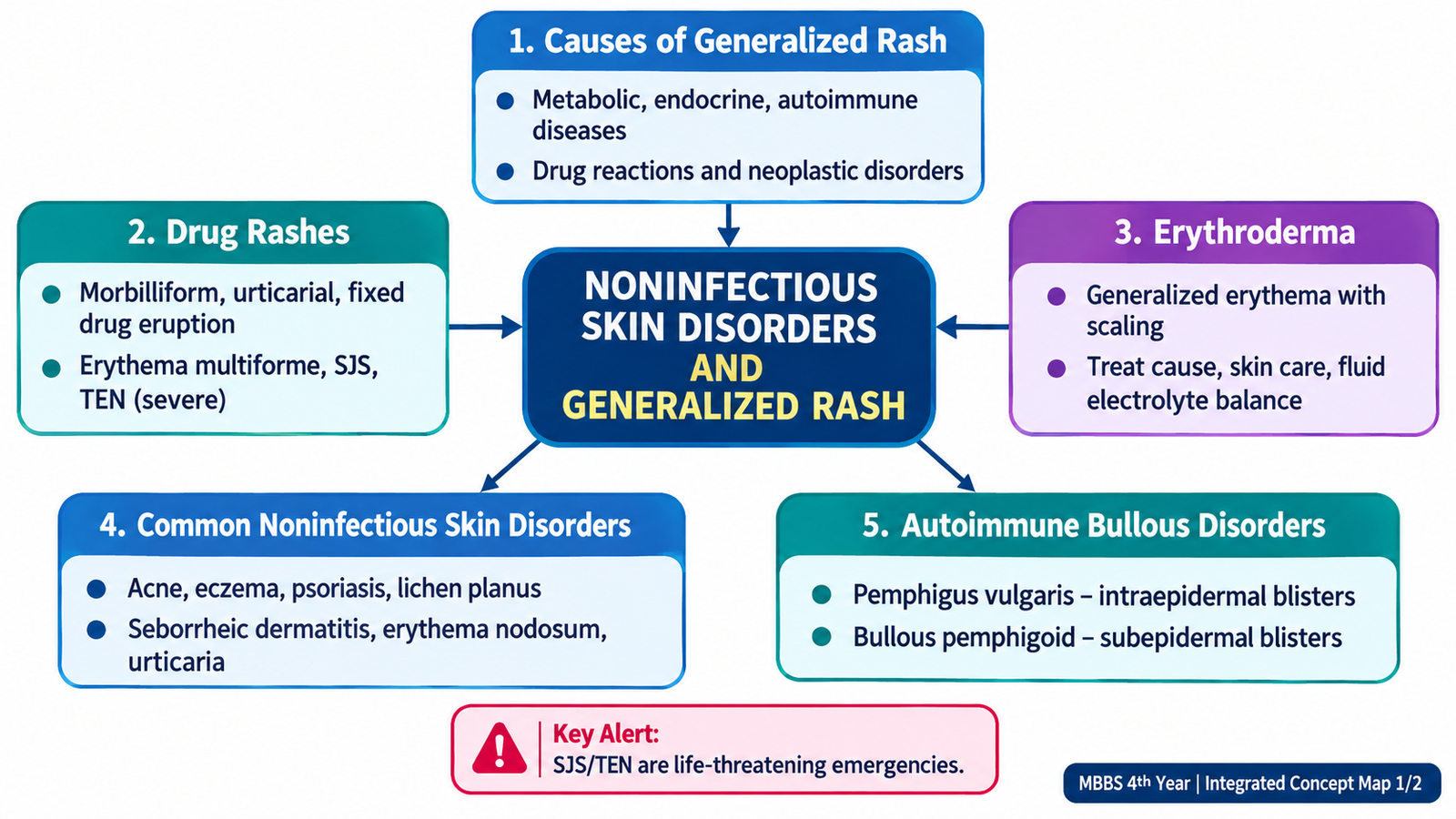
A. Clinical Approach to a Generalized Rash and Systemic Cutaneous Clues
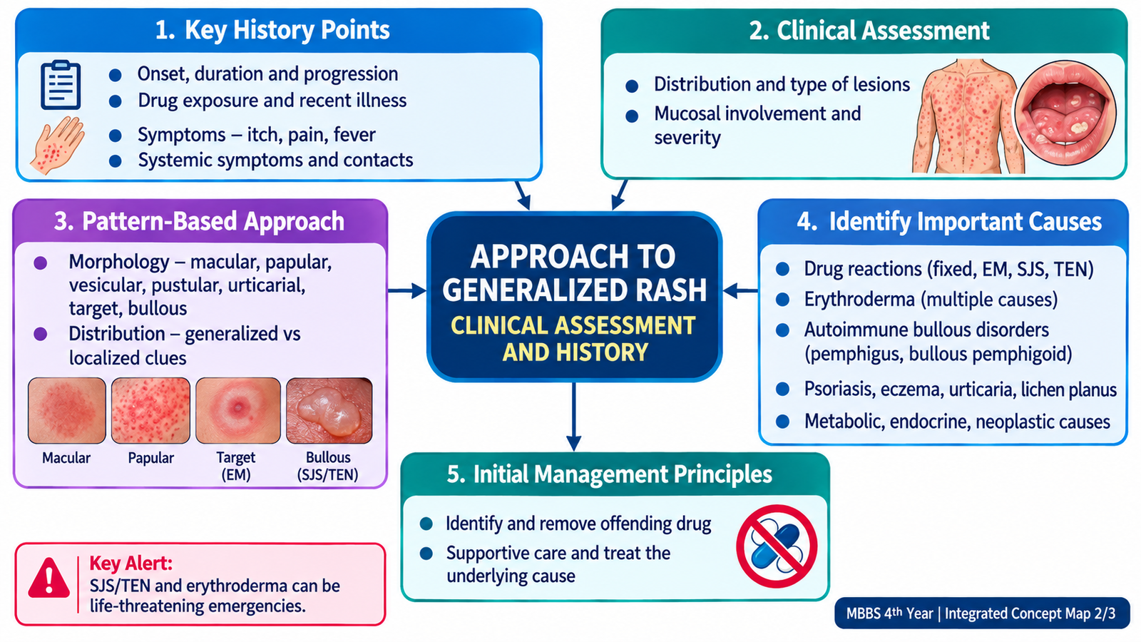
The first task in a patient with widespread skin lesions is to describe what is actually present before attaching a disease label. Dermatological diagnosis depends heavily on morphology—the type of primary lesion—and distribution. The same-looking eruption can have very different significance depending on whether the patient is otherwise well, has recently started a medicine, has mucosal involvement, or shows evidence of systemic disease.
Focused history
History should establish the chronology of the rash and look for exposures or systemic clues that change the differential diagnosis. Important areas include:
- Onset, duration, progression and first site involved.
- Itching, pain, burning, blistering or skin tenderness.
- Fever, malaise, joint symptoms or other systemic complaints.
- Recent prescription, over-the-counter or other medication exposure.
- Previous similar episodes and known skin disease.
- Personal or family history of atopy or psoriasis where relevant.
- Features suggesting endocrine, metabolic, autoimmune or neoplastic disease.
Examine the pattern, not just the lesion
Identify the primary lesion—such as macule, papule, plaque, wheal, nodule, vesicle, bulla or pustule—and then assess colour, scale, crusting, target-like morphology, erosion and secondary change. Note whether lesions are flexural, extensor, seborrheic, acral or truly generalized. Examine the scalp, nails and mucous membranes when clinically relevant.
Skin as a clue to systemic disease
Cutaneous findings may be the first visible sign of disease outside the skin. The important principle is to connect an unexplained skin change with the appropriate systemic context rather than treating every lesion as an isolated dermatological disorder.
- Metabolic disease: diabetes may be associated with characteristic skin changes and recurrent or poorly healing cutaneous problems.
- Endocrine disease: disorders of thyroid or adrenal function can alter skin texture, pigmentation, hair and sweating patterns.
- Autoimmune disease: inflammatory or photosensitive eruptions may accompany systemic autoimmune disorders.
- Neoplastic disease: an unusual, persistent or unexplained cutaneous manifestation may occasionally provide a clue to an underlying malignancy.
The clinical lesson is not to diagnose a systemic illness from one nonspecific rash. Instead, a compatible cutaneous finding should prompt focused history, examination and investigation for the suspected underlying disorder.

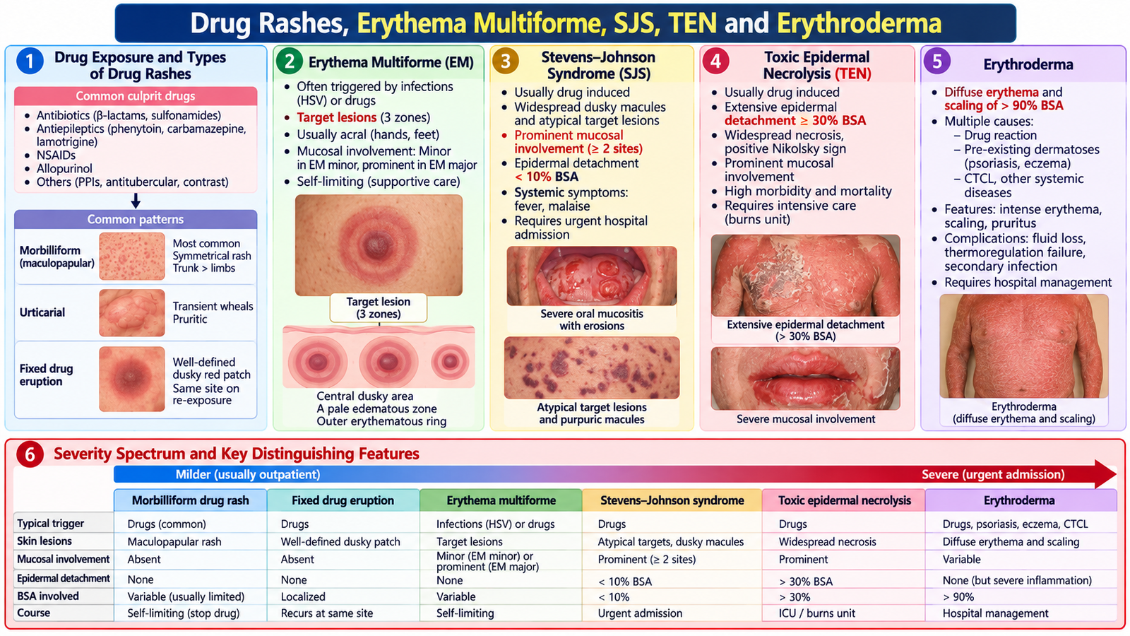
B. Drug Rashes, Erythema Multiforme, SJS, TEN and Erythroderma
Drug eruptions range from relatively mild exanthematous reactions to severe mucocutaneous disease. The key clinical skill is therefore not merely to recognize “a drug rash,” but to decide whether the eruption is uncomplicated or contains danger signs requiring urgent withdrawal of the suspected drug and escalation of care.
Classification and assessment of drug eruptions
Drug rashes can present in several morphological patterns, including morbilliform or maculopapular eruptions, urticarial reactions, fixed drug eruptions, erythema multiforme-like lesions and severe blistering/mucocutaneous reactions. Assessment begins by constructing a medication timeline: which drug was introduced, when the eruption appeared, whether mucosal or systemic features developed, and whether there was previous exposure.
Erythema multiforme
Erythema multiforme (EM) is an acute immune-mediated eruption characterized by typical target lesions, particularly on the extremities. It is clinically distinguished from the more severe spectrum of SJS/TEN by its characteristic lesion pattern and generally more limited epidermal injury.
Stevens–Johnson syndrome and toxic epidermal necrolysis
Stevens–Johnson syndrome (SJS) and toxic epidermal necrolysis (TEN) are severe mucocutaneous reactions with epidermal necrosis and detachment. Medication exposure is an important clinical context. Patients may develop fever and malaise followed by painful skin lesions, blistering, erosions and prominent mucosal involvement.
Skin pain, epidermal detachment, widespread blistering, oral/ocular/genital mucosal involvement or systemic illness after a new medication should be treated as a potentially serious drug reaction.
SJS and TEN are distinguished principally by the extent of epidermal detachment, with TEN representing much more extensive involvement. At undergraduate level, the essential management principle is to stop the suspected offending drug immediately, assess severity and systemic involvement, provide intensive supportive care appropriate to the patient’s condition, and obtain urgent specialist care.
Erythroderma
Erythroderma is extensive erythema and scaling involving most of the skin surface. It may arise from pre-existing inflammatory dermatoses, drug reactions or other underlying disease. Because extensive skin inflammation disrupts normal barrier and physiological functions, the patient may develop fluid and temperature disturbances and systemic deterioration.
Assessment should identify the underlying cause while simultaneously determining whether the patient is systemically compromised. Management therefore combines withdrawal or treatment of the precipitating cause with skin care, fluid and temperature support where needed, monitoring and specialist assessment.
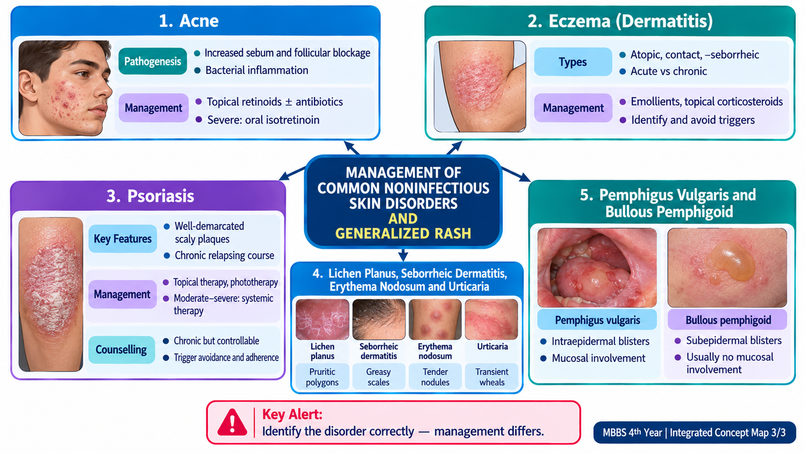
C. Acne and Eczema
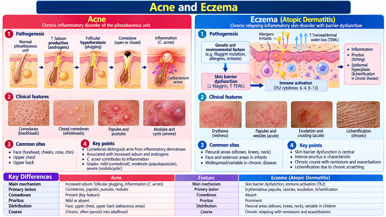
Acne and eczema are common but mechanistically different inflammatory skin disorders. Acne is centred on the pilosebaceous unit, whereas eczema is characterized by inflammation associated with impaired skin-barrier function and a tendency to pruritic dermatitis. Understanding this difference makes their clinical appearance and management easier to remember.
Acne
Acne vulgaris develops through interaction between increased sebum production, follicular keratinization, proliferation of Cutibacterium acnes and inflammation. Obstruction of the pilosebaceous follicle produces comedones; increasing inflammation produces papules, pustules and deeper lesions.
Clinically, acne may be predominantly comedonal, inflammatory or nodulocystic/severe. The face, upper chest and back are typical sites because these areas contain many sebaceous glands. Severity matters because deeper inflammation increases the risk of scarring. Management is guided by lesion type and severity. Topical therapies are appropriate for many mild cases, while more extensive inflammatory disease may require systemic treatment. Severe, scarring or treatment-resistant acne warrants specialist-directed therapy. The patient should understand that improvement is gradual and that manipulating lesions can worsen inflammation and scarring. Mild acne is treated with topical benzoyl peroxide and/or a retinoid such as adapalene. Moderate inflammatory acne may require oral doxycycline combined with appropriate topical therapy; antibiotic monotherapy should be avoided. Severe nodulocystic or scarring acne may require oral isotretinoin under dermatological supervision, with strict pregnancy prevention precautions.
Eczema
Eczema (dermatitis) is an inflammatory reaction pattern characterized clinically by itching and inflammation. Acute lesions may be erythematous and vesicular or weeping, while chronic scratching and inflammation lead to scaling, thickening and lichenification.
Important clinical forms include atopic dermatitis, contact dermatitis and seborrheic dermatitis. Management depends on the cause but commonly includes avoidance of identified triggers or irritants, restoration of the skin barrier with emollients, and appropriate anti-inflammatory treatment. Regular emollients form the basis of eczema treatment. Flares are managed with topical corticosteroids of appropriate potency, such as hydrocortisone for mild disease or sensitive areas and stronger preparations for thicker skin where indicated. Tacrolimus or pimecrolimus may be used as steroid-sparing treatments, particularly at sensitive sites.
D. Psoriasis, Lichen Planus and Seborrheic Dermatitis
These inflammatory dermatoses can all produce erythematous or scaly lesions, but their morphology and preferred distribution differ. Recognizing those patterns is more useful than memorizing disconnected lists.
Psoriasis
Psoriasis is a chronic immune-mediated inflammatory skin disease associated with accelerated epidermal turnover. The classic pattern is a well-demarcated erythematous plaque with silvery scale, commonly affecting extensor surfaces and the scalp. Nail changes may occur, and some patients develop associated inflammatory arthritis.
Management depends on extent and severity. Localized disease is commonly managed with topical treatment, while more extensive or difficult disease may require phototherapy or systemic therapy under specialist supervision. Associated joint symptoms should not be overlooked. Localized plaque psoriasis is commonly treated with topical corticosteroids and a vitamin D analogue such as calcipotriol, often in combination. Moderate-to-severe disease may require phototherapy or systemic treatment such as methotrexate under specialist supervision.
Counselling a patient with psoriasis
Patients should understand that psoriasis is a chronic inflammatory condition with periods of improvement and recurrence. It is not contagious. Treatment aims to control rather than permanently “cure” the tendency to disease. Explain the importance of adherence, appropriate skin care, follow-up, recognition of joint symptoms and the psychosocial effect of visible lesions. Counselling should avoid blame or stigma.
Lichen planus
Lichen planus classically produces pruritic, purple, polygonal, flat-topped papules or plaques. The wrists and ankles are typical sites, and mucosal disease may occur. The characteristic morphology helps separate it from psoriasis and eczema.
Seborrheic dermatitis
Seborrheic dermatitis affects sebaceous areas such as the scalp, eyebrows, nasolabial folds and other oily regions. It typically produces erythema with greasy scale. Management is directed at controlling scale, inflammation and the associated seborrheic process. Lichen planus is usually managed with topical corticosteroids; extensive or persistent disease requires dermatological assessment. Seborrheic dermatitis is treated with topical antifungals such as ketoconazole cream or shampoo, with short courses of low-potency topical corticosteroids for significant inflammation.
| Feature | Psoriasis | Lichen planus | Seborrheic dermatitis |
|---|---|---|---|
| Typical lesion | Well-defined scaly plaque | Purple flat-topped papule | Erythema with greasy scale |
| Distribution clue | Extensors and scalp | Wrists/ankles; mucosa possible | Sebaceous areas |
| Useful associated clue | Nails or inflammatory arthritis | Marked pruritus | Scalp/facial involvement |

A 28-year-old man has recurrent, sharply demarcated scaly plaques over both elbows and knees and similar lesions on the scalp. He is embarrassed by the appearance and asks whether he can transmit the condition to his family. He also mentions intermittent stiffness and swelling of several fingers.
- Which morphology and distribution make the skin diagnosis likely?
- Why are the joint symptoms clinically important?
- What key message should be included in counselling?
E. Urticaria and Erythema Nodosum
Urticaria and erythema nodosum illustrate why identifying the anatomical level of inflammation helps explain the appearance of a lesion. Urticaria is characterized by superficial dermal edema producing transient wheals, whereas erythema nodosum is a deeper inflammatory process involving subcutaneous tissue and therefore presents as tender nodules.
Urticaria
A wheal is a raised, edematous, usually pruritic lesion. Histamine and other mediators released from mast cells increase vascular permeability, producing localized dermal edema. Individual lesions are transient, although new wheals may continue to appear.
Assessment should identify possible triggers and, most importantly, determine whether the reaction is limited to the skin or associated with airway, breathing or circulatory compromise. Uncomplicated urticaria is managed by removing an identifiable trigger where possible and using antihistamine therapy. Features of anaphylaxis require emergency management rather than routine treatment of a rash. Second-generation H1 antihistamines such as cetirizine, loratadine or fexofenadine are first-line treatment for uncomplicated urticaria. Suspected anaphylaxis requires immediate intramuscular adrenaline (epinephrine) and emergency resuscitation; antihistamines must not delay adrenaline administration.
Erythema nodosum
Erythema nodosum presents as tender erythematous subcutaneous nodules, classically over the shins. It is best understood as a reaction pattern rather than a single disease. Assessment should therefore look for an associated trigger or systemic condition when clinically indicated. Management includes addressing the underlying cause and providing appropriate symptomatic care.

F. Pemphigus Vulgaris and Bullous Pemphigoid
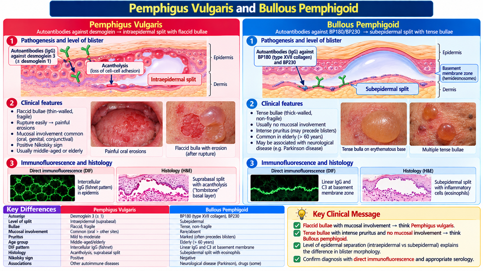
When a patient presents with blisters, the level at which the epidermis separates strongly influences the clinical appearance. This provides a useful mechanism for distinguishing pemphigus vulgaris from bullous pemphigoid.
Pemphigus vulgaris
Pemphigus vulgaris is an autoimmune blistering disease in which antibodies disrupt adhesion between epidermal keratinocytes. Loss of cell-to-cell adhesion—acantholysis—creates a relatively superficial intraepidermal split. Because the blister roof is thin, blisters are typically flaccid and rupture easily, leaving painful erosions. Oral mucosal involvement is an important clinical clue.
Bullous pemphigoid
Bullous pemphigoid is also autoimmune, but the separation occurs deeper at the epidermal–dermal junction. The deeper blister roof is stronger, producing tense bullae. It typically affects older adults, and prominent mucosal involvement is less characteristic than in pemphigus vulgaris.
| Feature | Pemphigus vulgaris | Bullous pemphigoid |
|---|---|---|
| Level of split | Intraepidermal | Subepidermal |
| Blister | Flaccid, easily ruptured | Tense |
| Mucosal involvement | Common and important | Less prominent |
| Typical context | Painful erosive blistering disease | Older patient with tense bullae |
Both conditions require confirmation and specialist management. Pemphigus vulgaris requires systemic treatment, commonly rituximab with corticosteroids, under specialist care. Bullous pemphigoid is often treated with a potent topical corticosteroid such as clobetasol; systemic prednisolone may be required in selected patients. Supportive wound care and monitoring for secondary infection are important in both conditions. Treatment aims to suppress the autoimmune inflammatory process while providing skin and wound care and monitoring for complications of disease and therapy. At Final Year level, the crucial reasoning link is level of blister formation → strength of blister roof → clinical morphology.
Visual placement: skin layers showing superficial pemphigus split versus deeper pemphigoid split and resulting blister appearance.
G. Connecting Recognition to Management
The safest way to approach these disorders is to let the clinical pattern determine the next step. A stable patient with a familiar chronic inflammatory dermatosis needs accurate recognition, severity assessment and appropriate treatment. A patient with widespread painful skin loss, mucosal involvement or systemic compromise requires a different priority: recognize danger first, remove a suspected precipitant where appropriate and escalate care.
A practical clinical sequence
Localized/common inflammatory disease: identify the likely condition, assess severity and complications, and use appropriate topical or systemic treatment principles. Generalized eruption: review medications, mucosa and systemic symptoms and consider whether the skin finding reflects a systemic disease. Blistering disease: determine whether the morphology suggests superficial fragile blisters or deeper tense bullae, and arrange appropriate confirmation and specialist treatment. Potential emergency: painful widespread eruption, extensive detachment, major mucosal disease or systemic instability changes the priority from routine dermatology to urgent supportive and specialist care.
Integrated Mechanism Flow
AIM High-Yield Review
- A generalized rash should be approached through morphology, distribution, chronology, drug exposure and systemic clues.
- ⭐ Painful skin with mucosal erosions and epidermal detachment after a medication exposure should raise concern for SJS/TEN.
- Erythroderma is extensive erythema and scaling and may cause clinically important systemic disturbance.
- Acne results from abnormalities of the pilosebaceous unit; comedones are a major diagnostic clue.
- Chronic eczema may become thickened and lichenified because persistent inflammation and scratching remodel the skin.
- ⭐ Psoriasis classically produces sharply defined scaly plaques on extensor surfaces and may involve the nails and joints.
- Psoriasis is chronic and inflammatory but not contagious; counselling should address recurrence, adherence, joint symptoms and psychosocial concerns.
- Lichen planus is suggested by pruritic purple, polygonal, flat-topped lesions; seborrheic dermatitis favours oily/sebaceous areas.
- Urticaria produces transient pruritic wheals through mediator-induced dermal edema; airway or circulatory involvement changes the problem to an emergency.
- Erythema nodosum produces tender subcutaneous nodules and should prompt consideration of an underlying trigger or systemic disorder.
- ⭐ Pemphigus vulgaris has a superficial intraepidermal split → fragile, flaccid bullae and erosions, with important mucosal involvement.
- ⭐ Bullous pemphigoid has a deeper subepidermal split → a stronger blister roof and tense bullae.
- Skin findings can signal metabolic, endocrine, autoimmune or neoplastic disease; interpret them within the whole patient rather than in isolation.
🎥 AIM Video Learning — Topic 10
Noninfectious Skin Disorders and Generalized Rash
Focus: autoimmune blistering disorders, clinical morphology, diagnosis and management.
📌 This verified working lecture covers pemphigus vulgaris including clinical presentation, biopsy, immunofluorescence and treatment.