
This chapter follows the KMU outcomes for burns from recognition and assessment to referral, management, complications and recovery. First understand the clinical sequence; use the High-Yield Review at the end for rapid revision.
Topic 12. Burns Assessment, Management and Recovery
MSK Module • Surgery / Plastic Surgery
A patient-centred approach to classifying burns, estimating burn size, assessing fluid needs, recognizing referral requirements, managing the acute and recovery phases, and anticipating complications.
Topic Introduction
A burn is tissue injury produced by thermal, chemical, electrical or other damaging energy. In Final Year, the important skill is not simply naming a burn: you must determine how deep it is, how extensive it is, whether the patient is physiologically threatened, whether special areas are involved, and what treatment is required next. A small superficial burn may need only local care, whereas an extensive or deep burn can cause major fluid loss, shock, infection, functional disability and long-term scarring. The safest approach is therefore sequential: assess the patient first, assess the burn second, estimate fluid requirements where appropriate, identify patients needing specialist care, and then plan wound healing and rehabilitation.
A. Understanding and Classifying Burns
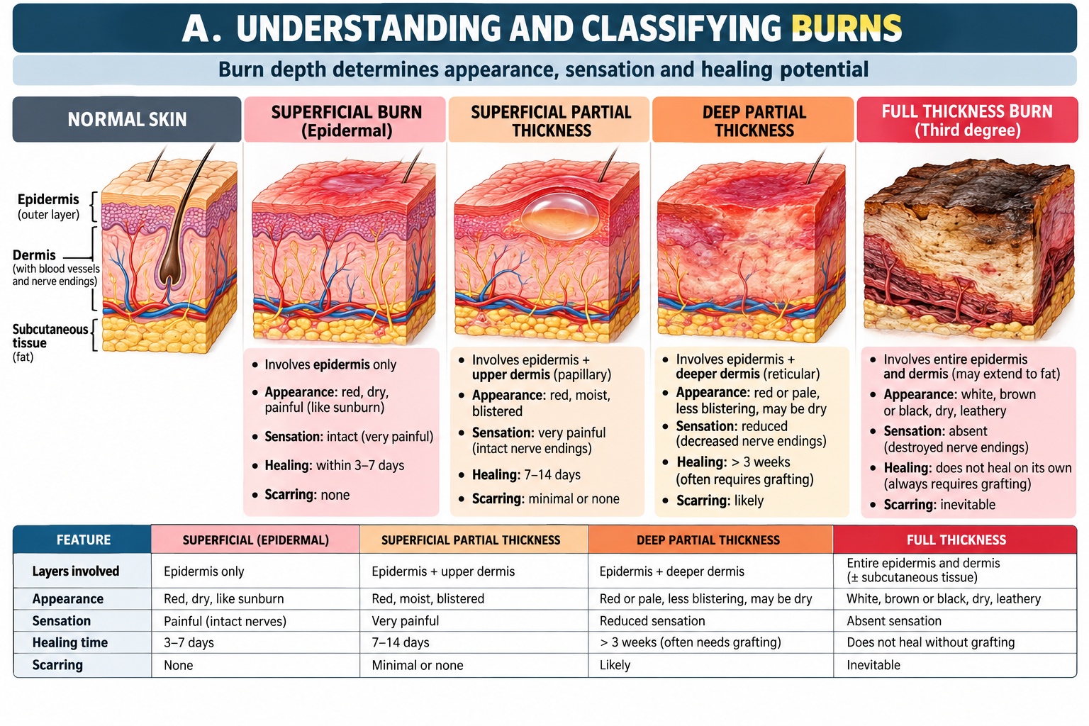
A burn should be described by its cause, depth and extent. Depth matters because it reflects how much of the skin has been destroyed and therefore influences pain, healing potential, scarring and the possible need for surgical treatment.
Classification by cause
- Thermal burns: caused by flame, hot liquids, steam or contact with hot objects.
- Chemical burns: result from tissue exposure to corrosive substances; ongoing contact may continue tissue injury until decontamination occurs.
- Electrical burns: may produce relatively small external wounds despite deeper tissue injury.
Classification by depth
Superficial burns involve the epidermis. They are typically red and painful but do not blister and generally heal without scarring.
Partial-thickness burns extend into the dermis. Superficial partial-thickness injury is typically moist, blistered and very painful because viable dermal structures and sensory endings remain. Deeper partial-thickness injury extends farther into the dermis, may appear paler and has reduced sensation and slower healing.
Full-thickness burns destroy the entire epidermis and dermis. The surface may appear white, brown or charred and can be relatively painless centrally because sensory nerve endings have been destroyed. These burns have poor spontaneous epithelial regeneration and commonly require specialist surgical management.
| Depth | Typical appearance | Sensation | Healing implication |
|---|---|---|---|
| Superficial | Red, dry | Painful | Usually heals without scarring |
| Superficial partial thickness | Moist, pink/red, blistered | Very painful | Usually heals spontaneously |
| Deep partial thickness | Paler, less moist | Reduced | Slower healing; greater scar risk |
| Full thickness | White, brown or charred; leathery | Reduced or absent centrally | Usually needs specialist surgical care |
B. Initial Assessment, Burn Area and Fluid Assessment
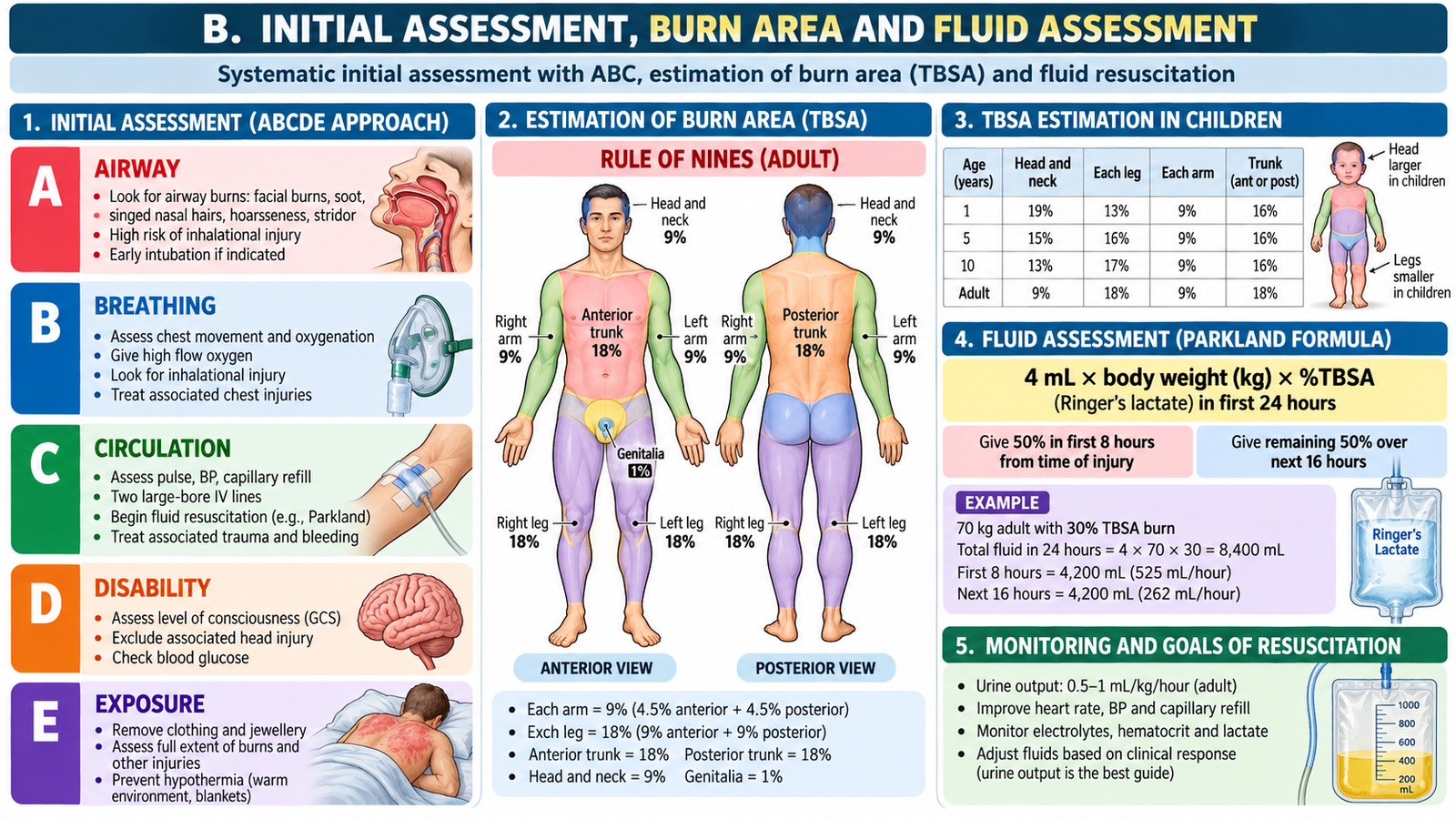
The first priority in a significant burn is the patient rather than the wound. A dramatic skin injury must not distract from airway compromise, respiratory injury or circulatory instability. Initial assessment therefore follows the familiar emergency sequence of airway, breathing and circulation before detailed burn assessment.
Immediate assessment
- Airway: assess patency and look for evidence suggesting inhalational injury, especially after burns in an enclosed space or burns involving the face and neck.
- Breathing: assess respiratory effort, oxygenation and chest movement.
- Circulation: assess pulse, blood pressure, peripheral perfusion and evidence of shock.
- Identify associated trauma and other immediately threatening injuries.
- Once stable, determine burn depth, total body surface area and anatomical sites involved.
Estimating total body surface area
The percentage of total body surface area (TBSA) burned helps determine severity, fluid requirements and referral. Superficial erythema alone is not counted when calculating the area requiring burn resuscitation.
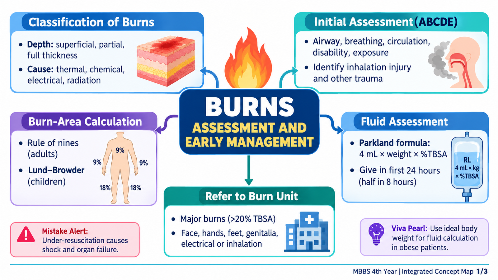
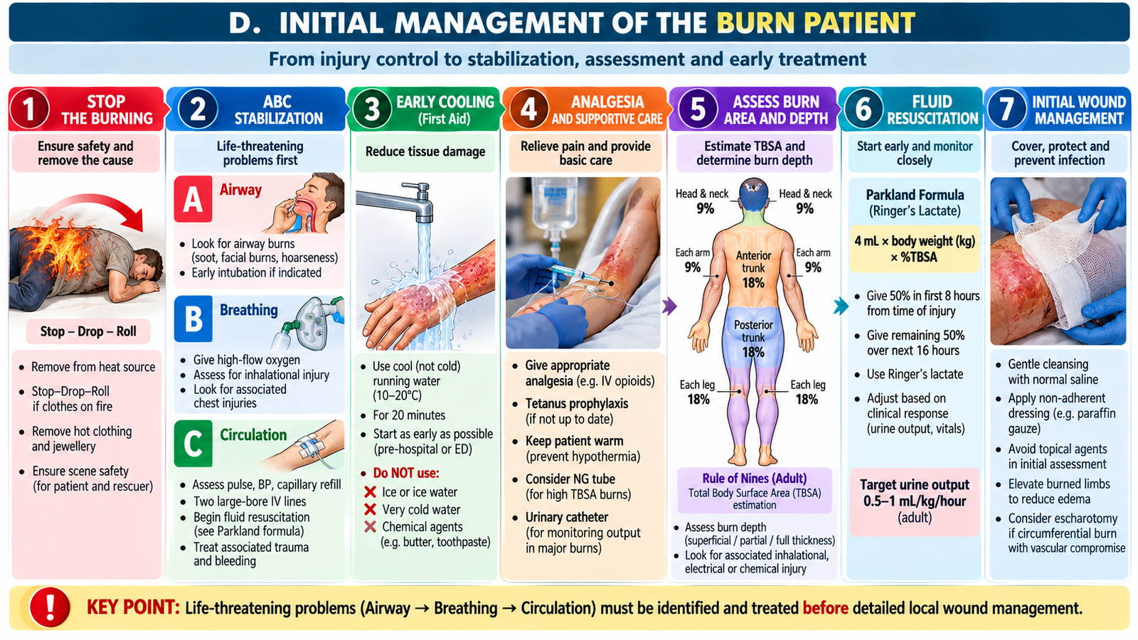
In adults, the Rule of Nines provides a rapid estimate: the head and neck represent 9%, each upper limb 9%, the anterior trunk 18%, posterior trunk 18%, each lower limb 18%, and the perineum approximately 1%.
For children, body proportions differ from adults—particularly the relatively larger head and smaller legs—so an age-adjusted chart such as the Lund–Browder chart provides a more accurate estimate. For small scattered burns, the patient’s palm including fingers can provide a rough estimate of approximately 1% TBSA.
Fluid assessment
Major burns increase capillary permeability. Plasma fluid moves from the intravascular compartment into injured and surrounding tissues, producing edema and reducing effective circulating volume. Extensive burns can therefore cause burn shock unless circulating volume is restored.
Major burn → increased capillary permeability → intravascular fluid loss → reduced tissue perfusion → fluid resuscitation with ongoing reassessment.
The traditional Parkland formula calculates 4 mL × body weight (kg) × %TBSA of crystalloid over 24 hours, with half given during the first 8 hours from injury. However, current American Burn Association guidance recommends starting adult resuscitation for burns ≥20% TBSA at 2 mL/kg/%TBSA to reduce fluid overload. Lactated Ringer’s solution is commonly used, and the initial rate must be adjusted according to clinical response and urine output (adult target approximately 0.5 mL/kg/hour).
C. Identifying Patients Who Need Specialized Burn Care
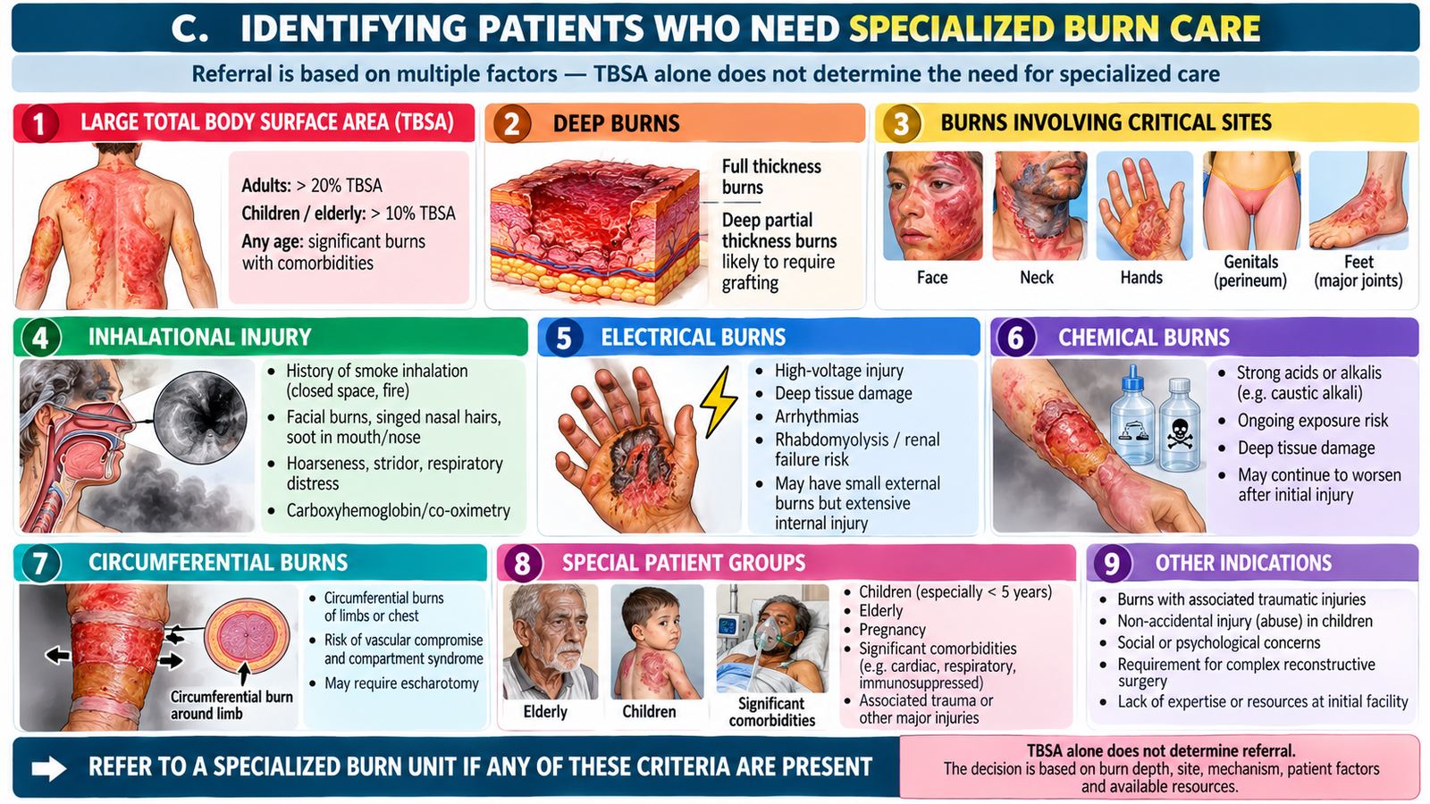
Burn severity cannot be judged from surface area alone. A relatively small burn may be serious because of its depth, anatomical location, mechanism, airway involvement, associated injury or patient factors. The purpose of referral assessment is to identify patients who may need specialist wound care, surgery, intensive monitoring or rehabilitation.
Specialist burn-unit assessment should be considered particularly when there are:
- extensive burns;
- deep partial-thickness or full-thickness burns;
- burns involving functionally or cosmetically important sites such as the face, hands, feet, genital/perineal region or major joints;
- suspected inhalational injury;
- significant electrical or chemical burns;
- circumferential burns that may threaten circulation or ventilation;
- important associated trauma or serious comorbidity;
- children or other patients in whom specialized burn care is required.
The central principle is simple: depth + extent + site + mechanism + physiological condition determine the seriousness of a burn more accurately than TBSA alone.
A 35-year-old man is brought after a house fire. He has painful partial-thickness burns over the anterior trunk and one upper limb. His face is singed and his voice has become hoarse. He is anxious and tachycardic.
Think clinically:
- Which problem must be prioritized before calculating the burn area?
- Which findings suggest that this patient may deteriorate despite apparently adequate breathing initially?
- How will burn extent help guide subsequent fluid and referral decisions?
D. Initial Management of the Burn Patient
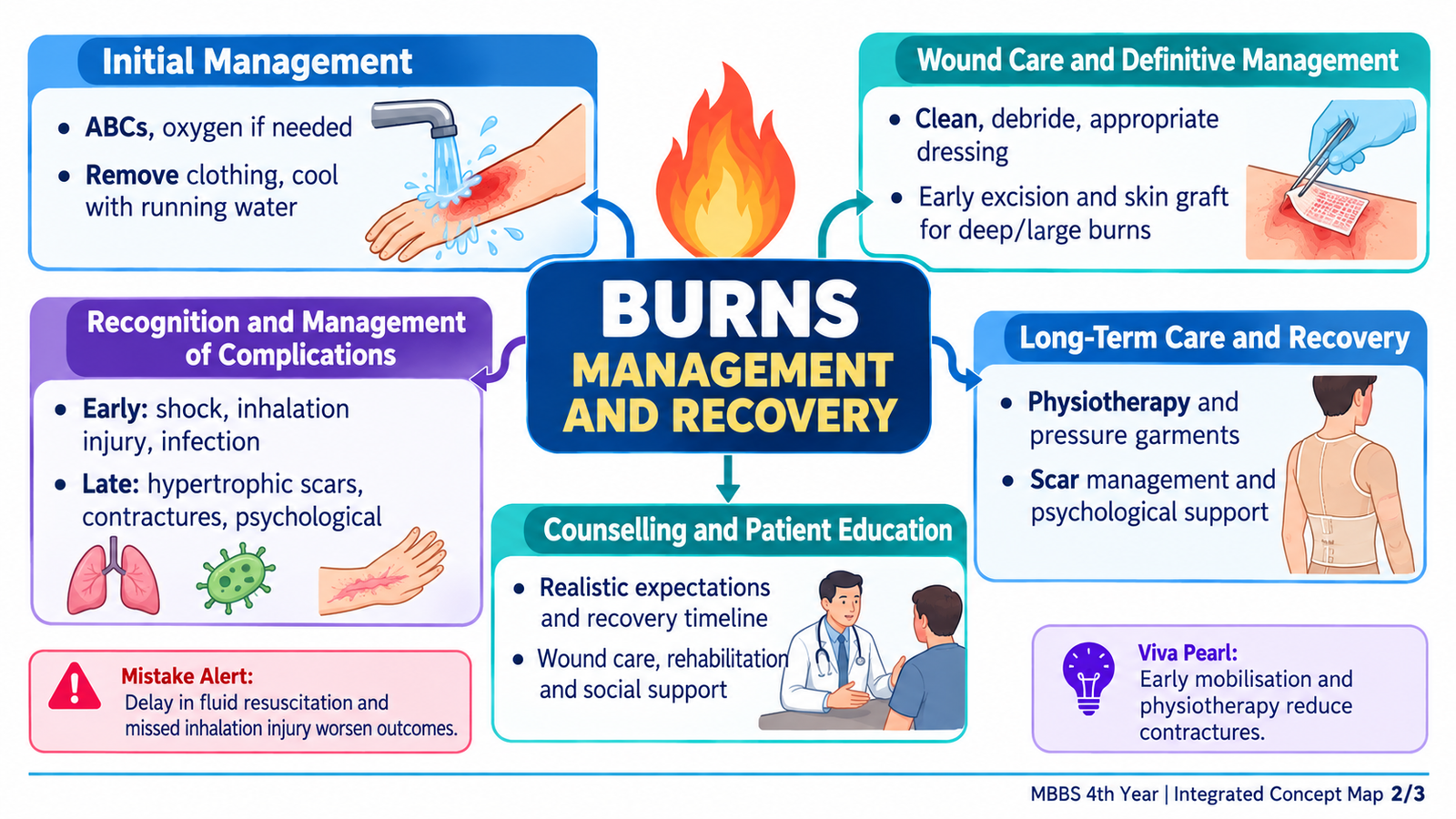
Early burn management has two simultaneous goals: protect life and limit further tissue injury. The sequence depends on severity. An unstable patient requires resuscitation first; local wound care becomes important once immediate threats have been addressed.
Immediate priorities
- Stop the burning process and remove the patient from the source of injury.
- Assess and protect the airway, particularly when inhalational injury is suspected.
- Support breathing and circulation as required.
- Remove constricting clothing and jewellery where safe because progressive edema can make them dangerous.
- Cool a recent thermal burn appropriately with cool running water; avoid ice and measures that produce hypothermia.
- Provide appropriate analgesia.
- Assess tetanus immunization status and provide tetanus prophylaxis when indicated. Cool recent thermal burns under cool running water for approximately 20 minutes while preventing hypothermia, especially in children and patients with extensive burns.
- Estimate burn depth and TBSA after initial stabilization.
- Begin fluid resuscitation when indicated and monitor the patient’s physiological response.
- Cover the burn with an appropriate clean dressing and protect the patient from heat loss.
Local wound management
Burn wounds require assessment, cleaning and appropriate dressings. The goal is to promote healing, reduce contamination, control pain and preserve function. Deeper burns require early specialist assessment because devitalized tissue may not heal adequately without surgical management.
Full-thickness and selected deep burns may require excision of nonviable tissue and skin grafting. The undergraduate principle is to recognize when spontaneous healing is unlikely and specialist surgical treatment is required rather than to memorize operative technique.
Special mechanisms
Chemical burns require prompt removal of the offending substance and appropriate decontamination to stop continuing tissue injury. Electrical burns deserve careful assessment because the visible skin lesion may underestimate deeper tissue damage.
E. Ongoing and Long-Term Management
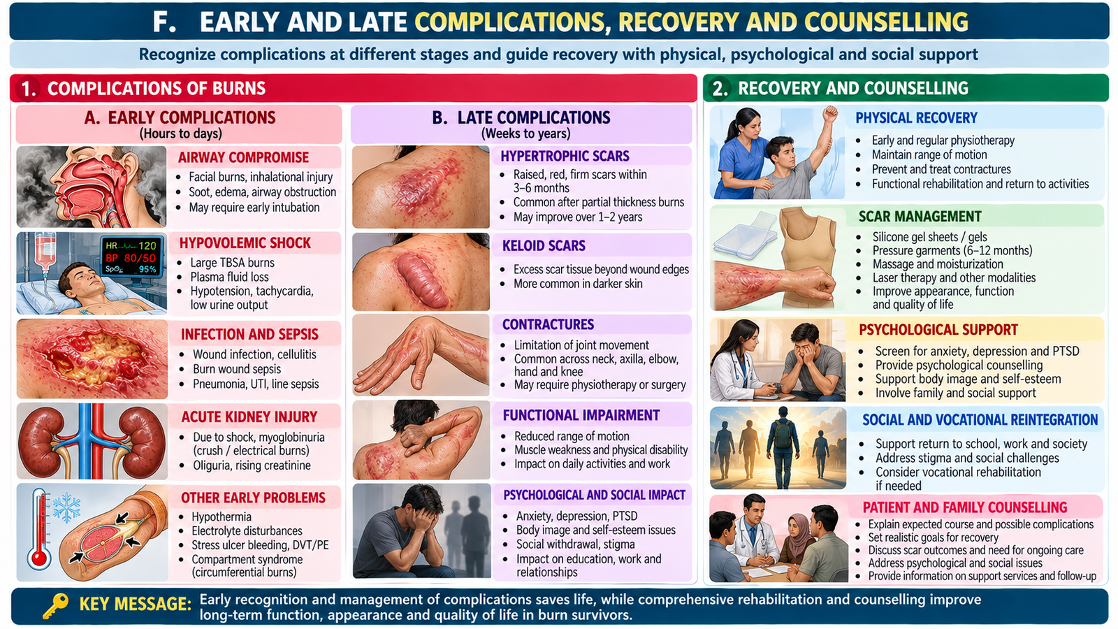
Burn treatment does not end after resuscitation. Once the patient is stable, care shifts toward wound healing, infection surveillance, nutrition, preservation of movement, scar control and restoration of function. These components are particularly important in extensive or deep burns because recovery may continue for weeks or months.
Wound healing and infection surveillance
The damaged skin barrier makes the wound vulnerable to microbial colonization and infection. Wounds therefore require repeated clinical assessment. Increasing pain, spreading erythema, purulent discharge, systemic deterioration or failure to progress as expected should prompt reassessment.
Nutrition
Major burns produce a marked metabolic stress response and increase nutritional requirements. Adequate nutritional support helps wound healing and recovery and becomes increasingly important as burn size and severity increase.
Surgery and wound closure
Deep wounds that cannot epithelialize adequately may require excision and grafting. Timely wound closure reduces the period during which the patient has an open wound and supports subsequent rehabilitation.
Rehabilitation
Healing across joints and mobile areas can produce stiffness and contracture. Positioning, physiotherapy and range-of-motion rehabilitation therefore aim to preserve movement and function. Scar management is important when hypertrophic scars or contractures develop.
Recovery also has psychological and social dimensions. Visible scarring, prolonged hospitalization, pain and functional limitations can affect confidence, family relationships and return to normal activities. Burn rehabilitation should therefore be patient-centred rather than wound-centred.
F. Early and Late Complications, Recovery and Counselling
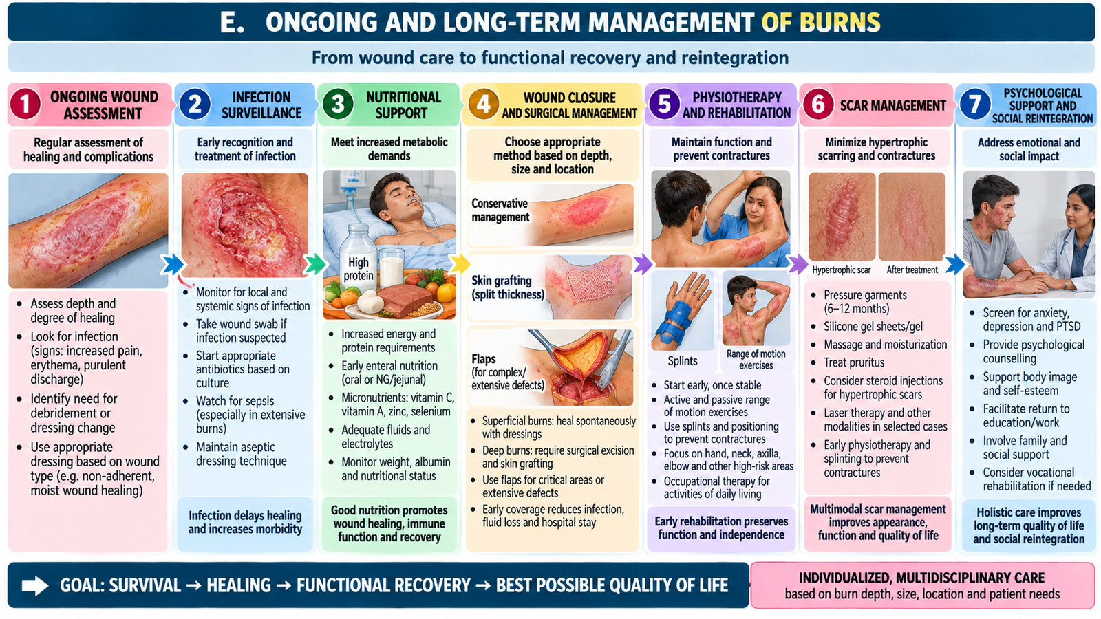
Complications are easier to understand when linked to the stage of burn injury. Early problems mainly result from airway injury, fluid loss, tissue damage and systemic stress. Late problems are more closely related to wound healing, scarring, loss of movement and psychosocial recovery.
Important early complications
- Airway compromise: edema after inhalational or facial injury can progressively narrow the airway.
- Hypovolemic burn shock: extensive capillary leak reduces effective circulating volume.
- Respiratory complications: inhalational injury can significantly worsen morbidity.
- Acute kidney injury: may follow inadequate tissue perfusion in severe burns.
- Wound infection and sepsis: loss of the protective skin barrier increases susceptibility to infection.
- Circulatory compromise: a tight circumferential burn can restrict perfusion as tissue edema develops.
Important late complications
- Hypertrophic scarring.
- Contractures and restriction of joint movement.
- Functional impairment, particularly after burns of the hands or across major joints.
- Persistent cosmetic concerns and altered body image.
- Psychological and social difficulties during recovery.
Counselling the patient and family
Counselling should explain the expected recovery process in understandable language. The patient and family should understand that treatment may involve repeated wound assessment, dressings, possible surgery for deeper injury, pain control, nutrition and rehabilitation.
They should also understand that healing does not necessarily mean the end of treatment. Follow-up may be required for scar maturation, movement, function and psychological recovery. Families should know which signs—such as systemic deterioration, worsening wound changes or increasing functional limitation—require reassessment.
The discussion should be realistic but supportive. Prognosis depends on factors such as burn depth, extent, site, complications and the patient’s general condition. Avoid giving false reassurance or unnecessarily alarming predictions.
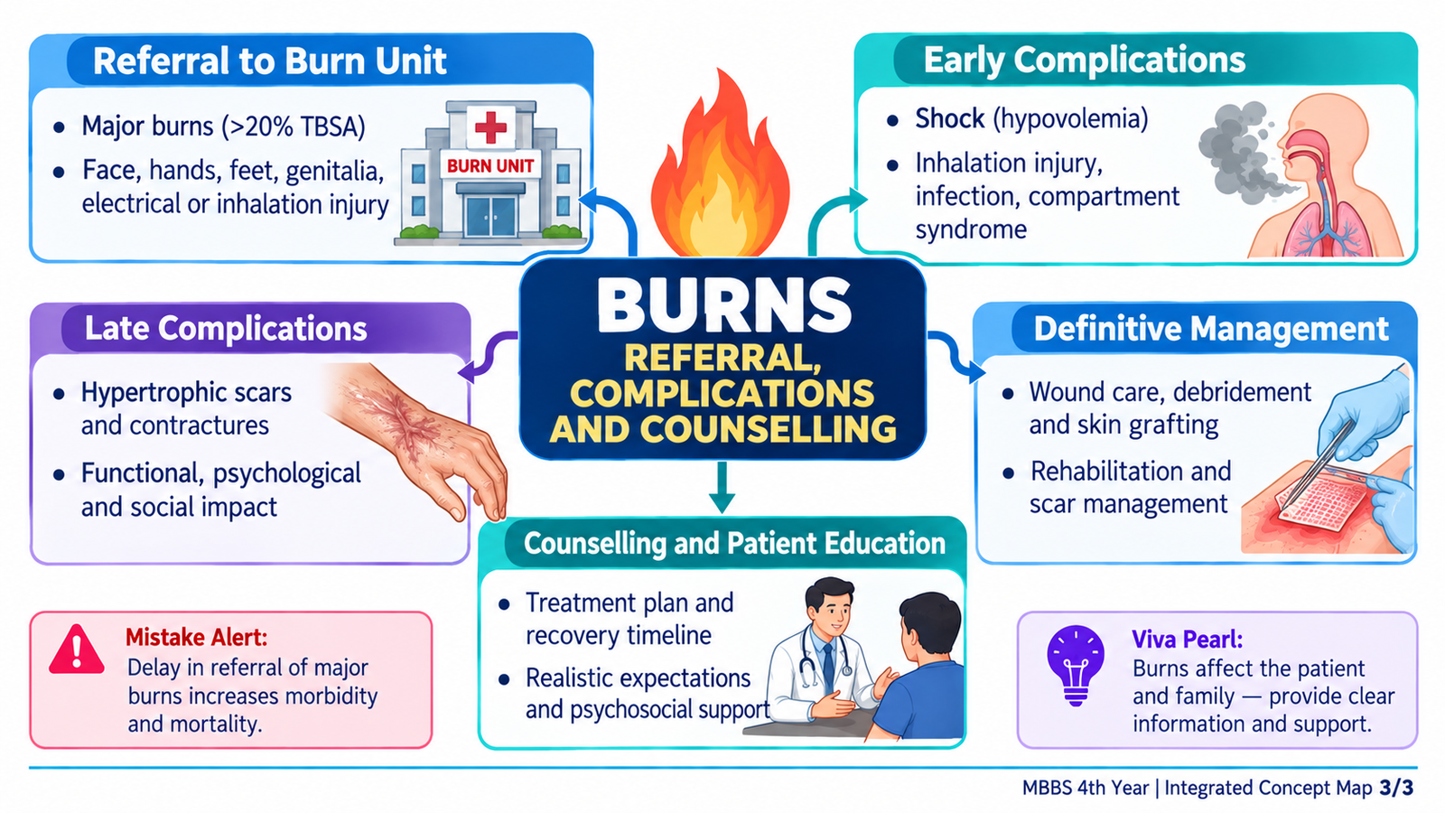
Integrated Mechanism Flow
Important Comparison — Early vs Late Burn Complications
| Early complications | Late complications |
|---|---|
| Airway compromise / inhalational injury | Hypertrophic scarring |
| Burn shock | Contractures |
| Acute kidney injury | Restricted movement |
| Infection / sepsis | Functional impairment |
| Circulatory compromise in circumferential burns | Psychological and cosmetic consequences |
AIM High-Yield Review
- Burn assessment is based on depth, TBSA, site, mechanism and the patient’s physiological condition.
- Superficial partial-thickness burns are typically moist and very painful; full-thickness burns may have markedly reduced sensation.
- ⭐ In a major burn, ABC assessment comes before detailed wound assessment.
- Facial/neck burns, hoarseness and an enclosed-space fire should raise concern for inhalational injury.
- The Rule of Nines rapidly estimates adult TBSA; the Lund–Browder method is more appropriate for age-related body proportion differences in children.
- ⭐ Superficial erythema is not included when calculating TBSA for burn fluid resuscitation.
- Extensive burns cause capillary leak, intravascular volume loss and potentially burn shock.
- ⭐ Parkland calculation provides an initial fluid estimate; the first 8 hours are counted from the time of injury.
- Fluid calculations guide initial therapy but clinical response and urine output guide continuing resuscitation.
- Critical-site, deep, extensive, inhalational, electrical and significant chemical burns may require specialist burn-unit care.
- Deep burns may require excision and skin grafting because spontaneous epithelial healing is inadequate.
- Early complications include airway compromise, shock, infection/sepsis and organ dysfunction.
- Late complications include hypertrophic scars, contractures and functional impairment.
- Burn recovery includes wound healing, nutrition, rehabilitation, scar management and patient-family counselling—not simply closure of the skin wound.
🎥 AIM Video Learning — Burns Assessment & Fluid Resuscitation
Review burn surface-area estimation, the Rule of Nines, Lund–Browder method, burn pathophysiology and principles of fluid resuscitation.