
This chapter follows the KMU outcomes for viral, bacterial and fungal skin infections. First learn how the morphology and distribution of lesions point toward the cause; use the High-Yield Review only after you understand the clinical patterns.
Topic 11. Viral, Bacterial and Fungal Skin Infections
Musculoskeletal Module • Dermatology
A clinical approach to recognizing common infectious skin lesions, distinguishing similar presentations and applying appropriate undergraduate management principles.
Topic Introduction
Infectious skin diseases become easier when you learn to recognize three things: what the lesion looks like, where it occurs and how the infection spreads. Viral infections commonly produce characteristic vesicles, papules or proliferative lesions; bacterial infections range from superficial crusting to deeper follicular disease; and fungal infections often produce scaling or involvement of warm, moist areas. This chapter connects these visible patterns with their causes, transmission, important differential diagnoses and management. It also covers cutaneous tuberculosis, leprosy and syphilis, where skin findings may reflect a more persistent systemic infection, and the important skin manifestations that may suggest HIV/AIDS. The goal is not to memorize a long list of rashes, but to develop a structured clinical approach to infectious skin disease.

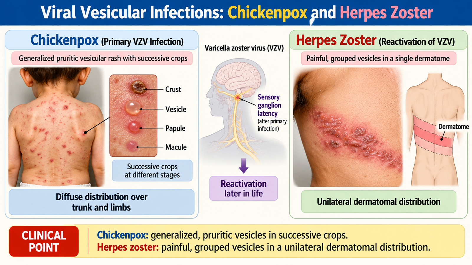
A. Viral Vesicular Infections: Chickenpox and Herpes Zoster
Chickenpox and herpes zoster are caused by the same virus, varicella-zoster virus (VZV), but represent different biological events. Chickenpox follows primary infection. After recovery, VZV remains latent in sensory ganglia. Later reactivation produces herpes zoster, in which the virus travels along a sensory nerve to the skin.
Primary VZV infection → viremia → widespread skin lesions → latency in sensory ganglia → later reactivation → spread along sensory nerve → dermatomal herpes zoster
Chickenpox
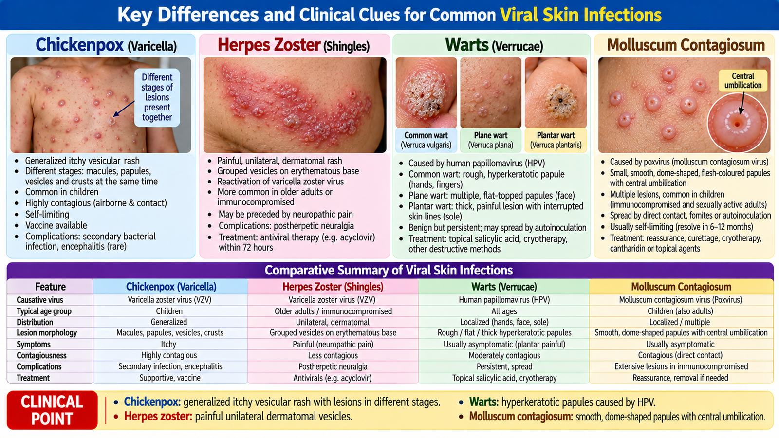
Chickenpox typically produces fever and a generalized itchy eruption. Lesions appear in successive crops, so macules, papules, vesicles and crusted lesions can be present at the same time. The eruption is commonly more prominent on the trunk than the distal limbs.
The presence of lesions at different stages is clinically useful. Important alternatives include other vesicular eruptions, insect-bite reactions and disseminated herpes infections, but the combination of systemic symptoms, successive crops and generalized distribution supports chickenpox.
Management: uncomplicated disease is mainly managed with symptomatic care, skin care and measures to reduce transmission. Antiviral therapy is considered when disease is severe or the patient has increased risk of complications. Secondary bacterial infection of scratched lesions is an important complication to recognize.
Treatment: Paracetamol and antihistamines provide symptomatic relief. Avoid aspirin in children. Oral acyclovir may be considered in high-risk patients, ideally within 24 hours of rash onset. Severe or complicated varicella requires hospital assessment and IV acyclovir.
Herpes zoster
Herpes zoster classically begins with localized pain, burning or abnormal sensation followed by grouped vesicles in a unilateral dermatomal distribution. The rash generally does not cross the midline because reactivation occurs within a particular sensory ganglion and its corresponding dermatome.
- Key clue: painful grouped vesicles confined to a dermatome.
- Important complication: persistent neuropathic pain after the eruption, especially in older patients.
- Important site: involvement of the ophthalmic division of the trigeminal nerve may threaten the eye and requires urgent assessment.
- Management principle: antiviral therapy is most useful when started early, together with appropriate analgesia and lesion care. Treatment: Oral acyclovir, valacyclovir or famciclovir should preferably be started within 72 hours of rash onset. Paracetamol or appropriate analgesics relieve acute pain; gabapentin or pregabalin may be used for persistent neuropathic pain.
B. Viral Papular and Proliferative Lesions: Warts and Molluscum Contagiosum
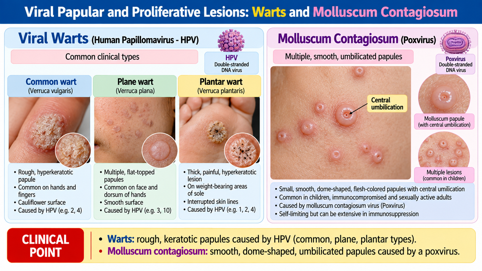
Warts and molluscum contagiosum are both transmissible viral skin diseases, but their morphology is quite different. Recognizing the surface and central structure of the lesion is therefore more useful than simply remembering that both are viral.
Warts
Warts are caused by human papillomavirus (HPV), which infects keratinocytes and promotes localized epidermal proliferation. Transmission occurs through direct contact and autoinoculation and is facilitated by small breaks in the skin.
Clinically, warts may be classified according to their morphology and location:
- Common warts: rough, hyperkeratotic papules, commonly on hands and fingers.
- Plane warts: smoother, flatter papules.
- Plantar warts: lesions on pressure-bearing plantar skin that may become painful.
- Anogenital warts: lesions occurring in the genital or perianal region.
A plantar wart can resemble a corn. A useful distinction is that a wart disrupts normal skin lines and may show tiny thrombosed capillaries, whereas a corn is primarily related to pressure and retains a different keratin pattern.
Management depends on site, symptoms and extent. Some warts regress spontaneously. Common undergraduate treatment principles include keratolytic therapy such as salicylic acid and destructive methods such as cryotherapy. Genital lesions require appropriate assessment because transmission is sexual and certain HPV types have important oncogenic associations.
Molluscum contagiosum
Molluscum contagiosum is caused by a poxvirus and spreads through direct skin contact, contaminated objects and, in adults, sometimes sexual contact. The classic lesion is a smooth, firm, pearly or flesh-coloured papule with central umbilication.
In otherwise healthy children, lesions are usually self-limited. Treatment may therefore be unnecessary unless lesions are troublesome, extensive or complicated. Physical methods can be used when treatment is indicated.
C. Cutaneous Manifestations of HIV/AIDS
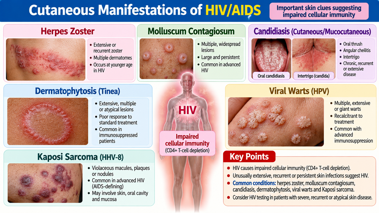
The skin can provide important clues to impaired immunity. HIV infection progressively affects cell-mediated immune function, so infections that are normally mild may become recurrent, extensive, persistent or unusually severe. The important clinical lesson is not that one skin lesion diagnoses HIV; rather, an unusual pattern or combination of conditions may indicate underlying immunosuppression.
Important manifestations that may be encountered include:
- extensive or recurrent herpes zoster;
- numerous or unusually large molluscum contagiosum lesions;
- persistent or extensive candidiasis;
- severe or recurrent dermatophyte infection;
- extensive viral warts;
- seborrheic dermatitis that is unusually severe;
- Kaposi sarcoma presenting as violaceous cutaneous lesions.
These findings should be interpreted in context. A common fungal infection or a single episode of zoster does not by itself establish HIV infection. The clue is the severity, recurrence, distribution and accompanying clinical context. When HIV is suspected, the patient requires appropriate counselling and diagnostic evaluation rather than diagnosis from appearance alone.
D. Common Pyogenic Bacterial Skin Infections
Impetigo, folliculitis, furuncles and carbuncles can be understood as infections occurring at progressively different anatomical levels. This anatomical approach helps explain their appearance: superficial epidermal infection produces crusting, infection centered on a hair follicle produces a pustule, and deeper extension around follicles produces painful nodules or interconnected abscesses.
Superficial epidermis → impetigo
Hair follicle → folliculitis
Deep follicle/perifollicular tissue → furuncle
Multiple interconnected furuncles → carbuncle
Impetigo
Impetigo is a superficial bacterial infection, commonly caused by Staphylococcus aureus and/or Streptococcus pyogenes. Non-bullous impetigo classically develops superficial vesicles or pustules that rupture and leave honey-coloured crusts. It is particularly common in children and is contagious.
Localized uncomplicated disease can be treated with appropriate local therapy, while more extensive disease may require systemic antibiotics according to clinical circumstances and local antimicrobial practice. Hygiene and avoidance of sharing contaminated personal items help limit transmission.
Treatment: Localized non-bullous impetigo can be treated with topical hydrogen peroxide 1%, or fusidic acid 2% or mupirocin 2% when a topical antibiotic is indicated. Widespread or bullous impetigo generally requires an oral antibiotic such as flucloxacillin, according to local guidelines.
Folliculitis, furunculosis and carbuncles
Folliculitis is inflammation and infection centered on hair follicles and appears as small follicular pustules. When infection extends deeply into the follicle and surrounding tissue, a painful inflammatory nodule or abscess forms—a furuncle. A carbuncle is a deeper interconnected collection involving several adjacent follicles.
S. aureus is an important cause. Recurrent disease should prompt consideration of predisposing factors such as persistent colonization, diabetes or impaired immunity where clinically appropriate.
Management depends on depth and extent. Mild superficial folliculitis may respond to local measures. A fluctuant furuncle or carbuncle represents a collection of pus, so drainage becomes an important management principle; systemic antibacterial treatment is added when clinically indicated.


🩺 AIM Clinical Bridge
A 10-year-old child develops several superficial facial lesions that rupture and leave golden-yellow crusts. His younger sibling develops similar lesions several days later. In another patient, a painful fluctuant nodule develops around a hair follicle and contains pus.
Think clinically:
- Which lesion represents superficial epidermal infection?
- Which lesion indicates deeper follicular infection with abscess formation?
- Why would drainage become more important in the second patient?
E. Chronic Bacterial and Sexually Transmitted Skin Disease: Tuberculosis, Leprosy and Syphilis
Some bacterial skin diseases do not present as acute pustular infections. Cutaneous tuberculosis, leprosy and syphilis may produce chronic plaques, nodules, ulcers or other persistent lesions. Their diagnosis depends on combining morphology with sensory findings, systemic clues, exposure history and appropriate laboratory or histopathological assessment.
Cutaneous tuberculosis
Cutaneous tuberculosis is caused by Mycobacterium tuberculosis. Skin disease can result from direct inoculation, contiguous spread, hematogenous spread or an immune response associated with tuberculosis. The exact appearance therefore varies with the route of infection and host immunity.
Lupus vulgaris is an important chronic form and typically produces slowly progressive reddish-brown plaques. Scrofuloderma develops when tuberculosis in an underlying structure such as a lymph node extends directly into the skin, producing nodules, ulceration and sinus formation.
Diagnosis is supported by the overall clinical pattern together with appropriate testing for tuberculosis, histopathology and microbiological investigation where possible. Management follows established multidrug antituberculous treatment principles rather than isolated topical treatment.
Leprosy
Leprosy is caused by Mycobacterium leprae and has a special affinity for the skin and peripheral nerves. This explains one of its most important clinical clues: a chronic skin lesion associated with altered or reduced sensation.
The clinical spectrum reflects the patient’s cell-mediated immune response. At one end, tuberculoid disease tends to produce fewer, well-defined lesions with marked sensory loss and localized nerve involvement. At the other end, lepromatous disease tends to be more widespread and symmetrical with numerous lesions and greater organism burden.
Focused assessment therefore looks for:
- hypopigmented or erythematous patches or plaques;
- sensory impairment within lesions;
- peripheral nerve involvement;
- motor or autonomic consequences of neuropathy;
- features suggesting more widespread disease.
Management requires multidrug therapy appropriate to disease classification. Early recognition matters because nerve damage can lead to disability even when the skin lesions themselves appear relatively limited. WHO multidrug therapy: Rifampicin, dapsone and clofazimine are used for both paucibacillary and multibacillary leprosy. Treatment duration is 6 months for paucibacillary and 12 months for multibacillary disease.
Syphilis
Syphilis is caused by Treponema pallidum and is mainly sexually transmitted. Its skin findings change with the stage of infection. Primary syphilis classically produces a painless chancre at the site of inoculation. Secondary syphilis can produce a generalized eruption that characteristically may involve the palms and soles, together with systemic features and lymphadenopathy.
Diagnosis combines clinical suspicion with appropriate serological testing. Management is based on penicillin therapy according to the clinical stage, together with evaluation and management of sexual partners and appropriate assessment for other sexually transmitted infections. First-line treatment: Primary, secondary and early latent syphilis are treated with benzathine penicillin G 2.4 million units IM as a single dose in adults. Late latent syphilis or infection of unknown duration requires weekly injections for 3 weeks. Neurosyphilis requires IV aqueous crystalline penicillin G under specialist care

F. Superficial Fungal Infections: Pityriasis Versicolor, Dermatophytosis and Candidiasis
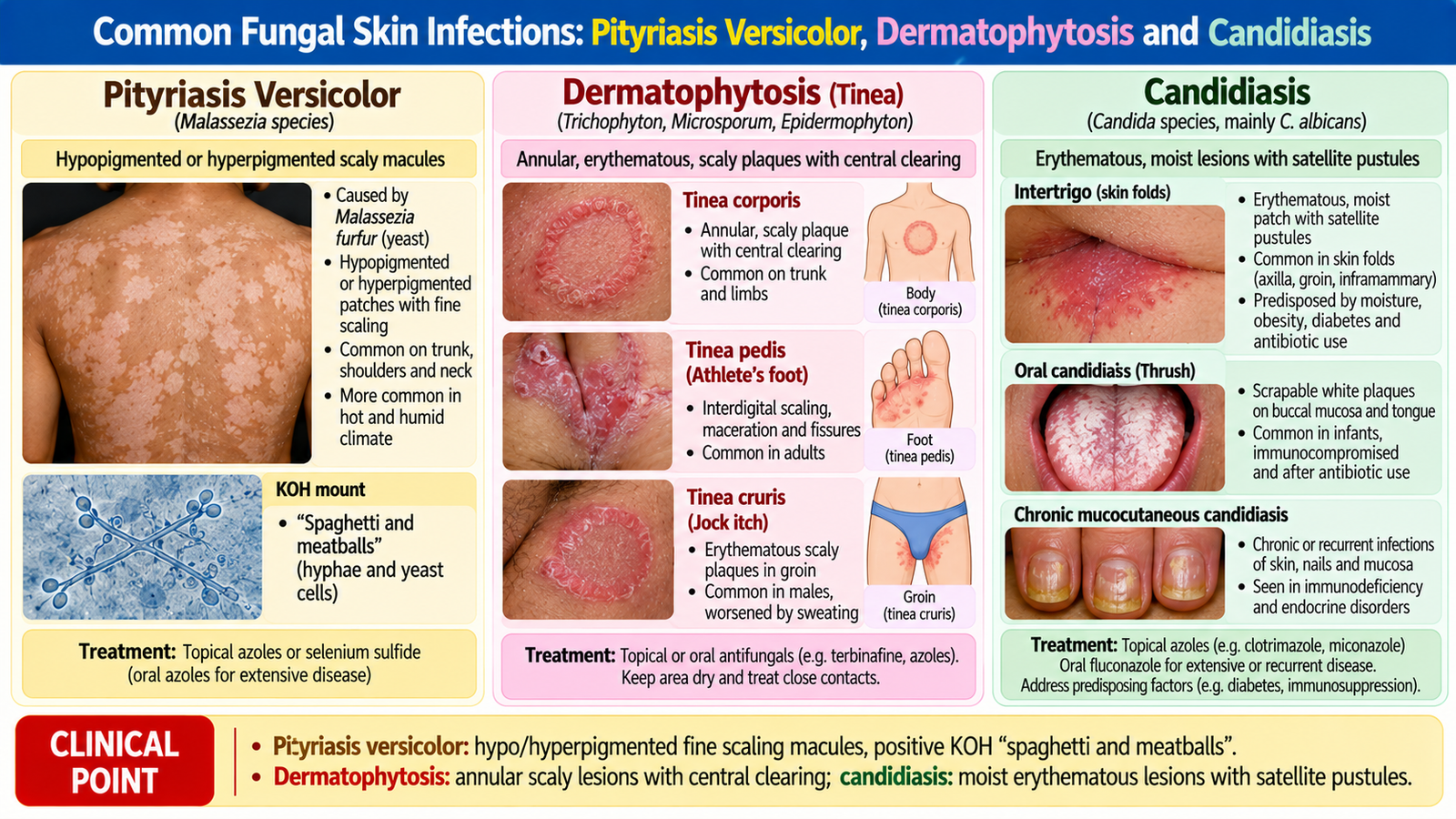
Superficial fungal diseases can be separated by understanding which organism is involved and which tissue environment it prefers. Pityriasis versicolor is associated with Malassezia in superficial skin; dermatophytes use keratin and therefore affect skin, hair or nails; and Candida particularly favors warm, moist environments and mucosal surfaces.
Pityriasis versicolor
Pityriasis versicolor is a superficial infection associated with Malassezia. It typically causes multiple hypo- or hyperpigmented macules with fine scale, especially over the upper trunk, shoulders and proximal upper limbs.
The colour change may persist temporarily even after the organism has been treated, so residual pigmentation does not necessarily indicate treatment failure. Diagnosis is commonly clinical and may be supported by examination of skin scrapings when needed. Topical antifungal treatment is appropriate for most localized disease.
Treatment: Topical ketoconazole or selenium sulfide is commonly used. Extensive or recurrent disease may require oral itraconazole or fluconazole.
Dermatophytosis
Dermatophytes infect keratinized tissues. Disease is commonly named according to anatomical location—such as tinea corporis on the body, tinea cruris in the groin and tinea pedis on the feet.
Tinea corporis classically produces an enlarging annular scaly plaque with a more active peripheral margin and relative central clearing. The advancing edge represents active fungal growth. Skin scrapings from the active margin can demonstrate fungal elements when diagnostic confirmation is needed.
Localized skin disease is generally treated with topical antifungal therapy. More extensive infection or involvement of hair or nails may require systemic therapy. Keeping affected areas dry and addressing sources of reinfection are useful management principles.
Treatment: Localized tinea corporis, cruris and pedis can be treated with topical terbinafine or clotrimazole. Tinea capitis requires systemic antifungal therapy, commonly oral terbinafine or griseofulvin. Avoid inappropriate topical steroid combinations.
Candidiasis
Candida can become pathogenic when local moisture, altered host defenses or disruption of normal barriers favors overgrowth. Cutaneous candidiasis commonly affects intertriginous areas and produces a red, moist eruption that may have satellite papules or pustules.
Clinical management includes antifungal treatment together with correction of predisposing local factors such as persistent moisture. Recurrent or unusually extensive candidiasis should prompt consideration of underlying factors such as diabetes or immunosuppression.Treatment: Localized cutaneous candidiasis is treated with topical clotrimazole or nystatin, together with keeping skin folds dry. Extensive or resistant infection may require oral fluconazole after clinical assessment.
G. Clinical Recognition and Diagnostic Reasoning
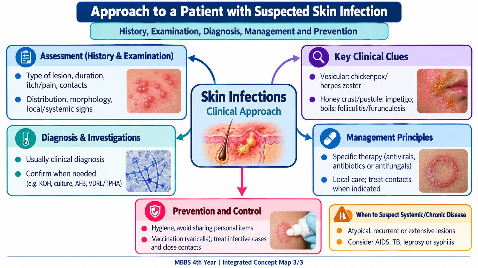
The safest way to approach an infectious skin lesion is to avoid diagnosing from colour alone. First identify the primary lesion, then determine its distribution, surface, symptoms, duration and relevant exposure or host factors. This turns a large list of infections into a manageable clinical pattern.
A practical diagnostic approach
- Vesicles: generalized lesions in different stages suggest chickenpox; painful unilateral dermatomal vesicles suggest zoster.
- Rough keratotic papules: consider viral warts.
- Umbilicated papules: suggest molluscum contagiosum.
- Honey-coloured crusts: suggest non-bullous impetigo.
- Follicular pustules: suggest folliculitis; deeper painful fluctuant lesions suggest furunculosis.
- Chronic patch with sensory loss: strongly suggests leprosy.
- Annular scaling with active margin: favors dermatophytosis.
- Moist flexural erythema with satellite lesions: favors candidiasis.
Next assess the host. Age, immune status, diabetes, recurrent infections, close contact, sexual exposure and the presence of systemic symptoms may change the interpretation of the same lesion. Investigations are most useful when they answer a specific clinical question—for example, fungal microscopy when the morphology is uncertain, serology when syphilis is suspected or microbiological/histological assessment when chronic infection requires confirmation.
Integrated Mechanism Flow
Important Comparison: Pattern Recognition
| Condition | Typical lesion | Distribution / clue | Key discriminator |
|---|---|---|---|
| Chickenpox | Vesicles and crusts | Generalized, trunk prominent | Different stages together |
| Herpes zoster | Grouped vesicles | Unilateral dermatome | Prominent pain |
| Molluscum | Smooth papules | Variable | Central umbilication |
| Impetigo | Superficial erosions/crusts | Often exposed skin | Honey-coloured crust |
| Leprosy | Chronic patch/plaque | Variable | Sensory impairment |
| Dermatophytosis | Annular scaly plaque | Keratinized tissues | Active peripheral edge |
| Candidiasis | Moist erythema | Skin folds | Satellite lesions |
AIM High-Yield Review
- Chickenpox: generalized lesions occur in successive crops, so different stages coexist.
- Herpes zoster: painful unilateral dermatomal vesicles represent reactivation of latent VZV.
- ⭐ Ophthalmic zoster may threaten vision and needs urgent assessment.
- Warts: HPV produces localized epidermal proliferation; plantar warts may mimic corns.
- ⭐ Central umbilication is the classic morphological clue to molluscum contagiosum.
- HIV/AIDS: think of underlying immunosuppression when common infections become unusually extensive, recurrent or persistent.
- ⭐ Honey-coloured crusting strongly suggests non-bullous impetigo.
- Folliculitis → furuncle → carbuncle reflects progressively deeper follicular infection.
- ⭐ A chronic skin lesion with sensory loss is a major diagnostic clue to leprosy.
- Secondary syphilis: remember the generalized eruption that may involve palms and soles.
- Pityriasis versicolor: pigmentary macules with fine scale commonly affect the upper trunk.
- ⭐ Dermatophytosis typically shows an active scaly margin because fungal growth advances peripherally.
- Candidiasis: moist intertriginous erythema with satellite lesions is highly suggestive.
- Clinical approach: morphology + distribution + symptoms + host factors usually narrows the diagnosis before investigations are ordered.
Viral, Bacterial and Fungal Skin Infections
Watch this video after completing the learning material to reinforce recognition and management of common bacterial, fungal and viral skin infections.