
This chapter follows the KMU learning outcomes and explains osteoarthritis and gout in a simple clinical sequence. First understand why each treatment is used and how complications develop; then use the High-Yield Review for rapid revision.
Topic 4. Osteoarthritis and Gout
Musculoskeletal Module • Management and complications of osteoarthritis, acute gout and gouty arthritis
Topic Introduction
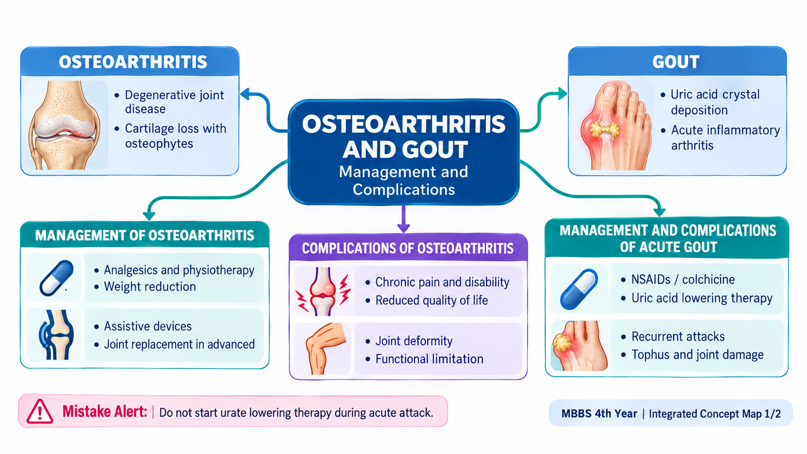
Osteoarthritis and gout are both common causes of joint pain, but they develop through very different processes and therefore require different treatment strategies. Osteoarthritis (OA) is mainly a degenerative joint disorder in which articular cartilage, subchondral bone and other joint structures gradually change, producing mechanical pain, stiffness and loss of function. Gout is an inflammatory arthritis caused by deposition of monosodium urate crystals in and around joints because of persistent hyperuricemia. This chapter focuses on the KMU-required areas: how osteoarthritis is managed, how its complications are limited, how an acute gout attack is treated, how recurrent gout is controlled and which important complications may occur.
A. Osteoarthritis — Understanding the Problem Before Management
Osteoarthritis is a chronic disorder of a synovial joint in which the normal balance between tissue breakdown and repair is lost. Articular cartilage becomes progressively damaged, the underlying subchondral bone becomes thicker and osteophytes may develop at the joint margins. The result is a joint that becomes painful during use, stiff after rest and gradually less mobile.
Why management is different from inflammatory arthritis
The main problem in osteoarthritis is not a primary systemic autoimmune process. Treatment therefore focuses on reducing mechanical stress, improving muscle support, controlling pain and maintaining function. Structural damage that has already developed cannot usually be reversed by simple drug therapy.
Main clinical problems that treatment targets
- Pain: typically worsens with activity or prolonged loading of the affected joint.
- Brief stiffness after rest: often improves once the joint begins moving.
- Reduced range of movement: develops because of pain, osteophytes and structural joint change.
- Loss of function: knee or hip disease may interfere with walking, climbing stairs and daily activities.
- Joint deformity: may appear in more advanced disease.

B. Management and Complications of Osteoarthritis
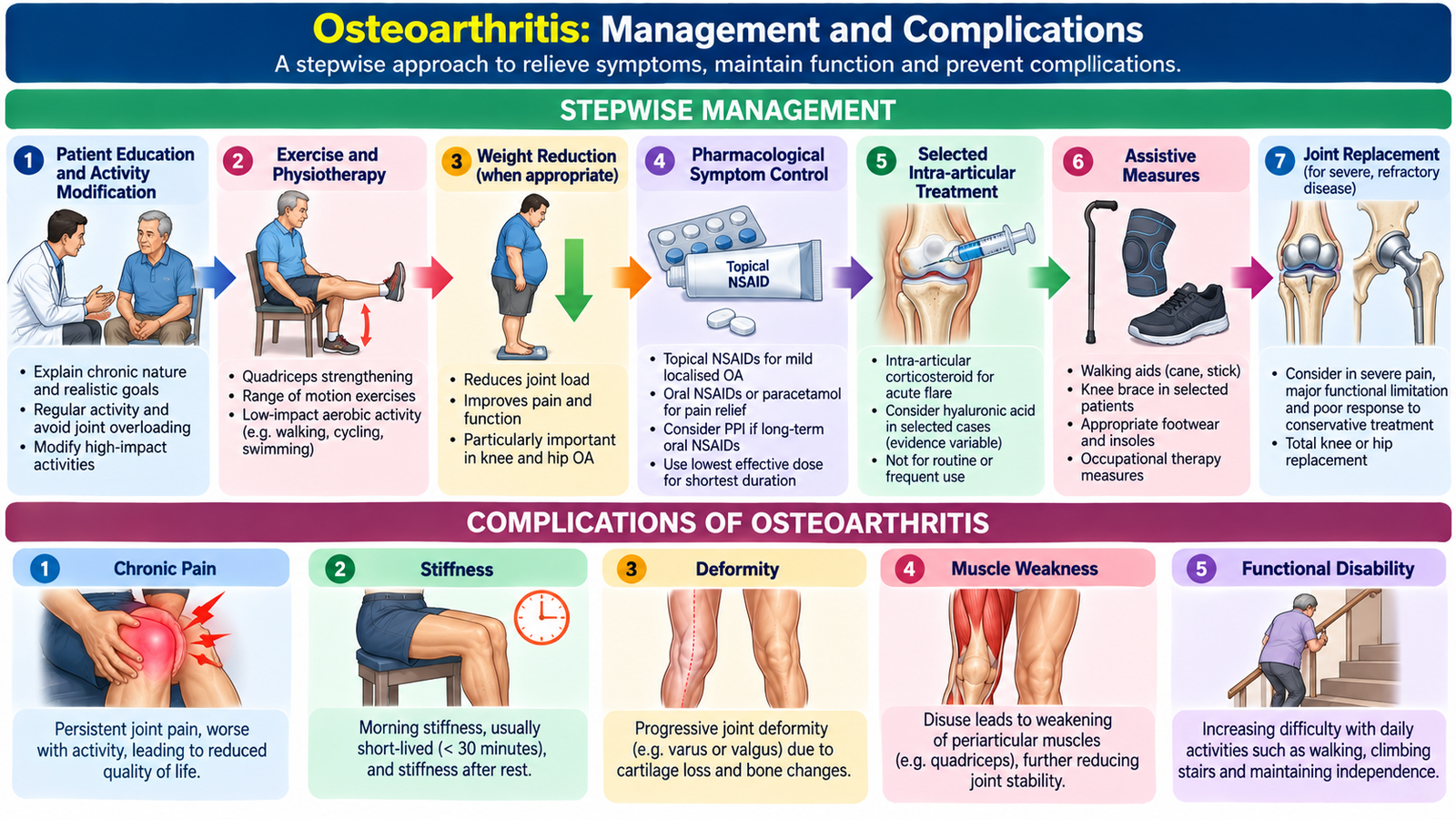
Osteoarthritis management is usually stepwise and individualized. The aim is not simply to prescribe an analgesic. Successful treatment combines education, physical measures, appropriate pain relief and, when necessary, surgical treatment. Management should be guided by the severity of symptoms and the effect of disease on daily function rather than by an X-ray appearance alone.
1. Education and activity modification
The patient should understand that regular movement is beneficial. Complete inactivity weakens the muscles that support the joint and may increase disability. Activities that repeatedly produce excessive impact or loading can be modified without stopping physical activity altogether.
- Encourage regular, tolerable physical activity.
- Avoid unnecessary repetitive high-impact loading of a painful joint.
- Plan periods of activity and rest according to symptoms.
- Use appropriate footwear when lower-limb joints are affected.
2. Exercise and physiotherapy
Exercise is a central part of osteoarthritis treatment because stronger periarticular muscles reduce stress on the affected joint and improve stability. Range-of-motion exercises also help prevent progressive stiffness.
- Muscle-strengthening exercise: especially useful around the knee and hip.
- Aerobic exercise: maintains general fitness and mobility.
- Range-of-motion exercise: helps preserve joint movement.
- Physiotherapy: may assist patients with weakness, gait difficulty or functional limitation.
3. Weight reduction when appropriate
Excess body weight increases the load passing through weight-bearing joints, particularly the knee. In an overweight patient, weight reduction decreases mechanical stress and may improve pain and mobility.
4. Analgesic and anti-inflammatory treatment
Drug therapy is used mainly to control symptoms so that the patient can remain active. Choice depends on the affected joint, severity of symptoms and the patient’s gastrointestinal, renal, cardiovascular and other clinical risks.
- Topical NSAIDs: useful particularly for localized knee or hand osteoarthritis and produce less systemic exposure than oral therapy.
- Oral NSAIDs: can reduce pain and inflammation when symptoms are more troublesome, but gastrointestinal, renal and cardiovascular risks must be considered.
- Paracetamol: may provide limited symptomatic benefit in selected patients, but it is generally less effective than NSAIDs for significant OA pain.
- Intra-articular glucocorticoid injection: may provide short-term relief in a painful symptomatic joint, particularly during an exacerbation.
5. Assistive measures
A walking aid may reduce the load transmitted through an affected hip or knee. Braces or other supportive devices may be useful in selected patients where instability or abnormal mechanical loading contributes significantly to symptoms.
6. Surgical management
Surgery is considered when pain and functional limitation remain severe despite appropriate conservative treatment and structural disease is advanced. Joint replacement, particularly of the hip or knee, can markedly reduce pain and restore function in appropriately selected patients.
Complications and consequences of osteoarthritis
Osteoarthritis is not usually associated with systemic organ damage. Its major complications arise from progressive structural joint disease and loss of mobility.
- Chronic pain that interferes with sleep or normal activities.
- Progressive stiffness and reduced range of movement.
- Joint deformity due to progressive structural change.
- Muscle weakness and wasting because pain leads to reduced use of the limb.
- Reduced walking ability and disability, particularly with hip or knee involvement.
- Reduced independence and quality of life in advanced disease.

C. Acute Gout — Why the Joint Becomes Acutely Inflamed
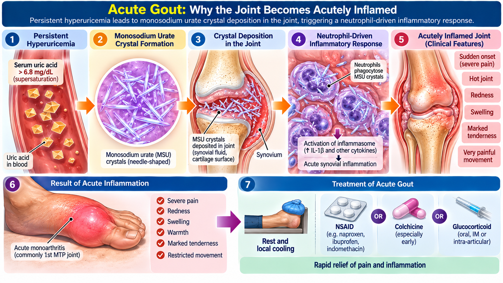
Gout is an inflammatory arthritis caused by deposition of monosodium urate crystals when serum urate has remained sufficiently high for crystal formation. Crystals may remain in joint tissues without symptoms, but when they are released into the joint space they trigger an intense inflammatory response.
This mechanism explains why treatment of an acute attack is directed primarily at suppressing inflammation quickly. Lowering the total body urate burden is important for long-term prevention, but it does not provide immediate relief from the inflammatory process already occurring inside the joint.
Typical acute attack
Acute gout usually produces rapid onset of severe pain, marked tenderness, swelling and warmth of one or a few joints. The first metatarsophalangeal joint is a classic site, although other joints can be affected. The intense tenderness occurs because crystal-triggered inflammation develops rapidly within the synovium and periarticular tissues.
D. Management and Complications of Acute and Recurrent Gout
Gout management has two distinct objectives. During an acute attack, the immediate aim is to stop crystal-induced inflammation and relieve pain. Between attacks, the aim is to reduce the body urate burden in patients who require long-term therapy so that future attacks, tophi and joint damage are prevented.
1. General measures during an acute attack
- Rest the affected joint during the period of severe pain.
- Local cooling with an ice pack may reduce pain and swelling.
- Begin anti-inflammatory treatment as early as possible when the diagnosis is established.
- Consider the patient’s renal function, gastrointestinal risk and other comorbidities before choosing medication.
2. NSAIDs
NSAIDs reduce prostaglandin-mediated inflammation and are effective for many patients with acute gout. They are particularly useful when started early in the attack. Their use may be limited by renal impairment, peptic ulcer disease, gastrointestinal bleeding risk or other important contraindications.
3. Colchicine
Colchicine suppresses the inflammatory response to urate crystals by interfering with microtubule-dependent neutrophil activity. It can therefore reduce the pain and inflammation of an acute attack, particularly when given early. Gastrointestinal adverse effects, especially diarrhea, abdominal discomfort, nausea and vomiting, are important limitations. Toxicity becomes more important in renal impairment and with certain interacting medicines, so treatment must be used appropriately rather than indiscriminately.
4. Glucocorticoids
Glucocorticoids provide strong anti-inflammatory action and are useful when NSAIDs or colchicine are unsuitable. Depending on the clinical situation, they may be given systemically or injected into an affected joint after infection has been reasonably excluded.
5. Long-term control of gout
Repeated attacks indicate that the underlying urate burden remains high. In patients who require long-term urate-lowering treatment, the aim is to maintain serum urate below the level at which crystals continue to accumulate. Existing crystal deposits can then gradually dissolve and future attacks become less likely. Long-term urate-lowering therapy is indicated particularly for recurrent gout attacks, tophi or gout-related joint damage. Allopurinol is generally the preferred first-line drug, started at a low dose and adjusted to achieve a serum urate target below 6 mg/dL. When initiating urate-lowering therapy, low-dose colchicine is commonly prescribed for 3–6 months to prevent treatment-associated gout flares.
Allopurinol
Allopurinol inhibits xanthine oxidase, reducing conversion of hypoxanthine and xanthine to uric acid. It is commonly used for long-term urate lowering and prevention of recurrent gout. It is not an analgesic and does not terminate an acute inflammatory attack by itself.
Febuxostat
Febuxostat also inhibits xanthine oxidase and lowers uric acid production. It is an alternative urate-lowering drug in selected patients when allopurinol is unsuitable or not tolerated.
Uricosuric therapy
Uricosuric medicines increase renal urate excretion and may be useful in selected patients with adequate renal function. They are not suitable for every patient and are generally avoided when urinary uric-acid stones are a major concern.
6. Lifestyle and contributing factors
Lifestyle measures support drug treatment but should not be presented as a substitute for indicated urate-lowering therapy. Helpful measures include maintaining a healthy body weight, avoiding excessive alcohol intake, maintaining adequate hydration and reviewing medicines or metabolic factors that may contribute to hyperuricemia when clinically appropriate.
Complications of gout
If hyperuricemia remains uncontrolled, repeated inflammation and continued crystal deposition can convert an episodic disease into a chronic destructive disorder.
- Recurrent acute attacks: attacks may become more frequent and may involve additional joints.
- Tophi: collections of urate crystals can develop in soft tissues and around joints.
- Chronic gouty arthritis: repeated crystal deposition and inflammation may produce persistent pain, erosive joint damage, deformity and reduced function.
- Renal calculi: uric-acid stones may develop in susceptible patients.
- Renal involvement: chronic hyperuricemia and associated conditions may coexist with renal impairment, making management more complex.

Integrated Management Flow
Mechanical/degenerative joint change ↓ Pain + stiffness + impaired movement ↓ Education + exercise + weight control ↓ Appropriate analgesic/anti-inflammatory therapy ↓ Assistive measures when required ↓ Joint replacement for severe refractory disease
Hyperuricemia ↓ Monosodium urate crystal deposition ↓ Acute inflammatory attack ↓ NSAID / colchicine / glucocorticoid ↓ Long-term urate reduction when indicated ↓ Prevention of recurrent attacks, tophi and joint destruction
Important Comparison — Osteoarthritis vs Gout
| Feature | Osteoarthritis | Gout |
|---|---|---|
| Main process | Degenerative/mechanical joint disease | Monosodium urate crystal-induced inflammation |
| Typical pain pattern | Usually worsens with joint use | Sudden, severe inflammatory pain during a flare |
| Immediate treatment aim | Reduce pain and improve function | Rapidly suppress acute inflammation |
| Key non-drug measure | Exercise, physiotherapy and weight control when required | Rest/cooling during attack and modification of contributing factors |
| Main long-term complication | Progressive pain, deformity and disability | Tophi, chronic erosive arthritis and recurrent attacks |
| Disease-control concept | Reduce mechanical stress and preserve function | Reduce urate burden when long-term therapy is indicated |
⭐ AIM High-Yield Review
- Osteoarthritis is primarily a degenerative/mechanical joint disorder; management aims to control pain and preserve function.
- Exercise is a core treatment in OA because stronger periarticular muscles improve joint support and function.
- Weight reduction decreases mechanical loading of weight-bearing joints in overweight patients.
- Topical or oral NSAIDs can reduce OA pain; systemic risks must be considered when oral NSAIDs are used.
- Intra-articular glucocorticoids may provide short-term relief in selected symptomatic OA joints.
- Severe OA with persistent pain and major functional limitation despite conservative treatment may require joint replacement.
- Major OA consequences are chronic pain, stiffness, deformity, reduced mobility, muscle weakness and disability.
- Gout results from monosodium urate crystal deposition associated with hyperuricemia.
- An acute gout flare is treated with anti-inflammatory therapy such as an NSAID, colchicine or a glucocorticoid.
- Colchicine reduces crystal-induced inflammation mainly by inhibiting neutrophil microtubule-dependent activity.
- Allopurinol lowers uric acid formation by inhibiting xanthine oxidase and is used for long-term urate reduction rather than immediate analgesia.
- Uncontrolled gout can cause recurrent attacks, tophi, chronic erosive arthritis, deformity and uric-acid renal stones.
- ⭐ A hot, acutely swollen joint should not automatically be labelled gout; septic arthritis must be considered where clinically appropriate.
Gout — Causes, Symptoms, Diagnosis & Treatment
Watch this video after completing the learning material to reinforce urate crystal formation, acute inflammation and the principles of gout management.