
Cerebrovascular Disorders: Ischemic Stroke, Hemorrhagic Stroke & SAH
Integrated Learning Material | Medicine | KMU-aligned
Topic Introduction
Stroke is a neurological emergency in which a sudden disturbance of brain circulation produces focal deficits or impaired consciousness. This chapter explains how to recognize stroke, assess severity, distinguish ischemia from bleeding on imaging, choose safe acute management and counsel patients about complications and prevention.

A. Understanding Stroke and Recognizing Its Presentation
Stroke is a sudden neurological deficit caused by interruption of cerebral blood flow (ischemia) or rupture of an intracranial vessel (hemorrhage). Brain cells need continuous oxygen and glucose; loss of perfusion rapidly impairs neuronal function. A transient ischemic attack (TIA) produces transient focal symptoms without evidence of acute infarction and is an urgent warning of future stroke. Ischemic stroke accounts for most strokes. It results from thrombosis in a diseased artery, embolism (for example, from atrial fibrillation), or reduced perfusion. Hemorrhagic stroke involves bleeding into brain tissue; subarachnoid hemorrhage (SAH) involves bleeding into the space between arachnoid and pia, often after aneurysm rupture. Recognize FAST: facial droop, arm weakness, speech difficulty, time to activate emergency services. Sudden visual loss, ataxia, vertigo with other focal signs, severe headache, or altered consciousness can also indicate stroke. Record last known well rather than the time symptoms were discovered. Localization: dominant-hemisphere cortical injury may cause aphasia; contralateral face/arm weakness suggests anterior circulation involvement. Posterior circulation injury may cause diplopia, dysphagia, ataxia, or impaired consciousness. Aphasia is a language disorder, whereas dysarthria is impaired articulation.

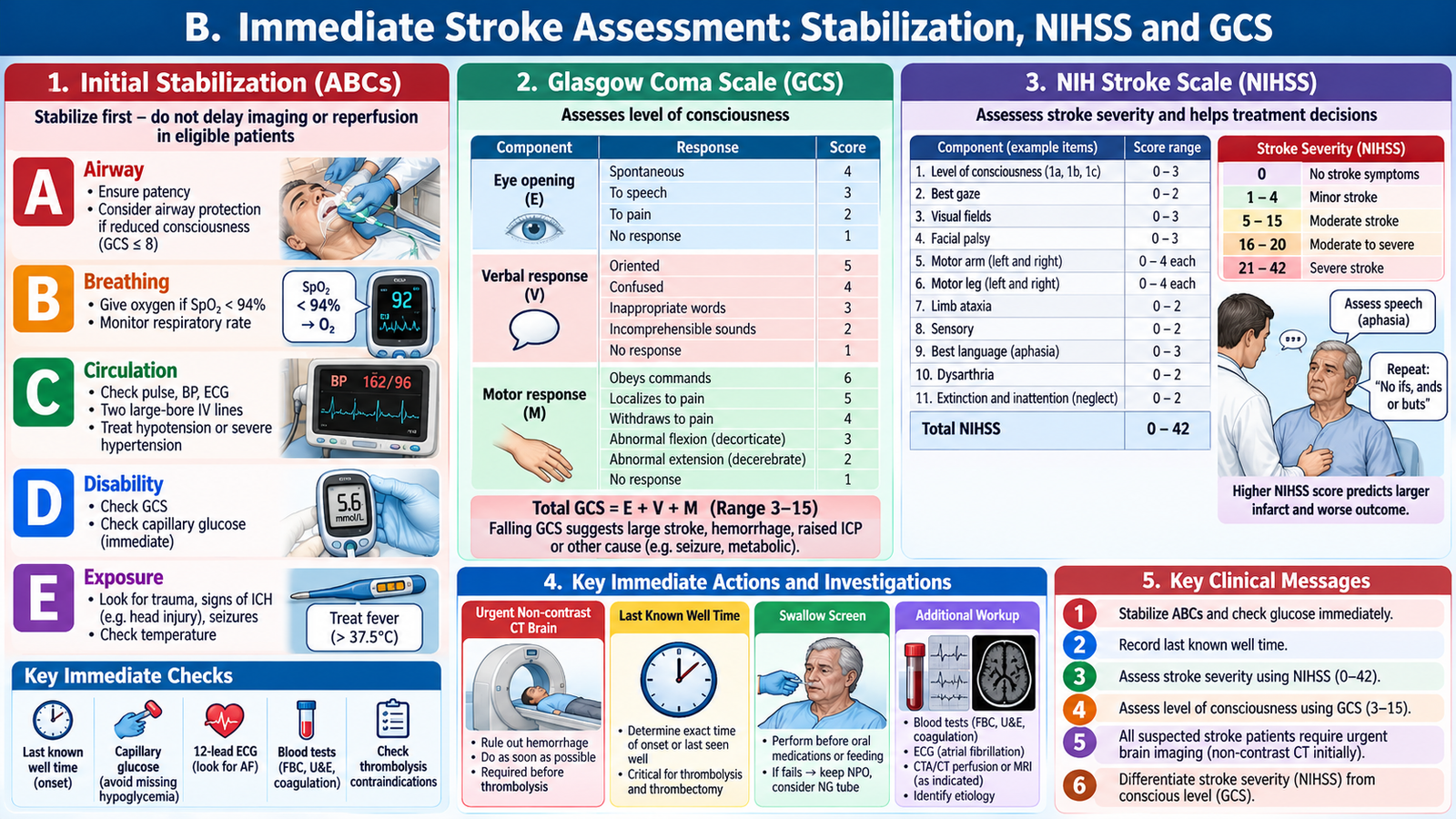
B. Immediate Stroke Assessment: Stabilization, NIHSS and GCS
Suspected stroke is a time-critical emergency. First assess airway, breathing and circulation, oxygen saturation, blood pressure, temperature, and capillary glucose. Treat hypoglycemia immediately because it can mimic stroke. Establish IV access, check symptom onset/last-known-well, medication history (especially anticoagulants), prior disability, seizure or trauma, and arrange urgent brain imaging. Keep the patient nil by mouth until swallowing is screened. Focused neurological examination: level of alertness, pupils and gaze, facial movement, speech and language, limb power, sensation, coordination, visual fields, and signs of brainstem dysfunction. Check for headache, vomiting, neck stiffness and progressive drowsiness suggesting hemorrhage or raised intracranial pressure. NIH Stroke Scale (NIHSS) is a standardized stroke-deficit score assessing consciousness, gaze, visual fields, facial weakness, limb motor function, ataxia, sensation, language, dysarthria and neglect. Scores range from 0 to 42; higher scores generally indicate greater neurological impairment. It supports baseline documentation, communication and monitoring, but a low score does not exclude a disabling or posterior-circulation stroke. Glasgow Coma Scale (GCS) evaluates eye opening (E1–4), verbal response (V1–5) and motor response (M1–6): total 3–15. Document components, e.g., E3 V4 M6 = 13/15. GCS measures consciousness, not focal stroke severity; it complements rather than replaces NIHSS. A falling GCS is a danger sign requiring urgent reassessment of airway, hemorrhage, edema or hydrocephalus.
C. Diagnostic Workup and CT/MRI Interpretation
Urgent non-contrast CT head is the initial imaging study in most acute stroke pathways because it rapidly identifies intracranial hemorrhage and guides eligibility for reperfusion. Early ischemic stroke can appear normal on CT. Subtle early findings include loss of gray–white differentiation, obscuration of the insular ribbon, sulcal effacement and a hyperdense vessel sign. Established infarction becomes a hypodense vascular-territory lesion. Acute intracerebral hemorrhage usually appears hyperdense (bright) on non-contrast CT, sometimes with surrounding edema, mass effect or intraventricular extension. SAH produces hyperdensity in basal cisterns, sulci or fissures. MRI diffusion-weighted imaging (DWI) is highly sensitive for acute ischemia, showing restricted diffusion (DWI bright with low ADC). MRI may be useful when CT is nondiagnostic, especially for small or posterior fossa infarcts, but must not delay emergency treatment. Further investigations are guided by suspected mechanism: CBC and platelets, electrolytes, renal function, glucose, coagulation studies where indicated, ECG for atrial fibrillation, and vascular imaging such as CT angiography when large-vessel occlusion or aneurysm is suspected. Carotid imaging and echocardiography help selected patients after stabilization. Imaging and clinical findings together establish the diagnosis; do not use a normal early CT to dismiss ischemic stroke. Practical distinction: ischemic infarct may initially be CT-occult; intracerebral blood is typically bright within parenchyma; SAH blood tracks along cisterns and sulci. Never give antiplatelets or thrombolysis before hemorrhage has been excluded.

D. Ischemic Stroke: Diagnosis and Acute Management
In ischemic stroke, vessel occlusion creates an infarct core surrounded by potentially salvageable hypoperfused tissue (penumbra). Rapid reperfusion can preserve function. Hemiparesis and aphasia strongly suggest a cerebral hemispheric stroke, but stroke mimics such as hypoglycemia, seizure with postictal weakness, and migraine must be considered without delaying urgent imaging.
Thrombolytic medicines: IV alteplase (0.9 mg/kg, maximum 90 mg, given over 60 minutes with 10% as an initial bolus) or tenecteplase (0.25 mg/kg, maximum 25 mg, single IV bolus) may be used in eligible patients according to the acute stroke protocol. Treatment must follow assessment of contraindications and intracranial bleeding risk.
Selected patients may qualify under advanced imaging protocols beyond standard time windows. Mechanical thrombectomy is considered for eligible large-vessel occlusion, generally within 6 hours and in selected cases up to 24 hours based on imaging and specialist criteria. Key safety principle: intracranial hemorrhage must be excluded before reperfusion. Blood pressure and bleeding risks require protocol-based assessment. Do not lower blood pressure aggressively in routine acute ischemic stroke: excessive reduction may worsen perfusion. For thrombolysis candidates, blood pressure must generally be below 185/110 mmHg before treatment and maintained below 180/105 mmHg afterward according to local stroke protocol. If thrombolysis is not given and hemorrhage is excluded, aspirin is generally started within 24–48 hours when appropriate. After IV thrombolysis, avoid antiplatelet/anticoagulant therapy during the first 24 hours and obtain follow-up imaging before starting it. Early anticoagulation is not routine treatment for acute ischemic stroke, even when atrial fibrillation is suspected; later initiation depends on infarct size, hemorrhagic risk and clinical assessment. Support oxygenation, temperature, hydration, glucose control, swallow safety, DVT prevention and early rehabilitation.


E. Intracerebral Hemorrhage: Diagnosis and Management
Intracerebral hemorrhage (ICH) is bleeding within brain parenchyma. Common causes include chronic hypertension, cerebral amyloid angiopathy in older adults, anticoagulant-associated bleeding and vascular abnormalities. The expanding hematoma damages tissue directly and can increase intracranial pressure, cause midline shift and reduce consciousness. Headache, vomiting, focal weakness and rapid deterioration may occur, but symptoms alone cannot reliably distinguish hemorrhagic from ischemic stroke. Confirm ICH using non-contrast CT, assess hematoma location, size, intraventricular extension, hydrocephalus and mass effect, and obtain coagulation tests and medication history. Repeat imaging is considered if neurological status worsens. Manage in an appropriately monitored stroke or critical-care setting with frequent neurological checks, airway protection when needed, head positioning and specialist consultation. Blood pressure management should be smooth and protocol-guided; for many patients with mild–moderate spontaneous ICH and presenting systolic pressure 150–220 mmHg, targeting around 140 mmHg while avoiding excessive lowering may be appropriate. Reverse anticoagulation urgently when relevant using the agent-specific protocol.
Anticoagulant reversal: Warfarin-associated intracerebral hemorrhage requires urgent four-factor prothrombin complex concentrate (4F-PCC) with IV vitamin K. Dabigatran can be reversed with idarucizumab, while factor Xa inhibitors such as apixaban or rivaroxaban require specialist-directed reversal according to agent availability and local protocol.
Manage fever, glucose abnormalities, seizures if present and raised intracranial pressure. Neurosurgical input is especially important for cerebellar hemorrhage with deterioration, hydrocephalus, brainstem compression or selected surgically accessible hematomas. Do not administer thrombolytics or routine antithrombotics during active intracerebral hemorrhage. Important complications are hematoma expansion, cerebral edema, herniation, hydrocephalus, aspiration and persistent neurological disability.


F. Subarachnoid Hemorrhage: Recognition and Emergency Care
Subarachnoid hemorrhage (SAH) is bleeding into the CSF-filled subarachnoid space. A ruptured saccular intracranial aneurysm is an important spontaneous cause. The characteristic presentation is a sudden, maximal-at-onset thunderclap headache, sometimes with vomiting, neck stiffness, photophobia, collapse or reduced consciousness. Some patients have a normal initial neurological examination: sudden severe headache still warrants urgent evaluation. Obtain urgent non-contrast CT. Blood may be visible in basal cisterns or cortical sulci. If CT is negative but clinical suspicion remains, further testing is required according to timing and local protocol: lumbar puncture for CSF evidence of hemorrhage may be appropriate, particularly after a negative CT obtained more than 6 hours after onset; specialist-directed vascular imaging may also be needed. CT angiography identifies a suspected aneurysm, and catheter angiography may be required for definitive vascular assessment. Manage as a neurosurgical emergency: stabilize ABCs, provide analgesia and antiemetics, monitor blood pressure without provoking hypotension, arrange urgent neurosurgical/neurointerventional assessment and secure the aneurysm by coiling or clipping when indicated. Nimodipine is used in aneurysmal SAH to improve neurological outcomes associated with delayed cerebral ischemia; monitor for hypotension. Watch for rebleeding, hydrocephalus, delayed cerebral ischemia/vasospasm, seizures and hyponatremia. A new decline in consciousness or new focal deficit requires urgent assessment. Do not perform lumbar puncture in an unstable patient or one with suspected dangerous mass effect without specialist assessment.

G. Stroke Complications, Rehabilitation and Prevention Counselling
Stroke recovery starts during acute care. Common early complications include aspiration pneumonia from dysphagia, venous thromboembolism due to immobility, pressure injury, urinary problems, delirium, falls, seizures and cerebral edema. Screen swallowing before oral intake, support safe positioning and mobility, and involve physiotherapy, occupational therapy, speech and language therapy, nursing and caregivers. Aphasia requires communication support, not an assumption of reduced intelligence. Secondary prevention depends on stroke mechanism. After non-cardioembolic ischemic stroke, antiplatelet therapy and statin-based lipid lowering are common. For atrial fibrillation-related ischemic stroke, long-term oral anticoagulation is generally preferred when safe; timing is individualized after the acute event. Selected symptomatic carotid stenosis may require vascular intervention. In hemorrhagic stroke, meticulous long-term blood pressure control and review of bleeding risk are especially important. Counsel on blood pressure control, diabetes management, smoking cessation, exercise, healthy diet, weight management, lipid control, medication adherence and follow-up. Explain recurrence warning signs using FAST and the need for immediate emergency response rather than waiting for symptoms to resolve. Discuss realistic recovery goals, fatigue, mood symptoms, cognition, caregiver burden and rehabilitation needs. Safety-net counselling: seek emergency care for new weakness, speech disturbance, sudden severe headache, reduced consciousness or seizures. Arrange follow-up for disability, medication monitoring, rehabilitation and modifiable risk factors. Prevention and functional recovery require coordinated, patient-centred long-term care.

AIM Clinical Bridge
A patient arrives with sudden right hemiparesis and inability to speak. Stabilize ABCs, check glucose, establish last-known-well, document NIHSS/GCS and obtain urgent non-contrast CT. A normal early CT does not rule out ischemia; visible blood changes management completely. Treatment decisions depend on time, disability, vessel status and contraindications.
AIM High-Yield Review
- Stroke is clinical; urgent CT first distinguishes hemorrhage from suspected ischemia.
- Record last known well for reperfusion decisions.
- NIHSS measures focal stroke deficits; GCS measures consciousness.
- Acute ischemic stroke: consider thrombolysis/thrombectomy only in eligible patients.
- Do not give thrombolysis in intracranial hemorrhage.
- ICH: stabilize, control BP appropriately, reverse anticoagulants and monitor for mass effect.
- SAH: thunderclap headache, cisternal blood, aneurysm assessment and urgent specialist care.
- Screen swallowing, prevent complications and start rehabilitation early.
- Secondary prevention must match stroke mechanism and individual bleeding risk.
AIM LEARNING CYCLE | FINAL YEAR MBBS
Video Learning — Cerebrovascular Disorders
Ischemic Stroke, Hemorrhagic Stroke & Subarachnoid Hemorrhage
Ischemic and Hemorrhagic Strokes | High Yield
Ninja Nerd | Approximately 39 minutes
This lecture explains the mechanisms, clinical presentations, diagnostic imaging and major treatment principles of ischemic and hemorrhagic strokes, including subarachnoid hemorrhage.
AIM — AI-Integrated Modular Learning | Neuroscience-III Module | KMU Final Year MBBS