
Movement Disorders, Parkinson’s Disease & Ataxia
Step 2 — Integrated Learning Material
Understanding the Clinical Problem
Normal movement requires coordinated activity between the motor cortex, basal ganglia, cerebellum, brainstem and spinal motor pathways. Disorders affecting these structures produce different patterns of abnormal movement, ranging from slowness and rigidity to involuntary movements and loss of coordination. The most useful clinical approach is to recognize the movement pattern first, identify the likely neurological system involved, and then select appropriate investigations and treatment. Core clinical question: Is the patient moving too little, moving involuntarily, or moving without proper coordination?
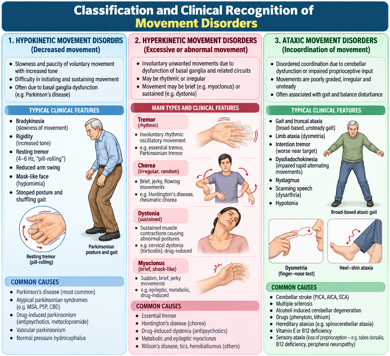
A. Classification and Clinical Recognition of Movement Disorders
Movement disorders are neurological conditions in which the speed, quantity, control or coordination of movement becomes abnormal. Classification according to the observed movement is the starting point for diagnosis.
1. Hypokinetic movement disorders
These disorders cause reduced spontaneous movement or difficulty initiating movement. Parkinsonism is the principal example.
- Bradykinesia: slowness of movement with progressive reduction in speed or amplitude during repeated actions.
- Akinesia: difficulty initiating voluntary movement.
- Rigidity: increased resistance to passive movement throughout the range.
- Reduced automatic movement: diminished arm swing, facial expression and blinking.
2. Hyperkinetic movement disorders
These conditions produce excessive involuntary movement.
- Tremor: rhythmic, oscillatory movement of a body part.
- Chorea: brief, irregular, unpredictable movements that appear to flow between body parts.
- Dystonia: sustained or intermittent muscle contractions causing twisting movements or abnormal postures.
- Myoclonus: sudden, brief, shock-like muscle jerks.
- Tics: repetitive movements or sounds that may be temporarily suppressed.
- Athetosis: slow, writhing movements, especially involving distal limbs.
3. Disorders of coordination
Ataxia is impaired coordination of voluntary movement. It commonly results from cerebellar dysfunction but may also arise from impaired proprioception or vestibular disease. Important distinction: Ataxia does not necessarily indicate muscle weakness. A patient may have preserved power but inaccurate, poorly coordinated movements.
| Movement pattern | Typical finding | Clinical association |
|---|---|---|
| Hypokinetic | Bradykinesia, rigidity | Parkinsonism |
| Hyperkinetic | Chorea, dystonia, tremor | Basal ganglia dysfunction or drug effects |
| Ataxic | Dysmetria, unsteady gait | Cerebellar or sensory pathway disorder |

B. Parkinson’s Disease: Pathophysiology, Clinical Features and Diagnosis
Parkinson’s disease is a progressive neurodegenerative disorder characterized by bradykinesia together with resting tremor and/or rigidity. It is the most important cause of degenerative parkinsonism.
1. Pathophysiology
The basal ganglia regulate the initiation, selection and smooth execution of movement. Dopamine released from the substantia nigra pars compacta modifies activity within basal ganglia motor circuits. In Parkinson’s disease, progressive degeneration of dopaminergic neurons reduces dopamine delivery to the striatum. This alters basal ganglia output and increases inhibition of thalamocortical motor activity, producing slowness and difficulty initiating movement. Lewy bodies containing aggregated alpha-synuclein are characteristic pathological findings, although diagnosis during life is primarily clinical.
2. Cardinal motor features
- Bradykinesia: slow finger tapping, reduced handwriting size (micrographia), difficulty with dressing and turning.
- Rest tremor: classically a 4–6 Hz pill-rolling tremor, commonly beginning asymmetrically and decreasing during voluntary movement.
- Rigidity: lead-pipe resistance or cogwheel rigidity when tremor is superimposed.
- Postural instability: impaired balance, often developing later in typical Parkinson’s disease.
3. Additional motor manifestations
Patients may develop a stooped posture, shuffling gait, reduced arm swing, freezing episodes, hypomimia, soft speech and difficulty turning in bed. Repeated movements become progressively smaller or slower.
4. Non-motor manifestations
Parkinson’s disease is not exclusively a motor disorder. Important non-motor manifestations include constipation, reduced sense of smell, depression, anxiety, sleep disturbances, autonomic dysfunction and cognitive impairment. Some non-motor symptoms may precede obvious motor manifestations and can significantly affect quality of life.
5. Clinical diagnostic criteria
The essential requirement for identifying parkinsonism is bradykinesia together with either rest tremor or rigidity. Parkinson’s disease is then considered when the history and examination support a typical degenerative pattern and alternative causes are less likely. Supportive features include asymmetric onset, progressive symptoms, characteristic rest tremor and a clear beneficial response to dopaminergic treatment. Early recurrent falls, severe early autonomic failure, prominent early cerebellar signs, vertical gaze palsy or rapid progression should prompt consideration of atypical parkinsonism or another diagnosis. Investigations: There is no routine blood test that confirms Parkinson’s disease. MRI brain is considered when the presentation is atypical or an alternative structural cause is suspected. Functional dopamine imaging may assist selected uncertain cases but is not routinely required.


C. Parkinson’s Disease: Pharmacological Treatment, Rehabilitation and Psychosocial Care
Treatment aims to improve movement, preserve independence, minimize complications and support the patient’s daily functioning. Current therapies provide symptomatic benefit but do not reliably stop neurodegeneration.
1. Pharmacological management
Levodopa with carbidopa: Levodopa crosses the blood–brain barrier and is converted into dopamine. Carbidopa inhibits peripheral conversion of levodopa, increasing central availability and reducing peripheral adverse effects. Levodopa is the most effective symptomatic treatment for bradykinesia and rigidity. It is particularly useful when motor symptoms interfere with daily activities. Important adverse effects include nausea, postural hypotension, hallucinations and, with longer-term treatment, motor fluctuations and dyskinesias. Dopamine agonists: Pramipexole and ropinirole directly stimulate dopamine receptors. They may be used in selected patients but can cause excessive daytime sleepiness, hallucinations, edema and impulse-control disorders. They require particular caution in older patients or those with cognitive impairment. MAO-B inhibitors: Selegiline and rasagiline reduce dopamine breakdown in the brain. They may provide modest symptomatic benefit in early disease or serve as adjunctive treatment. COMT inhibitors: Entacapone prolongs the effect of levodopa by reducing peripheral metabolism. It is mainly used as an adjunct when wearing-off fluctuations develop. Anticholinergic drugs: Selected drugs such as trihexyphenidyl may reduce tremor but are generally avoided in older adults because of confusion, urinary retention and other anticholinergic adverse effects.
| Drug class | Main action | Important caution |
|---|---|---|
| Levodopa/carbidopa | Restores dopamine availability | Dyskinesia, fluctuations |
| Dopamine agonists | Stimulate dopamine receptors | Sleepiness, impulse-control disorders |
| MAO-B inhibitors | Reduce dopamine degradation | Drug interactions |
| COMT inhibitors | Prolong levodopa action | Increased dyskinesia |
| Anticholinergics | Reduce cholinergic activity | Cognitive adverse effects |
2. Long-term motor complications
Wearing-off: The benefit of an individual levodopa dose becomes shorter, and symptoms return before the next dose. On–off fluctuations: Patients experience transitions between relatively good mobility and marked immobility. Dyskinesias: Involuntary movements may occur during dopaminergic treatment, particularly with long-term levodopa exposure. Medication timing, dose adjustment and adjunctive therapy are individualized under appropriate clinical supervision. Deep brain stimulation may be considered by specialists in carefully selected patients with disabling motor complications.
3. Rehabilitation and multidisciplinary management
- Physiotherapy: gait training, balance exercises, posture correction and fall prevention.
- Occupational therapy: adaptive equipment and techniques for dressing, eating and daily activities.
- Speech and swallowing therapy: management of hypophonia, communication difficulties and dysphagia.
- Nutrition and supportive care: attention to constipation, hydration, weight loss and swallowing safety.
4. Psychosocial care and counselling
Patients and families should understand that Parkinson’s disease is chronic and progressive, but treatment and rehabilitation can preserve meaningful function for many years. Discuss medication adherence, exercise, home safety, fall prevention, mood symptoms, sleep difficulties and caregiver support. Screen for depression, hallucinations and cognitive deterioration during follow-up. Sudden interruption of dopaminergic medication should be avoided because severe rigidity and potentially life-threatening deterioration may occur.

D. Clinical Assessment: History Taking and Neurological Examination in Parkinsonism
A structured neurological assessment establishes the movement disorder pattern, measures functional impairment and identifies findings suggesting an alternative diagnosis.
1. Focused history
Begin with the presenting complaint and its effect on daily activities. Ask about onset, duration, progression and whether symptoms started on one side.
- Motor symptoms: tremor at rest or during action, slowness, stiffness, handwriting changes, gait difficulty and falls.
- Non-motor symptoms: constipation, loss of smell, sleep disturbance, mood changes, hallucinations and autonomic symptoms.
- Medication history: antipsychotics, metoclopramide and other dopamine-blocking drugs that may cause parkinsonism.
- Functional history: ability to dress, eat, walk, work and perform household tasks.
- Family history: relatives with Parkinson’s disease or other movement disorders.
2. General observation
Observe the patient while seated and during spontaneous activity. Look for reduced facial expression, infrequent blinking, resting tremor, stooped posture and reduced spontaneous movements.
3. Examination of the upper limbs
Tremor: Examine hands at rest, with arms outstretched and during purposeful movement. Determine whether the tremor is resting, postural or kinetic. Rigidity: Passively flex and extend the wrist and elbow. Assess for uniform lead-pipe resistance or cogwheel rigidity. Bradykinesia: Ask the patient to perform repetitive finger tapping, hand opening and closing, and alternating movements. Observe progressive reduction in speed or amplitude.
4. Gait and balance examination
Assess rising from a chair, gait initiation, step length, arm swing, turning and posture. A parkinsonian gait may show short shuffling steps, reduced arm swing and multiple small steps while turning. Postural stability can be assessed using an appropriately supervised pull test, with the examiner prepared to prevent a fall.
5. Differentiating Parkinson’s disease from mimics
Essential tremor: Predominantly action or postural tremor, often bilateral, without the characteristic bradykinesia of Parkinson’s disease. Drug-induced parkinsonism: May follow dopamine-blocking medication and can be relatively symmetrical, although symmetry alone is not diagnostic. Atypical parkinsonism: Early falls, prominent autonomic dysfunction, gaze abnormalities or cerebellar signs suggest alternative neurodegenerative syndromes.

E. Cerebellar Disorders and Ataxia: Clinical Recognition, Investigation and Management
The cerebellum coordinates the timing, accuracy and smoothness of movement. Cerebellar damage causes poorly coordinated movement despite relatively preserved muscle strength.
1. Types of ataxia
Cerebellar ataxia: Caused by dysfunction of the cerebellum or its connections. Patients develop a broad-based gait, dysmetria and impaired coordination. Instability generally persists with eyes open and closed. Sensory ataxia: Results from impaired proprioceptive input. Patients depend heavily on visual guidance and become significantly less stable when they close their eyes. Vestibular ataxia: Results from vestibular dysfunction and commonly accompanies vertigo, nausea and imbalance.
2. Causes of cerebellar dysfunction
- Vascular: cerebellar infarction or hemorrhage.
- Toxic: alcohol intoxication, chronic alcohol-related injury or medication toxicity.
- Inflammatory/demyelinating: multiple sclerosis and related disorders.
- Structural: posterior fossa tumors or other lesions.
- Metabolic/nutritional: vitamin deficiencies and selected metabolic disorders.
- Degenerative/hereditary: progressive cerebellar ataxias.
3. Cerebellar clinical signs
Dysmetria: Inability to judge the distance or amplitude of movement, producing overshooting or undershooting during finger–nose testing. Dysdiadochokinesia: Impaired rapid alternating movements. Intention tremor: Tremor that becomes more prominent as the limb approaches a target. Nystagmus: Involuntary rhythmic eye movement that may accompany cerebellar dysfunction. Ataxic dysarthria: Irregular, poorly coordinated speech articulation, sometimes described as scanning speech. Gait ataxia: Broad-based, irregular walking with difficulty performing tandem gait.
4. Bedside neurological assessment
Assess gait and posture, finger–nose coordination, heel–shin testing, rapid alternating movements, speech and eye movements. Examine power, reflexes and sensory function to distinguish cerebellar dysfunction from weakness or sensory loss. Romberg test: A marked increase in instability after eye closure suggests sensory or vestibular dysfunction. Patients with significant cerebellar ataxia may already be unstable with eyes open.
5. Diagnostic workup
The investigation strategy depends on whether symptoms are acute, subacute or chronic.
- Sudden onset: consider posterior circulation stroke or hemorrhage and arrange urgent neuroimaging.
- Subacute progression: consider inflammatory, toxic, metabolic or structural causes.
- Chronic progression: assess for degenerative, hereditary, nutritional and medication-related causes.
Relevant investigations may include MRI brain, blood glucose, electrolytes, thyroid function, vitamin B12 and other tests guided by clinical findings. Medication and alcohol histories are essential.
6. Management principles
Management is directed at the underlying cause. Acute vascular lesions require urgent stroke evaluation. Drug-induced or toxic causes require identification and appropriate withdrawal or modification of the offending exposure. Nutritional deficiencies require replacement. Physiotherapy, balance training, occupational therapy, fall prevention and assistive devices are important for persistent ataxia. Patients with dysarthria or swallowing impairment may require speech and swallowing assessment.

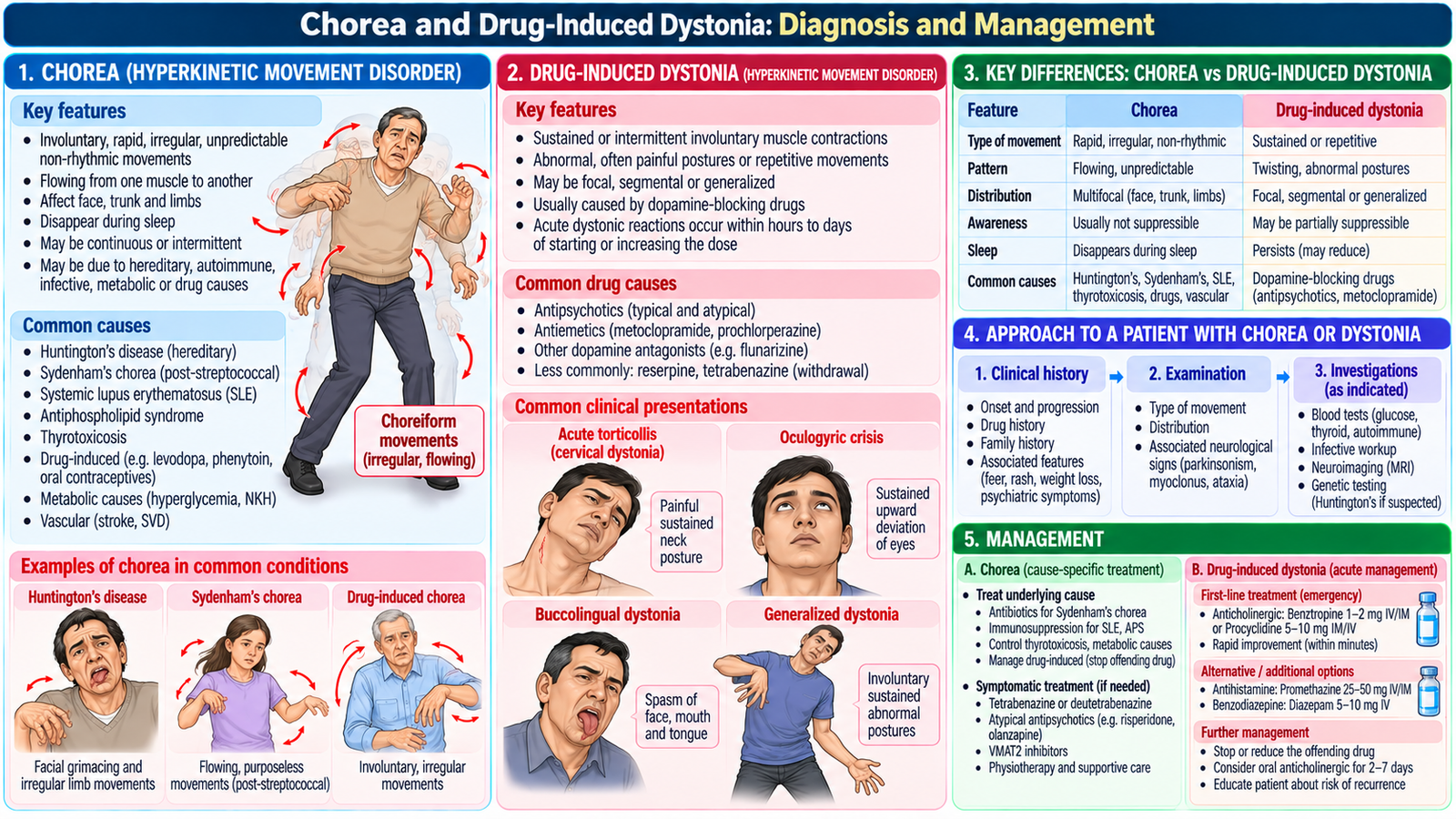
F. Chorea and Drug-Induced Dystonia: Diagnosis and Management
Chorea and dystonia are hyperkinetic movement disorders. Their recognition depends on the appearance of involuntary movements, the clinical history and the presence of medication exposure or associated neurological disease.
1. Chorea: mechanism and clinical presentation
Chorea consists of irregular, brief, non-rhythmic involuntary movements that flow unpredictably from one body region to another. Basal ganglia dysfunction, particularly altered regulation of motor inhibitory pathways, contributes to its development. Movements may resemble restlessness or purposeless gestures. Patients can have difficulty maintaining a sustained posture or grip.
2. Important causes of chorea
- Huntington’s disease: inherited autosomal dominant neurodegenerative disease associated with progressive chorea, cognitive decline and psychiatric manifestations.
- Sydenham chorea: immune-mediated movement disorder associated with rheumatic fever, particularly in children and adolescents.
- Drug-induced chorea: may occur with dopaminergic treatment or other medications.
- Metabolic and vascular causes: selected metabolic disturbances and basal ganglia vascular lesions can produce choreiform movements.
Huntington’s disease is caused by a CAG repeat expansion in the HTT gene, with prominent involvement of striatal neurons. A family history and progressive motor, behavioral and cognitive changes support clinical suspicion.
3. Diagnostic evaluation of chorea
History should establish onset, progression, family history, medication exposure, recent infection and associated cognitive or psychiatric changes. Examination assesses the distribution of involuntary movements, gait, coordination, tone and other neurological abnormalities. Investigations are selected according to the suspected cause. They may include glucose and metabolic testing, brain imaging, appropriate assessment for rheumatic fever, or genetic testing with counselling when Huntington’s disease is suspected.
4. Management of chorea
Management focuses on the underlying cause, functional disability and associated psychiatric or cognitive problems.
- Review and modify causative medications where appropriate.
- Treat identified metabolic or inflammatory causes.
- For disabling chorea in Huntington’s disease, specialist-directed symptomatic therapy may include vesicular monoamine transporter type 2 (VMAT2) inhibitors such as tetrabenazine.
- Provide physiotherapy, nutritional support, swallowing assessment and family counselling when required.
Tetrabenazine reduces presynaptic monoamine storage and dopamine availability. Important cautions include depression, suicidality, sedation and drug-induced parkinsonism.
5. Drug-induced acute dystonia
Acute dystonia is an important medication-related movement disorder caused by sustained involuntary muscle contractions and abnormal postures. It may develop within hours to several days after starting or increasing a dopamine receptor-blocking medication. Common precipitating medications include antipsychotics such as haloperidol and antiemetics such as metoclopramide. Blockade of dopamine D2 receptors in the nigrostriatal pathway produces a relative imbalance between dopaminergic and cholinergic activity, leading to abnormal muscle contractions.
6. Clinical manifestations of acute dystonia
- Torticollis: sustained abnormal neck rotation.
- Oculogyric crisis: involuntary sustained upward deviation of the eyes.
- Trismus: involuntary jaw muscle contraction.
- Opisthotonus: severe extension of the neck and back.
- Laryngeal dystonia: rare but potentially life-threatening airway involvement.
7. Management of drug-induced dystonia
First assess airway, breathing and circulation, particularly when stridor, breathing difficulty or severe neck and throat contractions are present. Stop or withhold the suspected causative drug and arrange appropriate clinical assessment. Acute symptoms usually respond rapidly to a parenteral anticholinergic such as benztropine or an antihistamine with anticholinergic action such as diphenhydramine, according to local availability and protocol.
Pediatric emergency treatment: Diphenhydramine 1 mg/kg IV or IM (maximum 50 mg) is an appropriate first-line treatment for acute drug-induced dystonia. Symptoms usually improve within minutes. A short oral course for 1–2 days may be needed to prevent recurrence. Stridor or respiratory compromise requires immediate airway management.
Observe for recurrence, review the need for the causative medication and document the adverse drug reaction. Subsequent prescribing should minimize recurrence risk.
Integrated Clinical Reasoning
Consider three patients presenting with abnormal movement: Patient 1: A 67-year-old develops progressive slowness, unilateral resting hand tremor, rigidity and reduced arm swing. This pattern suggests degenerative parkinsonism, particularly Parkinson’s disease. Patient 2: A 54-year-old develops sudden gait unsteadiness, vomiting and severe difficulty performing finger–nose testing. Acute cerebellar dysfunction is suspected, and posterior circulation stroke must be urgently excluded. Patient 3: A 23-year-old develops painful neck twisting and upward eye deviation shortly after receiving an antipsychotic. The temporal relationship and sustained muscle contractions suggest acute drug-induced dystonia. Integrated lesson: The movement pattern, onset, associated neurological findings and medication history often determine the diagnosis before advanced investigations are considered.
AIM Final High-Yield Revision
- Movement disorders are classified into hypokinetic, hyperkinetic and coordination disorders.
- Parkinsonism requires bradykinesia with rest tremor or rigidity.
- Parkinson’s disease involves degeneration of substantia nigra dopaminergic neurons.
- Levodopa/carbidopa is the most effective symptomatic pharmacological treatment for Parkinson’s motor features.
- Dopamine agonists may cause hallucinations, sleepiness and impulse-control disorders.
- Cerebellar ataxia produces dysmetria, intention tremor, dysdiadochokinesia and broad-based gait.
- Sensory ataxia worsens markedly when visual compensation is removed.
- Sudden ataxia may indicate a posterior circulation stroke.
- Chorea consists of irregular, flowing, non-rhythmic involuntary movements.
- Huntington’s disease combines progressive chorea with cognitive and psychiatric manifestations.
- Acute dystonia may follow dopamine-blocking drugs and responds to appropriate anticholinergic treatment.
- Laryngeal dystonia and respiratory compromise require emergency airway assessment.
Clinical Video Learning
Topic 5: Movement Disorders, Parkinson’s Disease & Ataxia
Video 1 — Parkinson’s Disease: Clinical Examination
Stanford Medicine 25 | Clinical demonstration
Learning focus: Bradykinesia, rigidity, resting tremor, gait abnormalities and postural stability.
Video 2 — Cerebellar Examination & Ataxia
Geeky Medics | Neurological examination
Learning focus: Gait ataxia, finger-to-nose test, dysmetria, dysdiadochokinesia, heel-to-shin testing, nystagmus and Romberg’s test.
Use these clinical demonstrations to connect the movement patterns and neurological examination findings with the diagnostic principles explained in your AIM Learning Material.