
📖 AIM Learning Material
TOPIC 11. Congenital Spinal Malformations, Vertebral Disorders & Kyphoscoliosis
Neuroscience Module • Pediatric Surgery & Orthopedics
Topic Introduction
The spinal column protects the spinal cord and supports movement and posture. Problems may begin before birth, as in spina bifida, or develop later through disc degeneration, injury, infection or abnormal spinal curvature. Their effects depend on whether the disorder changes spinal alignment, irritates a nerve root or damages the spinal cord. This chapter connects anatomy to the symptoms that matter clinically, then explains how to select investigations and plan treatment. It also covers the long-term needs of children with congenital spinal malformations and the assessment of kyphoscoliosis. The central task is to recognize neurological danger while supporting mobility and daily function.
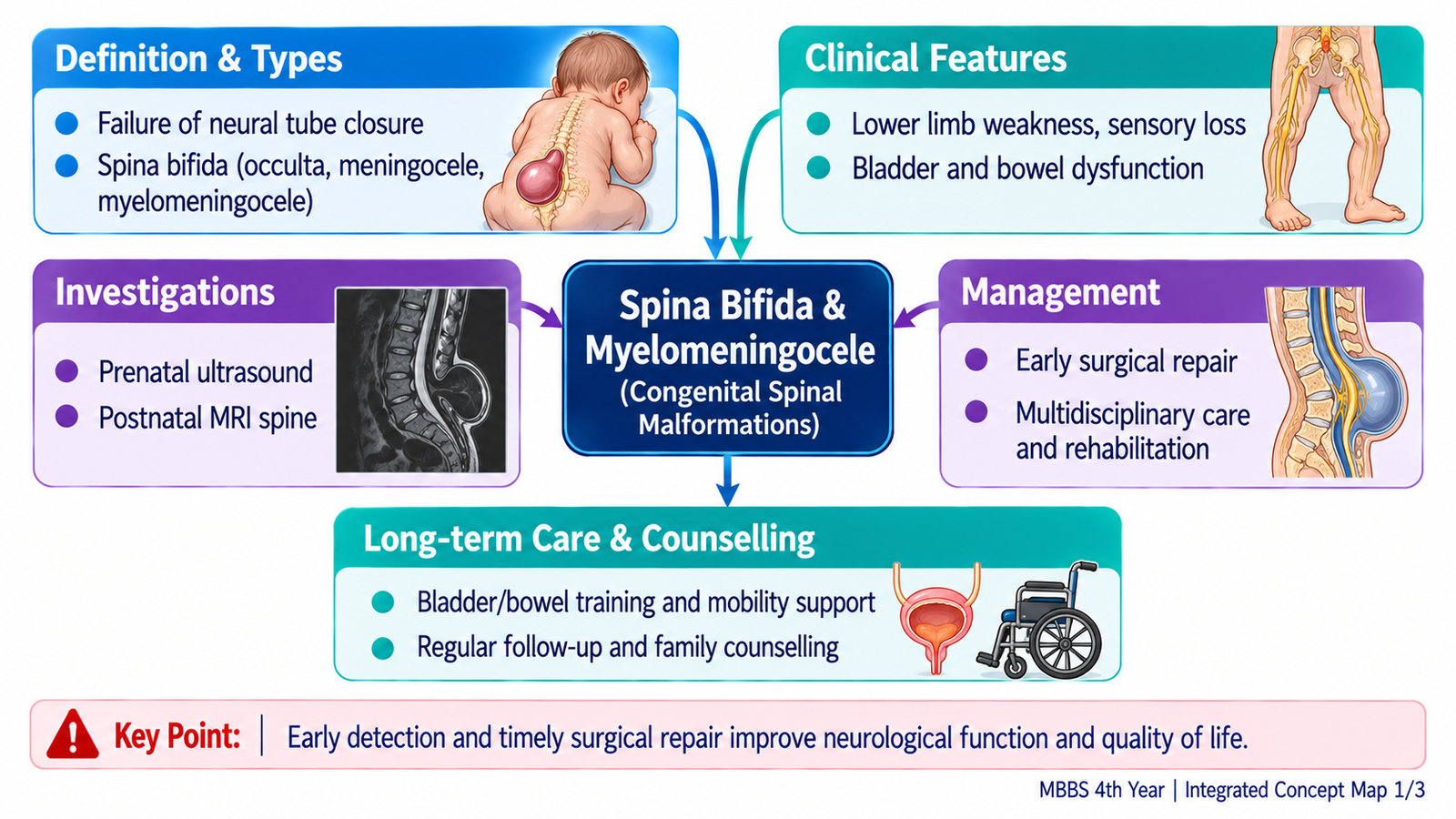
A. Understanding Congenital Spinal Malformations
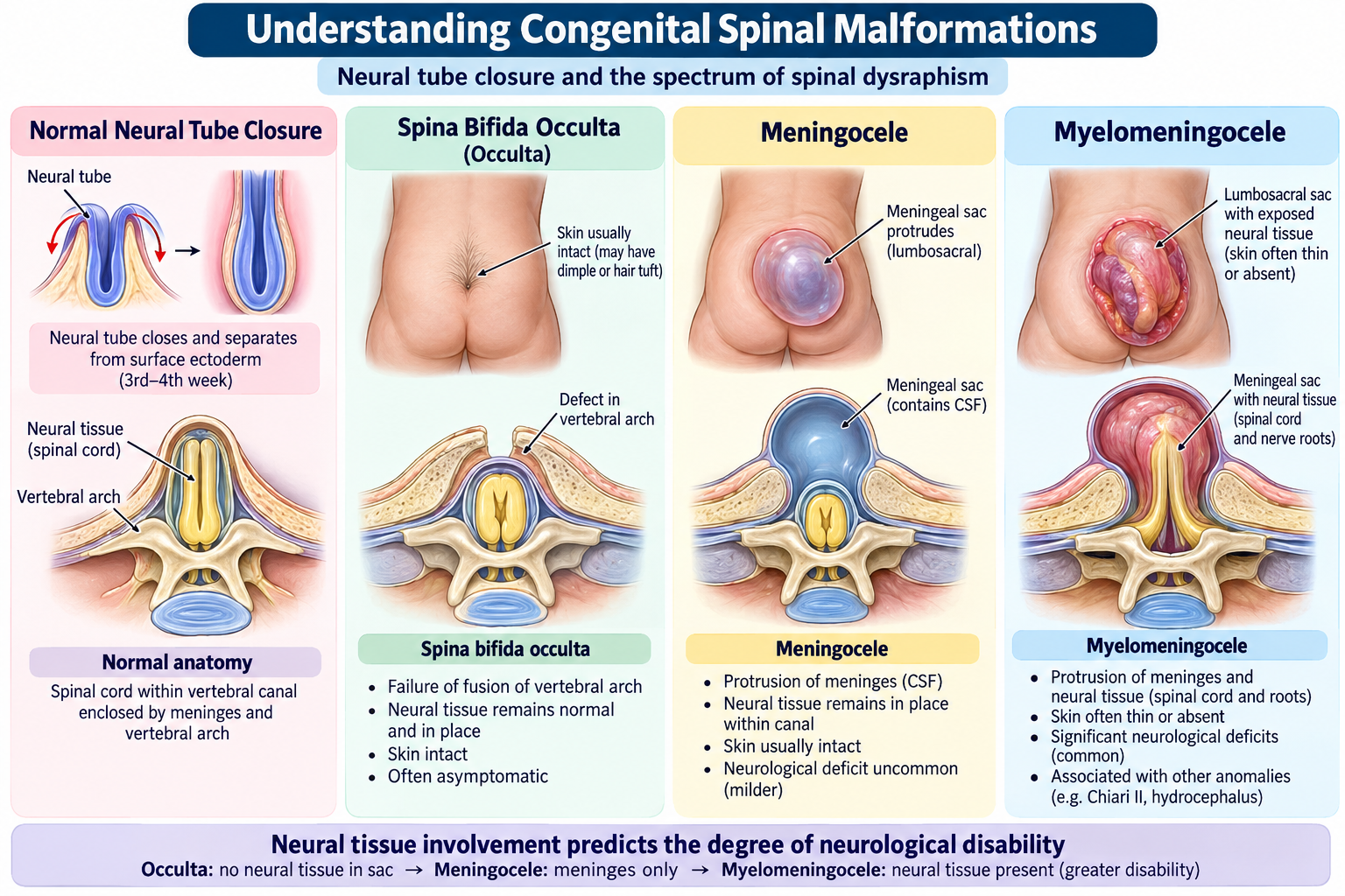
During embryonic development, the neural tube closes and the vertebral arches form around the spinal cord. Failure of normal closure produces spinal dysraphism. The clinical effect depends on whether neural tissue is involved and the level of the defect. A skin-covered or occult defect may be asymptomatic, whereas an open lesion exposes vulnerable neural tissue and is associated with substantial disability.
Important forms
- Spina bifida occulta: incomplete vertebral arch fusion, usually with intact skin; a tuft of hair, dimple or skin mark may indicate an underlying abnormality.
- Meningocele: meninges and cerebrospinal fluid protrude through a vertebral defect, but the spinal cord generally remains within the canal.
- Myelomeningocele: meninges and neural tissue protrude through the defect; motor, sensory and sphincter impairment are common.
Low maternal folate status increases neural-tube defect risk. Periconceptional folic acid supplementation reduces risk, although it does not prevent every case. The distinction between meningocele and myelomeningocele matters because neural tissue involvement strongly predicts functional impairment.
B. Spina Bifida and Myelomeningocele: Clinical Recognition
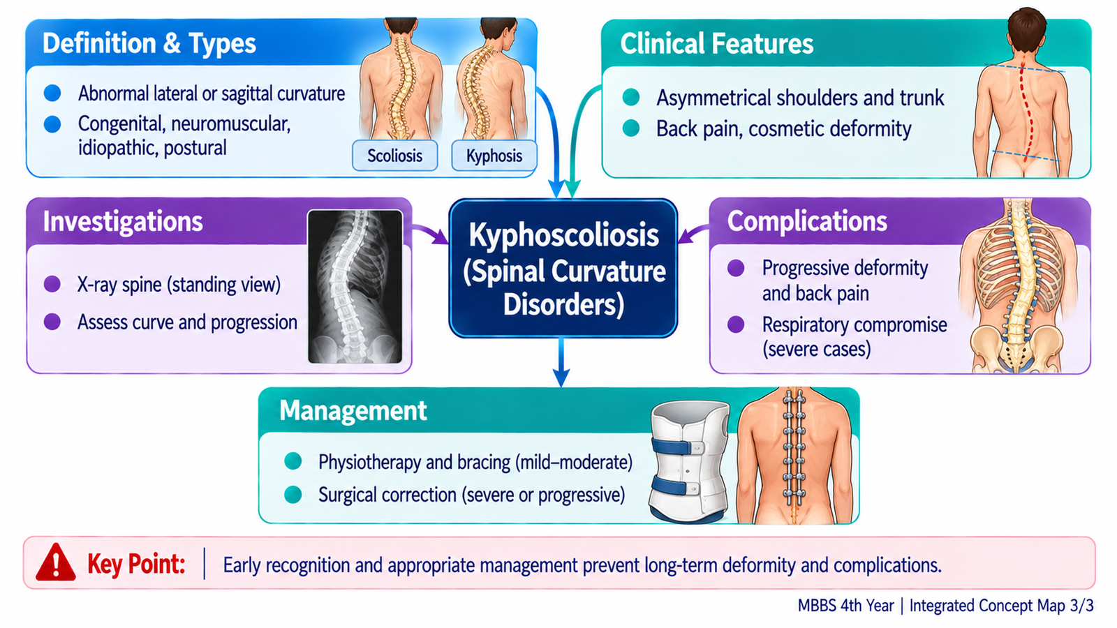
A newborn with myelomeningocele may have a visible lumbosacral sac or exposed neural placode. The lesion is not merely a skin problem: the neurological level determines which muscles function and whether the child can walk independently or requires assistive devices.
Focused clinical assessment
- Back: site, size, covering and integrity of lesion; look for leakage or inflammation without probing or compressing it.
- Lower limbs: spontaneous movements, muscle power, tone, reflexes, deformities and sensory responses appropriate to age.
- Bladder and bowel: urinary retention, incontinence, recurrent infections and constipation may reflect neurogenic dysfunction.
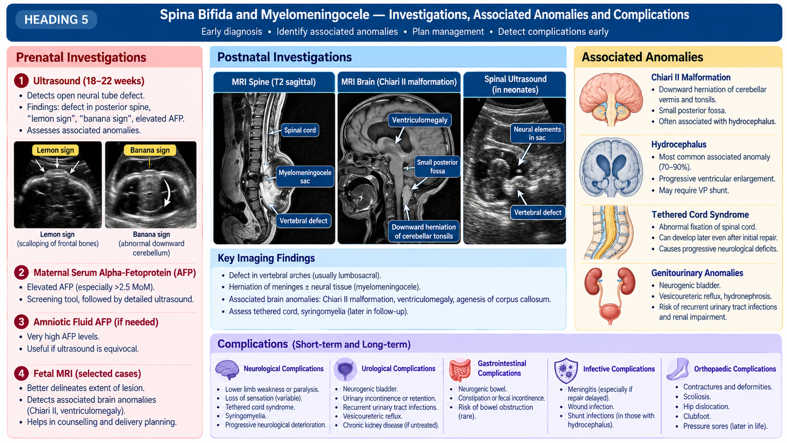
- Head and development: head circumference, fontanelle, vomiting, irritability and developmental progress; associated hydrocephalus and Chiari II malformation are important.
A child with an apparently minor cutaneous stigma but progressive weakness, abnormal gait or bladder symptoms also needs assessment for occult dysraphism or tethered cord. Clinical examination establishes function; imaging clarifies anatomy.
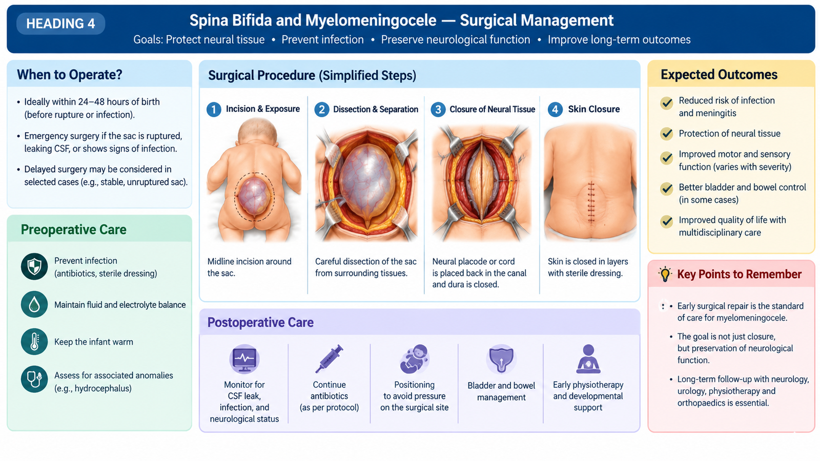
C. Investigations and Initial Management of Neural Tube Defects
Investigation should answer three practical questions: What structures are involved? Is there associated hydrocephalus? What neurological and urinary function is preserved? Spinal MRI delineates the cord, neural tissue and associated abnormalities when indicated; ultrasound may be useful in selected young infants with an intact posterior window. Cranial ultrasound or other brain imaging assesses ventriculomegaly when suspected. Renal/bladder ultrasound and specialist urinary evaluation help identify upper-tract risk.
Immediate priorities in an open lesion
Protect exposed neural tissue with a sterile, saline-moistened non-adherent dressing according to local neonatal protocol; position the infant prone or side-lying to avoid pressure. Maintain warmth, prevent contamination, assess neurological status and arrange urgent pediatric surgical/neurosurgical review. Do not attempt to push the sac back or rupture it.
Definitive treatment is timely surgical closure to protect neural tissue and reduce infection risk; it cannot reliably reverse neurological deficits already present. Associated hydrocephalus may require CSF diversion under specialist care. Antibiotics are guided by infection risk and local specialist protocols rather than replacing definitive evaluation.
Complications to recognize
- CSF leakage and meningitis
- Hydrocephalus and raised intracranial pressure
- Motor disability, contractures, foot or hip deformities
- Neurogenic bladder, recurrent urinary infection and renal damage
- Skin pressure injury and bowel dysfunction
D. Long-Term Care and Family Counselling
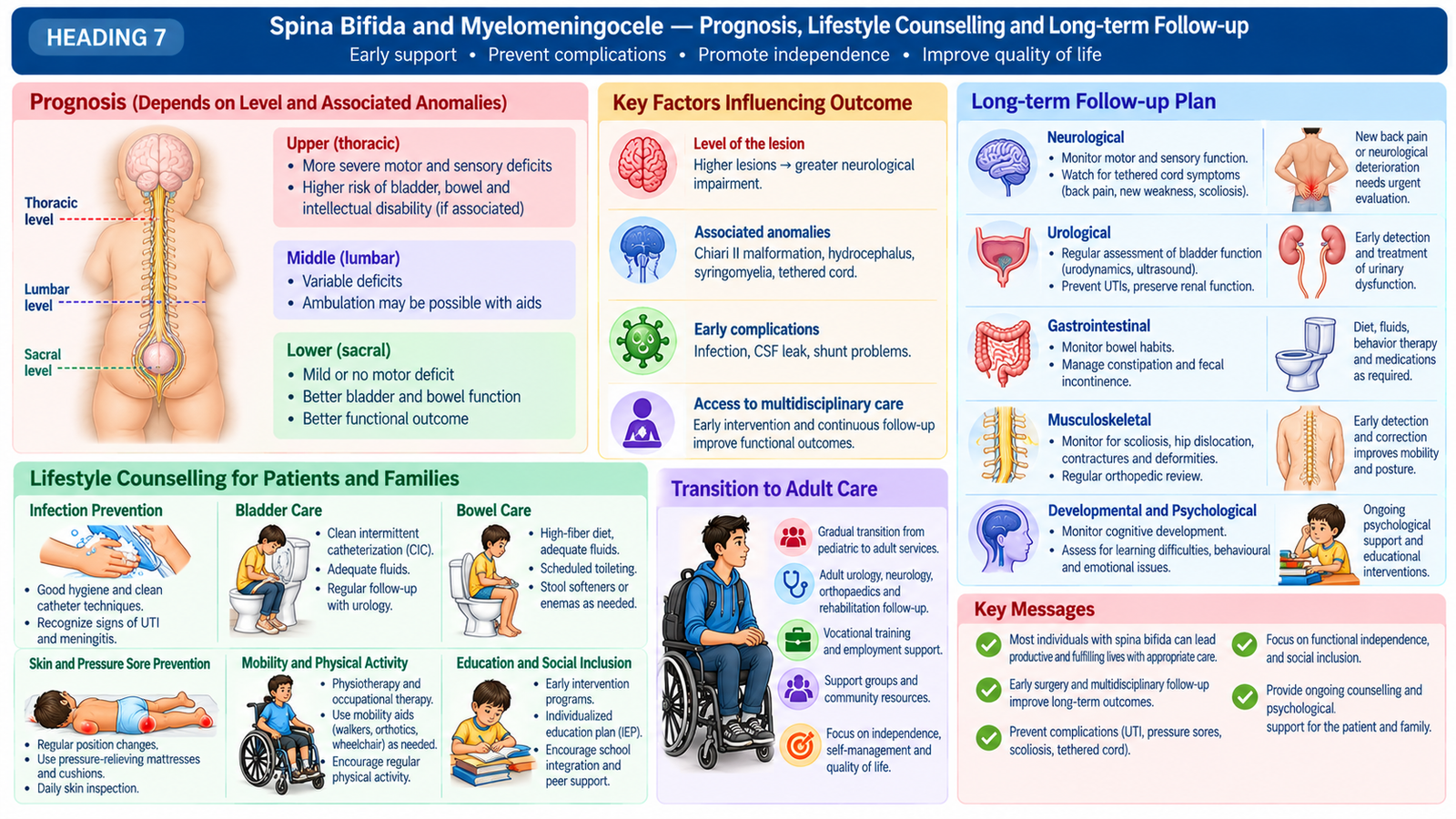
Care continues after closure because the underlying neurological deficit can affect mobility, continence, growth and participation in school. A multidisciplinary team commonly includes pediatric surgery/neurosurgery, pediatrics, orthopedics, physiotherapy, rehabilitation and urology. Goals should be individualized according to neurological level and functional ability.
Practical long-term plan
- Mobility: physiotherapy, stretching, orthoses, assistive devices and management of contractures or spinal deformity.
- Bladder protection: regular renal surveillance, continence planning and clean intermittent catheterization when prescribed by the urology team.
- Bowel care: scheduled toileting, dietary measures and an individualized bowel programme.
- Skin care: daily inspection of insensate areas and protection from pressure, heat and poorly fitting footwear.
- Development and wellbeing: monitor learning, independence, psychosocial needs and school participation.
Explain to families that surgery closes the defect but does not necessarily restore leg strength or bladder control. Seek prompt review for fever with urinary symptoms, new weakness, back pain, change in continence, headache, vomiting or progressive head enlargement; these may signal infection, tethered cord or shunt problems. Counsel on folic acid before future pregnancies, with higher-risk dosing determined by the treating clinician.
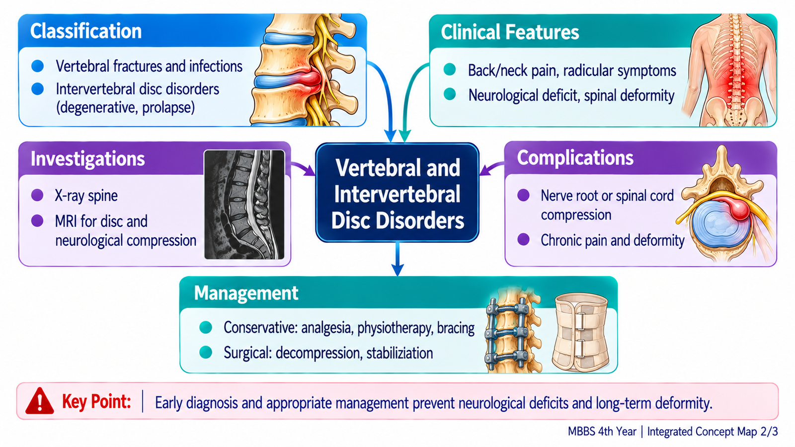
E. Classification and Clinical Features of Vertebral and Disc Disorders
Vertebral disease affects the bony column; disc disease affects the cushioning intervertebral discs and can irritate or compress nerve roots. A useful classification is degenerative, traumatic, infective, inflammatory, neoplastic and congenital/deformity-related. Disc disorders include degeneration, bulge, protrusion or herniation, and may coexist with spinal canal narrowing.
Recognizing the clinical pattern
- Mechanical back pain: linked to posture or activity, commonly without objective neurological loss.
- Radiculopathy: pain radiating along a nerve-root distribution with possible dermatomal sensory loss, myotomal weakness or reduced reflexes.
- Myelopathy: spinal cord dysfunction with gait difficulty, increased tone, brisk reflexes or sphincter disturbance; urgent evaluation may be needed.
- Spinal infection or tumor: persistent or progressive pain, systemic features, night pain or neurological deficit warrants investigation.
For example, a lumbar disc herniation compressing a nerve root may produce sciatica; a cervical lesion affecting the cord can cause hand clumsiness and spastic gait. Symptoms must be correlated with examination rather than attributed to an imaging abnormality alone.
F. Investigations, Complications and Management of Vertebral and Disc Disease
Start with history of onset, trauma, radiation of pain, fever, malignancy, functional loss and bowel/bladder symptoms. Examine posture, spinal movement, focal tenderness, lower-limb power, sensation, reflexes and gait. A straight-leg-raise test may support lumbar nerve-root irritation, but does not by itself establish the diagnosis.
Choosing investigations
- No red flags in uncomplicated acute mechanical pain: routine immediate imaging is often unnecessary.
- Plain radiographs: useful for alignment, deformity, fracture or destructive bony changes in selected cases.
- MRI: preferred for suspected cord or nerve-root compression, infection, tumor or cauda equina syndrome.
- CT: useful for detailed bony injury assessment when indicated.
- Blood tests: inflammatory markers and infection-directed tests when clinical findings suggest an inflammatory or infective cause.
Management and escalation
For uncomplicated degenerative pain, explain the condition, encourage graded activity, offer suitable analgesia and physiotherapy, and review function. Persistent disabling radicular symptoms or progressive deficits require specialist assessment; decompression or other surgery may be appropriate for selected compressive lesions. Fractures, infections and tumors require cause-specific treatment.
Pharmacological management: Oral NSAIDs such as ibuprofen or naproxen may be considered for uncomplicated mechanical low back pain, using the lowest effective dose for the shortest duration after assessing gastrointestinal, renal and cardiovascular risks. Routine gabapentinoids and oral corticosteroids are not recommended for sciatica. Persistent disabling radiculopathy despite conservative treatment may require specialist assessment for epidural injection or surgical decompression.
Complications include chronic pain, persistent neurological deficit, deformity, instability and, with severe compression, permanent sphincter dysfunction. Early recognition changes outcome.
G. Kyphoscoliosis: Causes, Assessment and Treatment
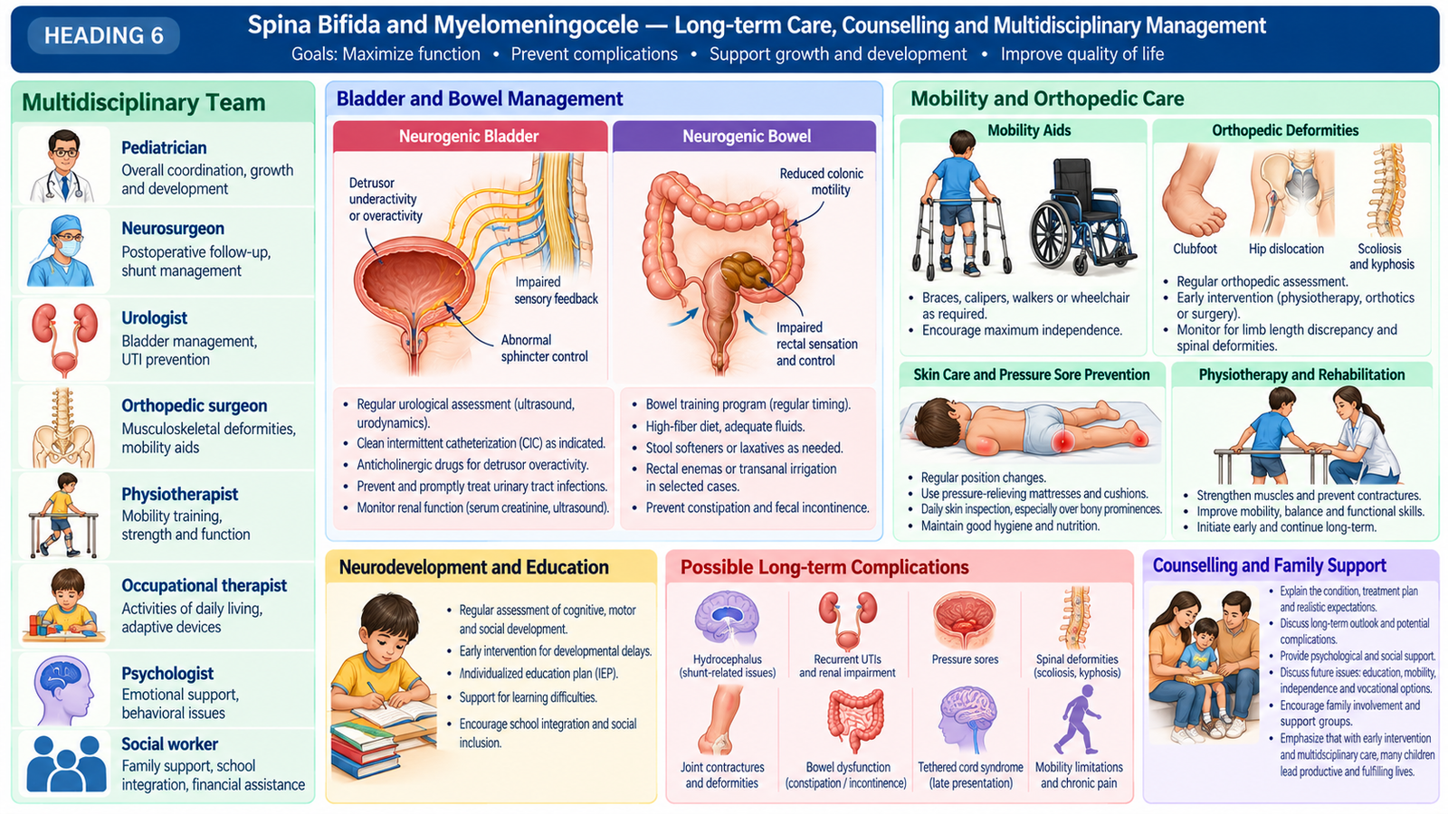
Kyphosis is excessive forward sagittal curvature; scoliosis is a lateral spinal curve with vertebral rotation. Kyphoscoliosis combines both deformities. Causes include congenital vertebral malformations, idiopathic scoliosis, neuromuscular disorders, postural or structural abnormalities and other acquired spinal disease.
Clinical assessment
Ask about age at onset, progression, growth, pain, functional limitation, neurological symptoms and breathing difficulty. Inspect standing alignment, shoulder and pelvic symmetry, rib prominence and sagittal profile. The Adams forward-bend test can reveal rotational rib or lumbar prominence. Perform a neurological examination and assess respiratory compromise in marked deformity.
Investigation and interpretation
Standing full-length spinal radiographs show the curve and help measure the Cobb angle, which quantifies scoliosis and assists monitoring. Skeletal maturity and serial changes help estimate progression risk. MRI is considered when neurological abnormalities, atypical curve features or suspected congenital neural-axis abnormalities are present. Pulmonary assessment may be needed in severe thoracic deformity.
Management principles
Mild, stable curves may be observed with scheduled review. Growing patients with selected progressive curves may benefit from bracing to limit progression; bracing does not reliably correct a fixed structural deformity. Physiotherapy supports posture, conditioning and function. Severe or progressive deformity, neurological compromise, major imbalance or cardiopulmonary effects may require specialist surgical correction and stabilization.
Complications include back pain, cosmetic and psychosocial effects, reduced function, progression during growth and restrictive respiratory impairment in severe thoracic deformity. Counselling should explain monitoring, brace adherence where prescribed, activity, realistic treatment goals and long-term follow-up.
Integrated Mechanism Flow
Neural tube closure failure → vertebral arch defect → meningeal ± neural tissue involvement → level-dependent weakness and sphincter dysfunction → imaging and functional assessment → surgical protection and long-term multidisciplinary care.
Important Comparison
| Condition | Key anatomy | Clinical implication |
|---|---|---|
| Spina bifida occulta | Bony arch defect, skin usually intact | Often asymptomatic; assess concerning skin or neurological signs |
| Meningocele | Meninges and CSF protrude | Neurological function may be relatively preserved |
| Myelomeningocele | Neural tissue and meninges protrude | Motor, sensory and bladder/bowel deficits common |
AIM High-Yield Review
- ⭐ Myelomeningocele contains neural tissue; neurological deficits depend on lesion level.
- A normal-looking skin surface does not exclude clinically significant occult dysraphism.
- Open spinal lesions require protection from trauma, drying and contamination before urgent specialist assessment.
- Hydrocephalus and Chiari II association should be considered in myelomeningocele.
- Surgical closure protects exposed tissue but may not reverse established paralysis.
- Neurogenic bladder surveillance is essential to prevent urinary tract and renal complications.
- Disc-related radiculopathy follows a nerve-root distribution; myelopathy produces spinal cord signs.
- ⭐ New urinary retention with saddle sensory loss is a cauda equina emergency.
- MRI is the key investigation for suspected neural compression or spinal infection.
- Uncomplicated mechanical back pain commonly begins with conservative treatment, not routine imaging.
- Scoliosis is a three-dimensional deformity; the Cobb angle is measured on standing radiographs.
- ⭐ Progressive severe kyphoscoliosis may impair respiratory function and require specialist correction.
AIM Video Learning — Topic 11
Congenital Spinal Malformations, Vertebral Disorders & Kyphoscoliosis
Spina Bifida & Myelomeningocele — Clinical Overview
Educational video by Osmosis from Elsevier. Focus on the developmental defect, clinical findings, investigations and management principles.