
📖 AIM Learning Material
Topic 8. Intracranial Space-Occupying Lesions, Pediatric Macrocephaly & Head Injury
Neuroscience-III • Neurosurgery and Pediatrics
Topic Introduction
Intracranial masses, an enlarging head in a child and traumatic brain injury can appear to be separate problems, but all involve the relationship between brain tissue, cerebrospinal fluid, blood and the space available inside the skull. This chapter explains how to recognise important symptoms, assess neurological danger, select appropriate imaging and understand treatment decisions. In infants, skull growth changes how raised pressure may present; after trauma, preventing secondary brain injury is especially important. The aim is a clear, safe clinical approach at Final-Year MBBS level.
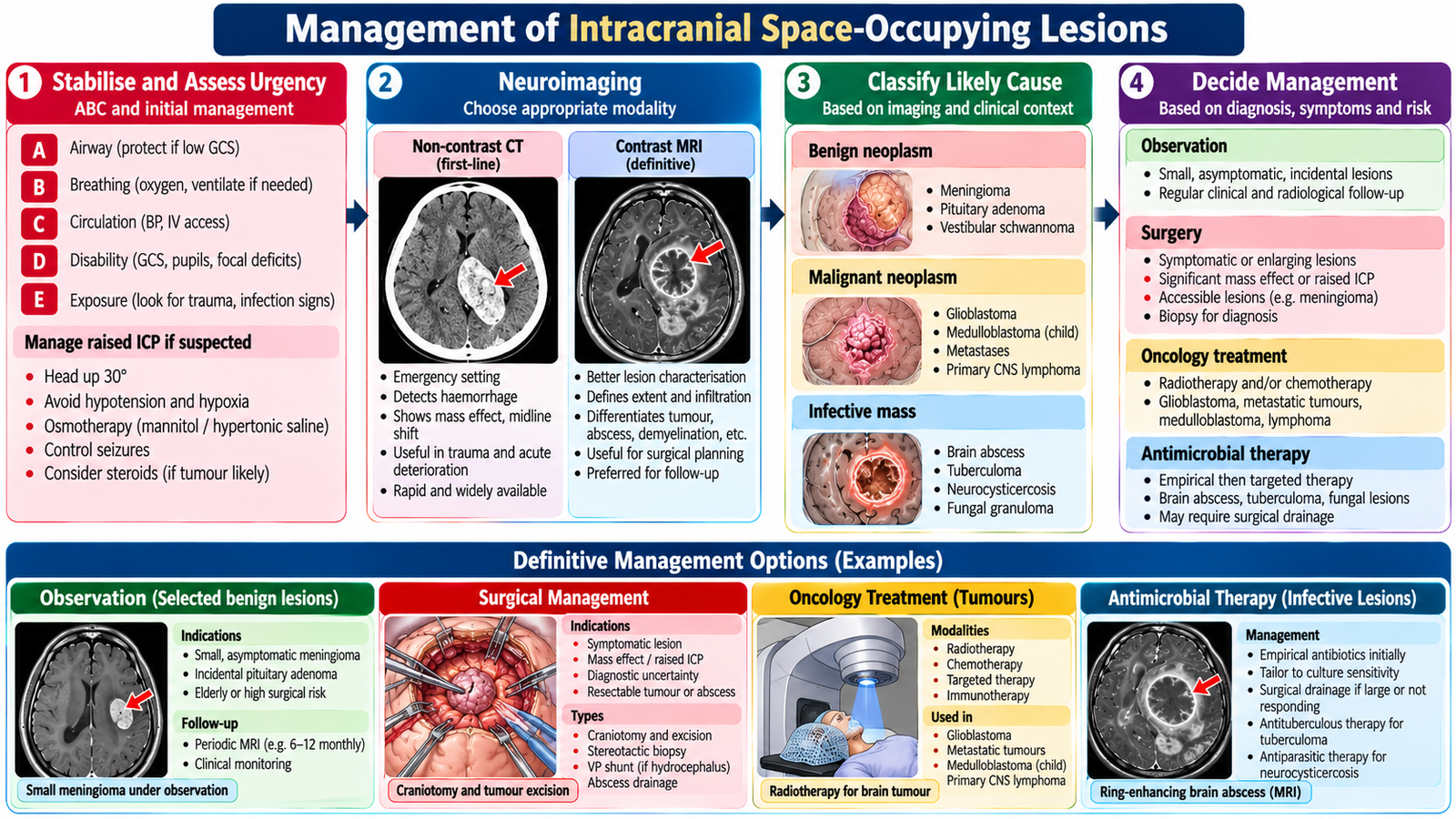
1. Classification of Intracranial Space-Occupying Lesions
Clinical classification
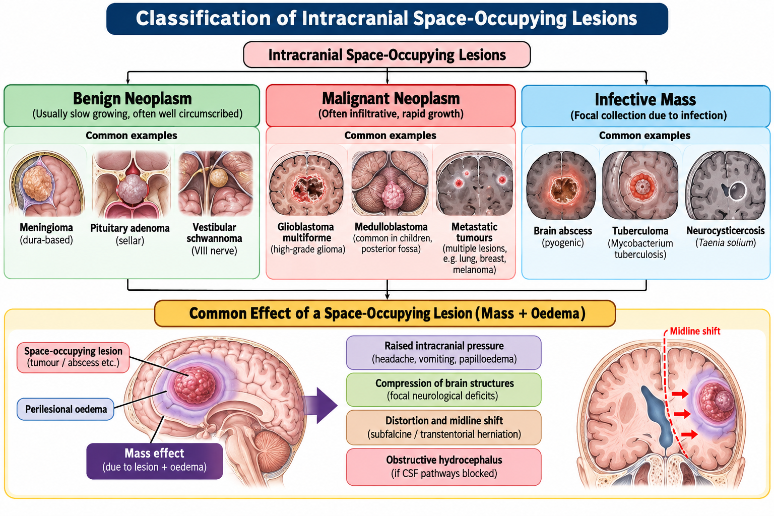
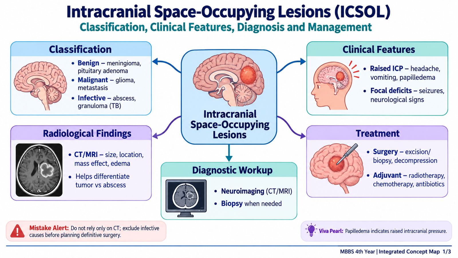
- Benign neoplastic: meningioma and some low-grade gliomas. Benign histology does not mean harmless: location, growth and compression determine danger.
- Malignant neoplastic: high-grade glioma, primary CNS lymphoma and brain metastases. These may infiltrate brain tissue or cause extensive surrounding oedema.
- Infective: pyogenic brain abscess and intracranial tuberculoma. Infections may produce focal deficits, seizures and raised ICP.
Useful distinction: A brain abscess contains a collection of pus; a tuberculoma is a granulomatous mass. Imaging can suggest the cause, but a ring-enhancing lesion is not specific for abscess and must be interpreted with clinical context.
2. Clinical Presentation and Focused Neurological Assessment
Raised intracranial pressure
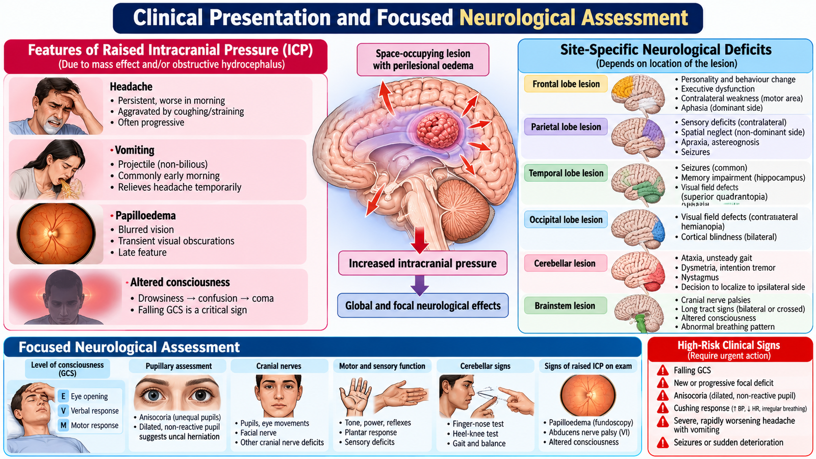
Progressive headache, vomiting, reduced alertness and papilloedema may occur. Headache can be worse on waking or with coughing. Papilloedema indicates optic disc swelling from raised ICP but may be absent early. Late hypertension, bradycardia and irregular respiration (Cushing response) suggest life-threatening intracranial hypertension.
Focal and generalised features
- Frontal: personality change, executive dysfunction, contralateral weakness.
- Temporal: seizures, memory or language disturbance in the dominant hemisphere.
- Parietal: sensory deficit or neglect.
- Occipital: visual-field defects.
- Cerebellum/posterior fossa: ataxia, imbalance, vomiting and possible obstructive hydrocephalus.
- Other: new-onset seizures, cranial nerve deficits and altered cognition.
Bedside assessment
Establish symptom progression, seizures, fever, recent infection, cancer history, immunosuppression and drug use. Record vital signs, Glasgow Coma Scale (GCS), pupils, limb power, speech, cranial nerves, gait when safe and fundoscopic findings. Falling GCS, unequal pupils, new focal deficit or repeated vomiting requires urgent escalation.
3. Neuroimaging and Diagnostic Interpretation
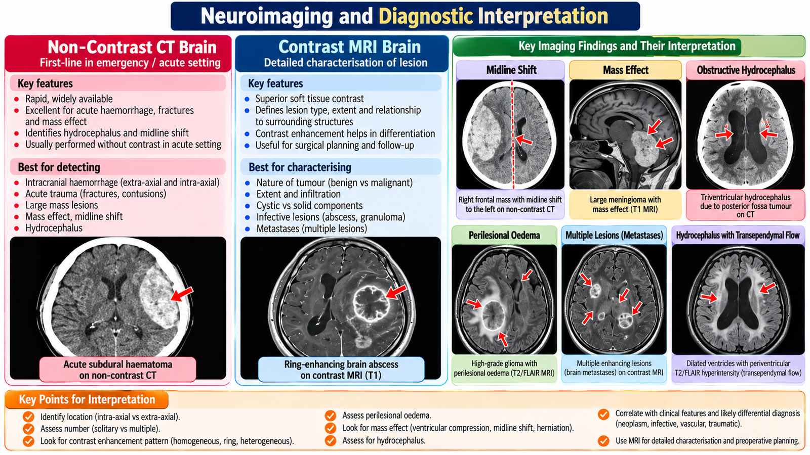
- Mass effect: compression of ventricles or displacement of the midline suggests pressure on adjacent structures.
- Vasogenic oedema: surrounding brain swelling contributes to symptoms and raised ICP.
- Contrast enhancement: reflects altered blood–brain barrier or vascularity; patterns are suggestive, not definitive.
- Hydrocephalus: enlarged ventricles from impaired CSF flow, particularly with posterior fossa lesions.
- Abscess: may show a ring-enhancing lesion with surrounding oedema; MRI diffusion restriction can support the diagnosis.
Imaging is interpreted alongside history and examination. Histopathology or microbiology may be needed for definitive diagnosis, selected by the specialist team. Do not perform lumbar puncture when a mass lesion with significant raised ICP or herniation risk is suspected; seek urgent imaging and specialist advice.
4. Management of Intracranial Space-Occupying Lesions
Cause-directed treatment
- Benign tumour: observation with interval imaging for selected stable lesions, or surgery when symptomatic, enlarging or compressive.
- Malignant tumour: specialist assessment for maximal safe resection or biopsy, followed by radiotherapy and/or systemic treatment depending on pathology.
- Brain abscess: prolonged appropriate intravenous antimicrobial therapy and, when indicated, neurosurgical aspiration or drainage.
- Tuberculoma: antituberculous treatment according to the treating team, with monitoring of clinical and radiological response.
Dexamethasone may reduce symptomatic vasogenic oedema around selected tumours; it is not routine for every SOL and is not a substitute for treating the cause. Antiseizure medicines are used for documented seizures or specific specialist indications rather than indiscriminately for every lesion. Surgical decisions depend on accessibility, diagnosis, mass effect, functional risk and patient condition.
5. Pediatric Macrocephaly: Assessment of an Enlarged Head
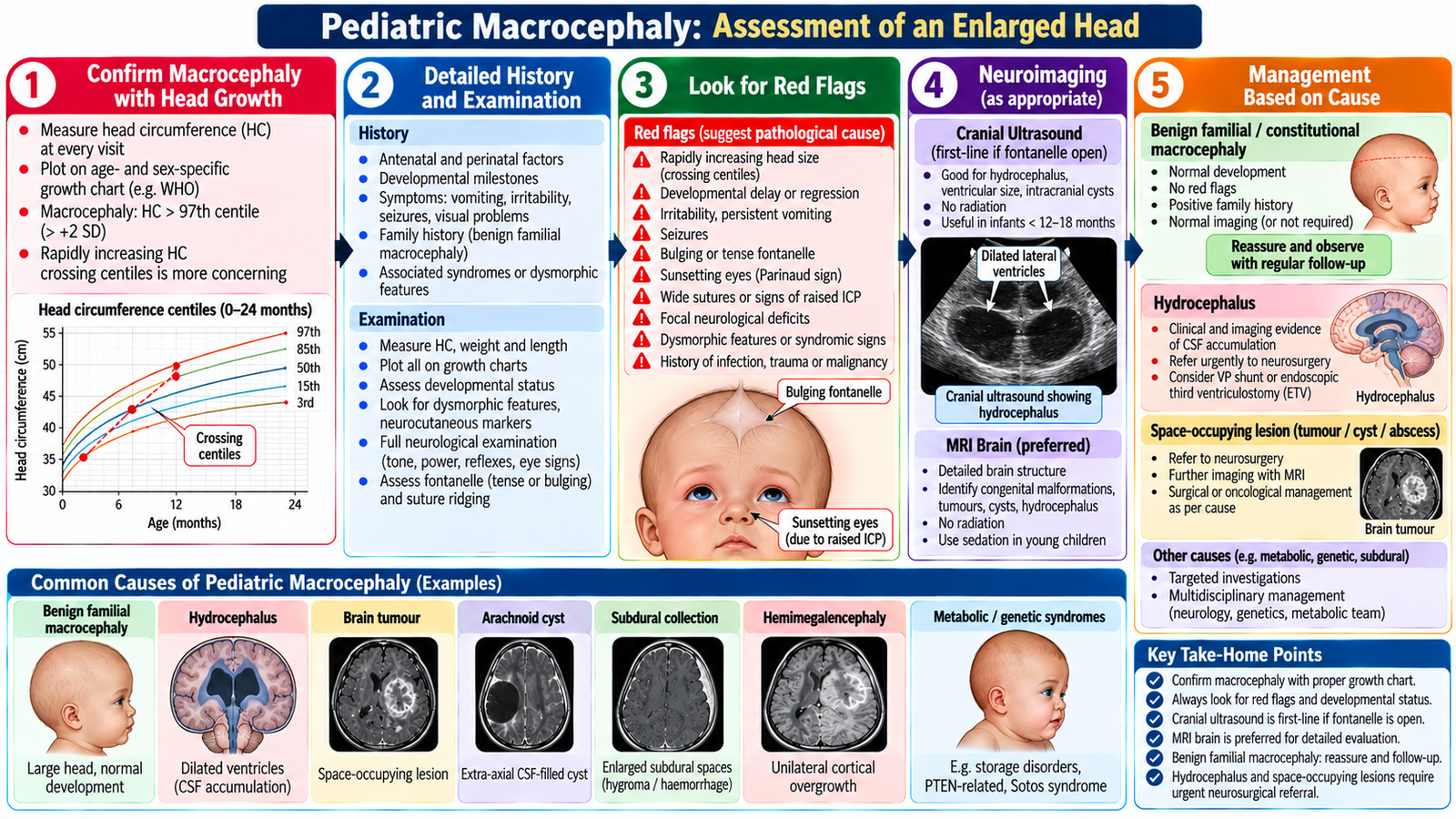
Important causes
- Familial benign macrocephaly: normal development, normal examination and large parental head circumference.
- Hydrocephalus: increased CSF within the ventricles due to obstruction or impaired absorption; may cause progressive head enlargement in infants.
- Benign enlargement of subarachnoid spaces: typically in otherwise well infants, diagnosed with clinical and imaging correlation.
- Intracranial mass or subdural collection: may increase head size or cause pressure signs.
- Other causes: megalencephaly or selected metabolic/genetic conditions, especially when development is abnormal.
History and examination
Plot head circumference on age- and sex-appropriate growth charts and compare previous values. Ask about family head size, pregnancy/birth history, vomiting, feeding, irritability, milestones, seizures and trauma. Examine fontanelle tension, scalp veins, eye position (including sunsetting eyes), tone, reflexes, development and signs of raised ICP.
Investigation and management
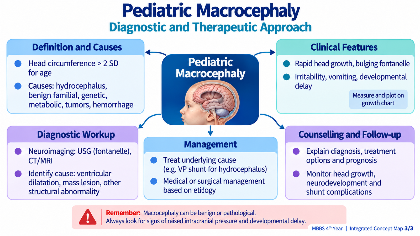
A well child with proportionate familial head size, normal development and stable growth may need observation and follow-up. Rapid crossing of centiles, bulging fontanelle, vomiting, lethargy, seizures or developmental regression requires urgent assessment. Cranial ultrasound is useful through an open fontanelle; MRI gives anatomical detail, while CT is reserved for urgent indications because of radiation. Treatment targets the cause: hydrocephalus may require neurosurgical CSF diversion, whereas benign cases need reassurance and growth/development monitoring.
6. Head Injury: Initial Assessment, Imaging and Treatment
Initial trauma approach
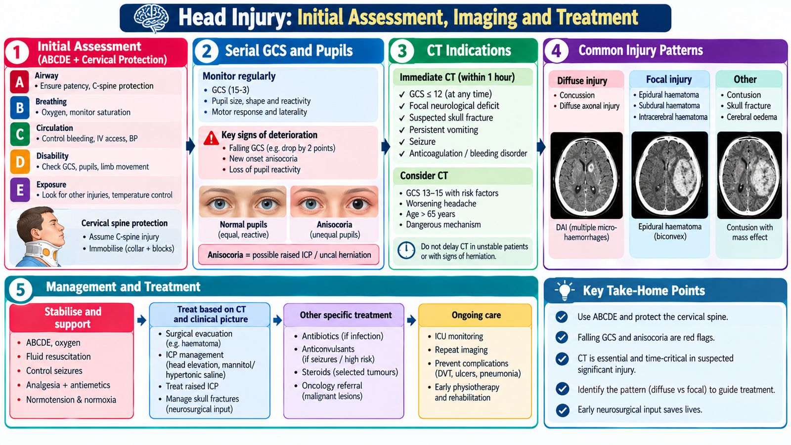
Use an ABCDE assessment with cervical-spine protection. Secure airway and oxygenation when necessary, control external bleeding, maintain circulation and check capillary glucose. Record GCS components, pupil size/reactivity, limb movement, seizures and vital signs; repeat observations because deterioration may be delayed.
Severity and imaging
GCS 13–15 indicates mild, 9–12 moderate and 3–8 severe TBI after initial stabilisation. Urgent CT is indicated for concerning features such as depressed consciousness, focal neurological deficit, suspected skull fracture, repeated vomiting, post-traumatic seizure or other validated head-injury risk criteria. A normal initial CT does not replace observation when symptoms or risk factors remain concerning.
Common intracranial injuries
- Epidural haematoma: classically biconvex/lens-shaped extra-axial collection, often related to skull fracture; may deteriorate rapidly.
- Subdural haematoma: crescent-shaped extra-axial collection, often due to bridging-vein injury.
- Contusion/intraparenchymal bleed: focal bruising or haemorrhage within brain tissue.
- Diffuse axonal injury: widespread shearing injury; severe impaired consciousness may occur despite relatively subtle CT findings.
Management principles
Mild uncomplicated injury may be managed with observation, clear return precautions and graded return to activity. Moderate or severe injury requires hospital care, frequent neurological review and specialist involvement. Significant haematoma, mass effect, depressed fracture or refractory raised ICP may require neurosurgical intervention. Avoid hypoxia, hypotension and unnecessary delays in referral.

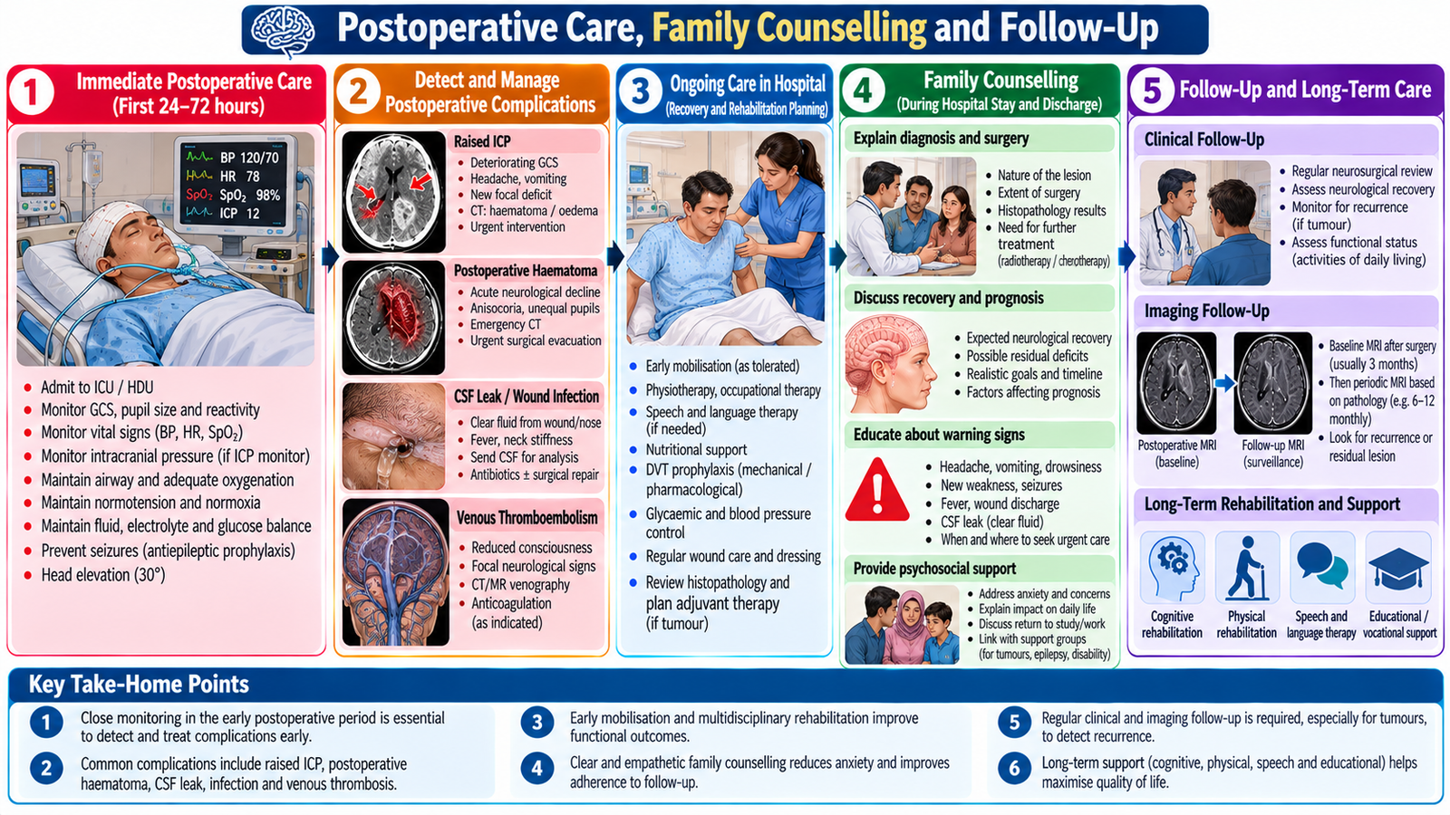
7. Postoperative Care, Family Counselling and Follow-Up
Integrated Mechanism Flow
Expanding mass or traumatic bleeding → increased intracranial volume → compensatory capacity exhausted → raised ICP and reduced cerebral perfusion → headache, vomiting, focal deficits or declining consciousness → urgent imaging and cause-directed treatment. Infant exception: open sutures may allow progressive head enlargement, so serial head circumference and development provide essential clues.
Important Comparison
| Feature | Epidural haematoma | Subdural haematoma |
|---|---|---|
| Typical vessel | Often arterial | Often bridging veins |
| CT appearance | Biconvex / lens-shaped | Crescent-shaped |
| Clinical concern | Rapid deterioration possible | Acute or delayed deterioration |
AIM High-Yield Review
- An intracranial SOL may be benign, malignant or infective; even benign lesions can cause dangerous mass effect.
- Progressive headache, vomiting, papilloedema and altered consciousness suggest raised ICP.
- A falling GCS or unequal pupils is an emergency warning sign.
- Non-contrast CT is valuable for urgent haemorrhage, mass effect and hydrocephalus; MRI better characterises many lesions.
- Avoid lumbar puncture when a mass lesion creates significant herniation risk.
- Brain abscess needs antimicrobial therapy and sometimes drainage; tumours need pathology-directed specialist care.
- Macrocephaly requires serial head circumference, developmental assessment and red-flag screening.
- Rapid head growth with bulging fontanelle or lethargy suggests urgent assessment for hydrocephalus or another intracranial cause.
- Head injury management starts with ABCDE, cervical-spine protection and prevention of hypoxia and hypotension.
- Epidural is classically lens-shaped; subdural is crescent-shaped on CT.
- Postoperative deterioration, seizures, fever or CSF leakage require urgent review.
AIM LEARNING CYCLE | FINAL YEAR MBBS
Video Learning — Topic 8
Intracranial Space-Occupying Lesions, Pediatric Macrocephaly & Head Injury
Neuroscience-III | KMU Curriculum
1. Intracranial Space-Occupying Lesions — Brain Tumors
Ninja Nerd: classification, clinical features, raised intracranial pressure, neuroimaging and treatment principles.
2. Head Injury — Epidural vs Subdural Hematoma
Learn Medicine Asap: anatomical location, mechanism and key clinical differences between two important traumatic intracranial hemorrhages.
3. Pediatric Macrocephaly & Hydrocephalus
UCSF Benioff Children’s Hospitals — pediatric neurosurgery teaching lecture covering enlarged head assessment, hydrocephalus, imaging and therapeutic principles.
This lecture opens on the hospital’s video page.
Use these videos alongside the AIM Learning Material. Focus on clinical presentation, CT/MRI findings, neurological deterioration and the major principles of neurosurgical management. The videos supplement rather than replace the KMU learning outcomes.