
📖 AIM Learning Material
Topic 9. Lower Limb Weakness, Multiple Sclerosis & Syringomyelia
Neuroscience · Medicine & Neurosurgery · Localization → investigation → patient-centred management
Topic Introduction
Lower limb weakness is a clinical presentation, not a final diagnosis. It can arise from the brain, spinal cord, nerve roots, peripheral nerves or muscles. Careful examination helps identify the affected level and determines which patients need urgent imaging or referral. Multiple sclerosis illustrates inflammatory damage to central nervous system myelin, whereas syringomyelia illustrates structural injury within the spinal cord. This chapter explains how to recognize important patterns, examine a patient with paraplegia, select investigations, plan treatment and counsel patients. The aim is to move from a symptom to a safe, reasoned clinical decision.
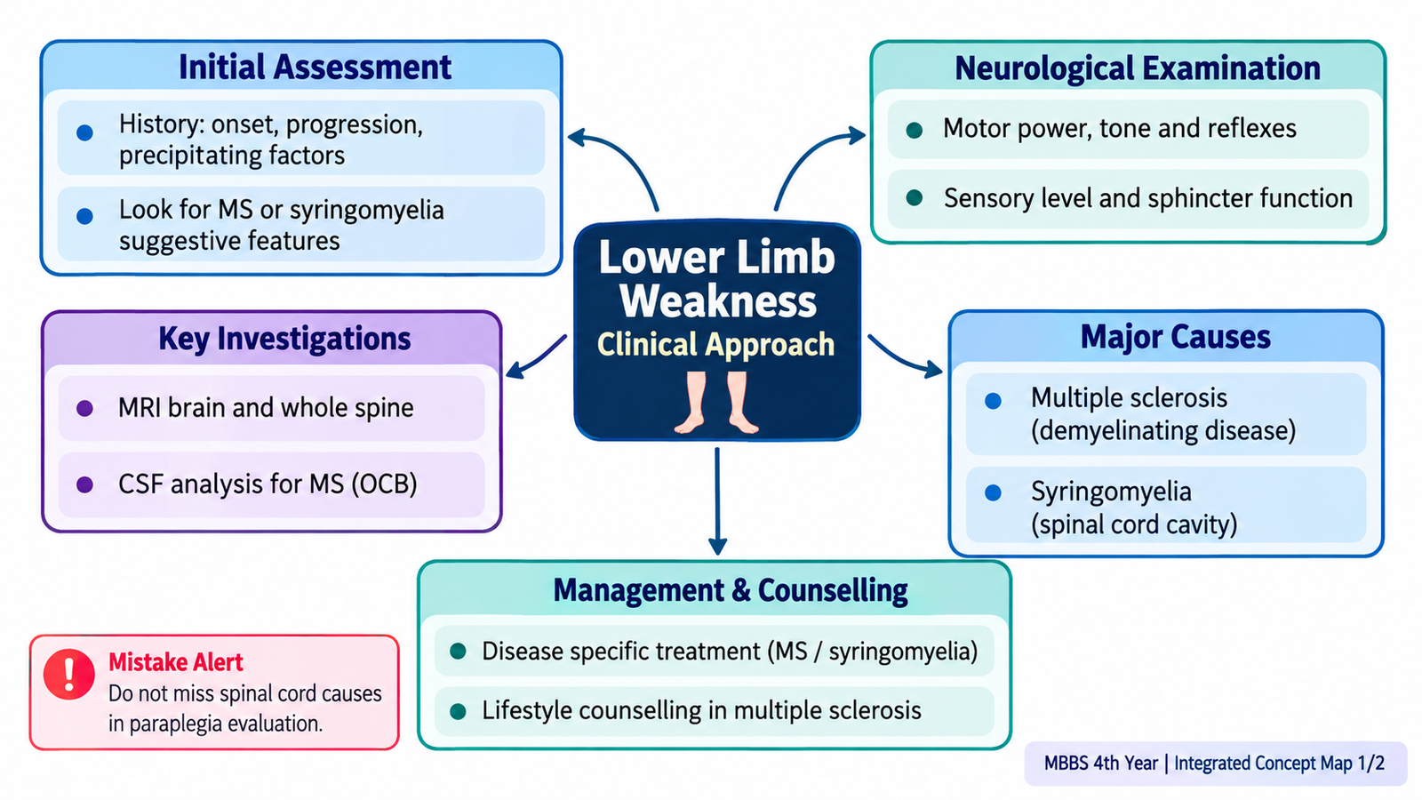
1. Understanding Lower Limb Weakness and Neuroanatomical Localization
The key principle
Weakness is reduced muscle power, not simply fatigue, pain or poor coordination. Paraparesis means weakness of both lower limbs; paraplegia indicates severe or complete paralysis. First establish onset, progression and whether symptoms are symmetrical.
Localize before naming a disease
A cerebral lesion may cause contralateral weakness, often with other focal signs. Spinal cord disease produces weakness below a neurological level and may disturb bladder function. Root disease causes radicular pain and segmental deficits; peripheral nerve disease follows nerve distributions; generalized neuropathy often causes distal symmetrical weakness.
Upper versus lower motor neuron patterns
An upper motor neuron (UMN) lesion causes increased tone, brisk reflexes, clonus and an extensor plantar response after the acute phase. A lower motor neuron (LMN) lesion causes reduced tone, reduced reflexes, wasting and sometimes fasciculations. Acute cord injury can initially produce flaccidity and areflexia (spinal shock), so timing matters.
Sensory clues
A definite sensory level suggests spinal cord pathology. Loss of vibration and joint position points toward posterior column involvement; loss of pain and temperature suggests spinothalamic pathway involvement. Saddle sensory loss and urinary retention suggest a cauda equina emergency.

2. Diagnostic Algorithm for a Patient With Lower Limb Weakness
Step 1 — identify urgency
Assess airway, breathing, circulation and vital signs when weakness is acute or rapidly progressive. Urgent warning signs include sudden onset, severe back pain with new neurological deficit, urinary retention, saddle anesthesia, fever with spinal tenderness, and rapidly ascending weakness or respiratory compromise.
Step 2 — take a targeted history
Ask about exact time of onset, progression, falls, gait change, back or neck pain, trauma, preceding infection, visual symptoms, numbness, sphincter symptoms, medication exposure and prior neurological episodes. Episodes separated in time and involving different CNS regions raise suspicion of demyelination.
Step 3 — examine and localize
Check cognition and cranial nerves where relevant; compare tone, power, reflexes and plantar responses; test pinprick, temperature, vibration and joint position; assess coordination and gait if safe. Map a sensory level and examine upper limbs when cord disease is suspected.
Step 4 — select investigations
Suspected acute stroke needs emergency brain imaging. Suspected spinal cord compression or inflammatory myelopathy needs urgent MRI of the appropriate spinal region. Basic blood tests (including glucose, electrolytes and blood count) address reversible contributors. Nerve conduction studies/EMG help when peripheral disease is suspected; lumbar puncture is selected after excluding contraindications.
Step 5 — act on localization
Do not delay referral for a suspected compressive cord lesion or cauda equina syndrome. A progressive or unexplained neurological deficit needs specialist assessment even when initial screening tests are normal.

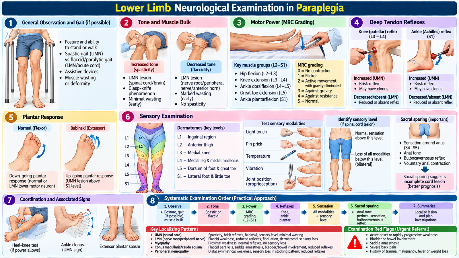
3. Lower Limb Neurological Examination in Paraplegia
Inspection and gait
Observe posture, muscle bulk, involuntary movements, deformity and pressure areas. If walking is safe, assess standing, walking, turning, heel/toe walking and need for support. A spastic, stiff-legged gait supports a pyramidal tract lesion.
Tone and power
With the patient relaxed, assess passive movement at hips, knees and ankles for spasticity or flaccidity. Test hip flexion, knee extension, ankle dorsiflexion and plantarflexion bilaterally using the MRC 0–5 power scale; compare sides and document function.
Reflexes and plantar response
Test knee (L3–L4) and ankle (S1) jerks; look for clonus when indicated. An extensor plantar response supports corticospinal tract involvement. Reflex loss in an acute presentation does not automatically exclude cord disease.
Sensation and coordination
Compare light touch and pinprick on both sides, seek a sensory level, and test vibration/proprioception at toes. Heel–shin testing is useful when power permits. Evaluate sacral symptoms and bladder/bowel function; detailed sacral examination belongs to supervised clinical assessment.
Document the clinical conclusion
Summarize distribution, MRC grade, tone, reflexes, plantar responses, sensory level and sphincter status. State the likely localization and urgency rather than writing only “paraplegia”.
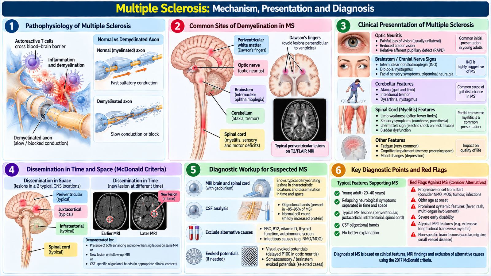
4. Multiple Sclerosis: Mechanism, Presentation and Diagnosis
Disease mechanism
Multiple sclerosis (MS) is an immune-mediated inflammatory demyelinating disease of the central nervous system. Myelin injury impairs saltatory conduction, producing neurological deficits; axonal injury contributes to persistent disability. Lesions commonly affect optic pathways, brainstem, cerebellum and spinal cord.
Clinical presentations
Optic neuritis may cause painful monocular visual loss. Spinal lesions may cause limb weakness, sensory change and urinary urgency. Brainstem or cerebellar involvement may produce diplopia, imbalance or ataxia. Fatigue and heat-related temporary worsening are common; fever or infection can transiently aggravate old symptoms.
Relapse and disease course
A true relapse is a new or worsening neurological symptom lasting at least 24 hours, without fever or infection, after a stable period. Relapsing–remitting disease has attacks and recovery; some patients later develop gradual progression. Progression can also occur from onset.
Diagnostic reasoning
Diagnosis requires evidence that characteristic CNS lesions are disseminated in space and time, with no better explanation. MRI brain and spinal cord (as clinically indicated) identifies characteristic lesions; gadolinium enhancement may help identify active inflammation. CSF oligoclonal bands can support diagnosis in selected cases. Apply formal diagnostic criteria through a neurologist; MRI lesions alone do not prove MS.
Differential diagnosis
Exclude other causes of optic-spinal symptoms and weakness, including compressive myelopathy, infection and relevant metabolic deficiencies. Atypical presentations require reassessment rather than automatic labeling as MS.
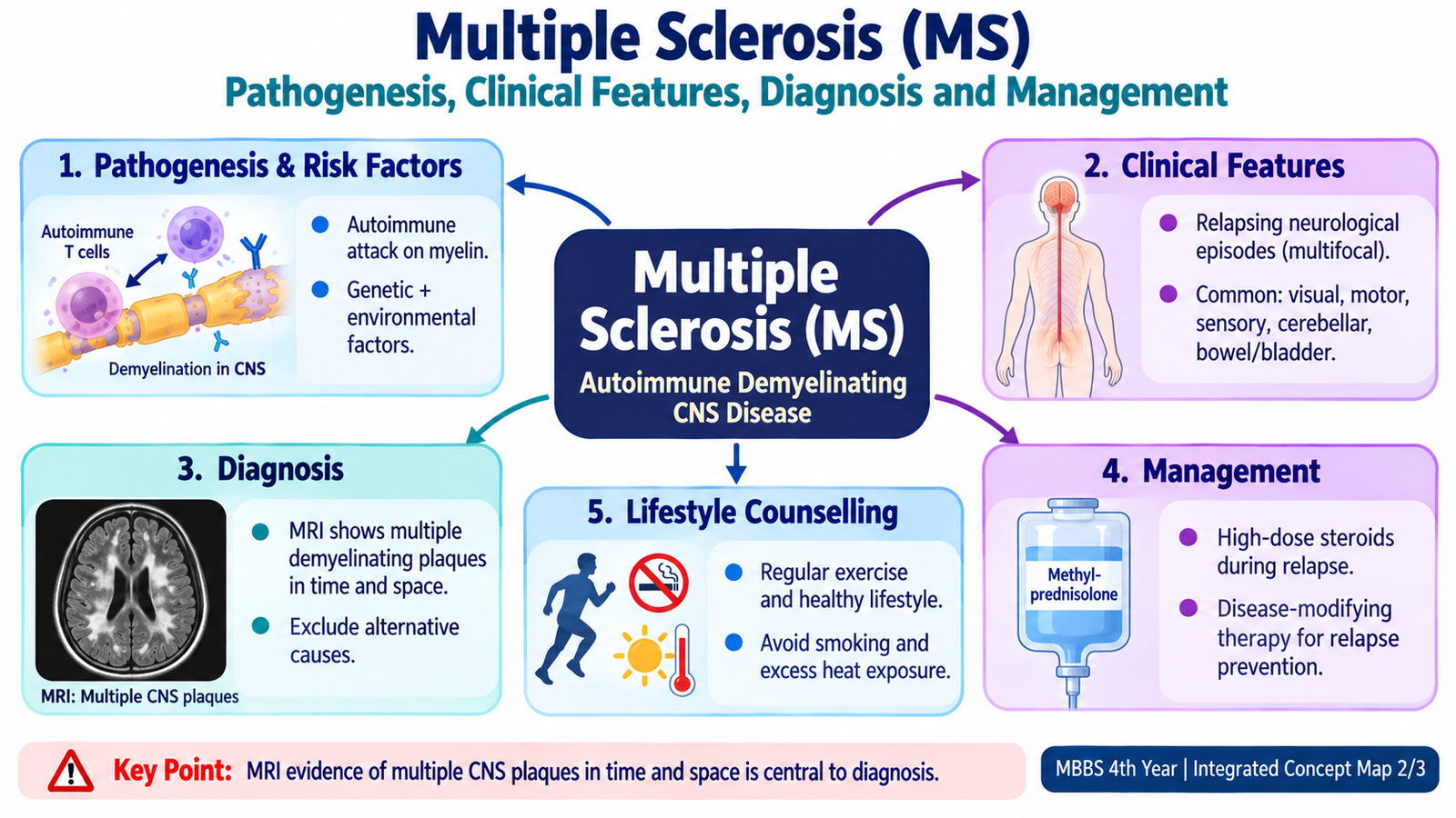
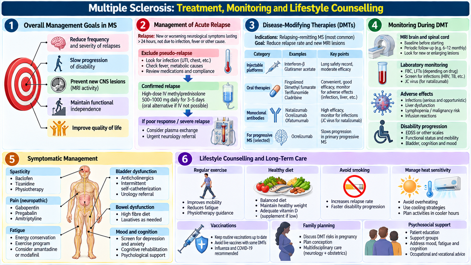
5. Multiple Sclerosis: Treatment, Monitoring and Lifestyle Counselling
Managing an acute relapse
Confirm a new inflammatory attack and look for infection or fever. A functionally significant relapse is commonly treated with high-dose corticosteroids under specialist supervision; steroids shorten recovery but do not replace long-term disease-modifying therapy. Assess walking, vision, bladder function and safety.
Acute relapse treatment: Oral methylprednisolone 500 mg daily for 5 days is recommended for a functionally significant relapse. IV methylprednisolone 1 g daily for 3–5 days may be used for severe relapses requiring hospitalization or when oral treatment is unsuitable.
Preventing future disease activity
Disease-modifying therapies (DMTs) reduce relapses and new inflammatory lesions in suitable patients; choice depends on disease activity, safety profile, pregnancy plans and monitoring requirements. A neurologist initiates and monitors treatment. Discuss adherence, infection screening and vaccination planning appropriate to the chosen therapy.
Examples of disease-modifying therapies: Interferon beta, glatiramer acetate and dimethyl fumarate are options for relapsing MS. Ocrelizumab is a higher-efficacy option for selected patients and may also be used in eligible early primary progressive MS. Treatment selection and monitoring require specialist supervision.
Symptom-directed care
Physiotherapy and graded exercise help mobility and endurance. Treat spasticity, pain, bladder dysfunction, fatigue and mood symptoms individually. Occupational therapy, fall prevention and assistive devices may improve independence.
Lifestyle counselling
Encourage smoking cessation, regular tolerable exercise, balanced nutrition, adequate sleep and avoidance of overheating when it worsens symptoms. Discuss vitamin D adequacy without presenting supplements as a cure. Advise prompt assessment of new deficits, infection-related deterioration and treatment adverse effects.
Follow-up and shared decisions
Review relapse history, disability, MRI activity and treatment tolerability. Discuss work, driving, family planning and mental well-being sensitively. MS is variable: explain that many people maintain function for years with coordinated care.
6. Syringomyelia: Onset, Clinical Features and Diagnostic Workup
Anatomy and mechanism
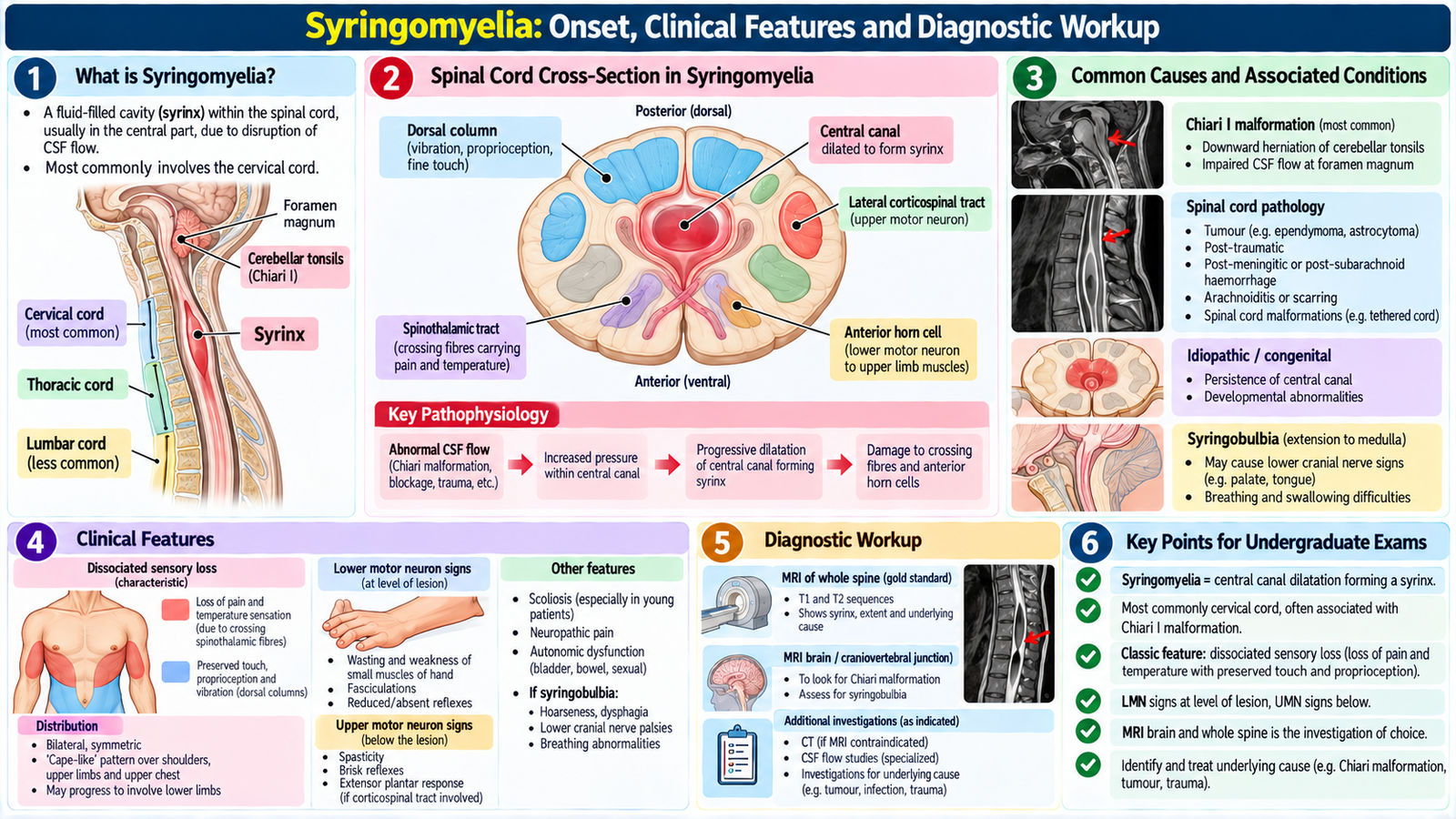
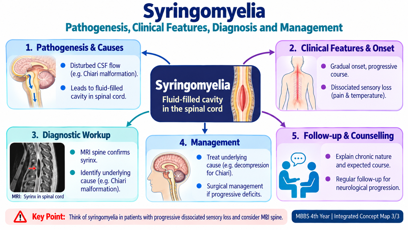
Syringomyelia is a fluid-filled cavity (syrinx) within the spinal cord, commonly cervical. It may be associated with Chiari I malformation, prior trauma, tumors or arachnoid scarring; some cases are idiopathic. Expansion damages crossing pain-temperature fibers near the central canal and may later affect anterior horn cells and long tracts.
Onset and symptoms
Symptoms often develop slowly over months or years: neck/shoulder pain, hand weakness or wasting, loss of pain and temperature across shoulders/arms, and later stiffness or weakness of the legs. Presentation varies with syrinx location and extent; post-traumatic cases may emerge long after injury.
Characteristic examination pattern
Segmental loss of pain and temperature with relative preservation of light touch and position sense is termed dissociated sensory loss. Cervical anterior horn involvement produces LMN signs in the hands, while descending corticospinal tract involvement may produce UMN signs in the legs. Absent protective pain sensation increases burn and injury risk.
Diagnostic workup
MRI of the spinal cord confirms a syrinx and assesses extent. MRI of the craniocervical junction helps identify associated Chiari malformation; contrast or additional imaging may be appropriate if tumor or another cause is suspected. Record neurological deficits and evaluate progression; consider alternative cord disorders if imaging and examination disagree.
7. Syringomyelia: Management, Referral and Long-Term Care
When observation is appropriate
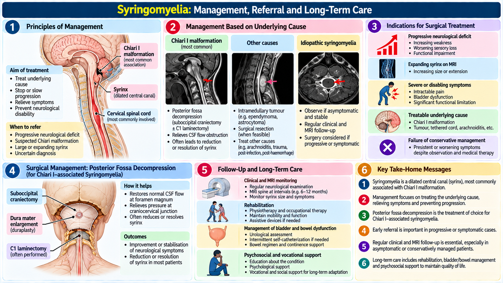
An incidental, small, stable syrinx without attributable progressive symptoms may be monitored clinically and with interval MRI under specialist care. Decisions depend on cause, imaging findings and neurological progression.
When surgery is considered
Progressive weakness, enlarging syrinx or a correctable obstruction of CSF flow warrants neurosurgical evaluation. Surgery aims to address the cause and restore CSF flow—for example, posterior fossa decompression for selected symptomatic Chiari-associated cases. Syrinx drainage/shunting is reserved for selected situations; procedures do not guarantee reversal of established cord damage.
Supportive care and safety
Provide rehabilitation for weakness, spasticity and functional limitations. Teach protection of areas with impaired pain/temperature sensation from burns and repeated trauma. Monitor gait, hand function, sensation and bladder symptoms; reassess promptly if neurological function worsens.
Counselling and prognosis
Explain that progression is variable and recovery depends partly on the duration of neurological injury. Follow-up MRI and examination help detect change. New rapidly progressive weakness or sphincter disturbance requires urgent assessment.
Integrated Mechanism Flow
MS: immune-mediated CNS inflammation → demyelination → impaired nerve conduction → optic, sensory or motor symptoms → MRI-supported diagnosis → relapse treatment and disease-modifying care.
Syringomyelia: disturbed spinal CSF dynamics / cord injury → central cavity expansion → crossing sensory fiber damage → dissociated pain-temperature loss → MRI confirmation → observation or cause-directed neurosurgical management.
Important Comparison: MS vs Syringomyelia
| Feature | Multiple sclerosis | Syringomyelia |
|---|---|---|
| Main pathology | Inflammatory CNS demyelination | Fluid-filled intramedullary cavity |
| Course | Relapses or progressive disability | Often slowly progressive |
| Clinical clue | Optic neuritis, multifocal deficits | Dissociated sensory loss, hand wasting |
| MRI | Disseminated characteristic CNS lesions | Syrinx within spinal cord |
| Main treatment | Relapse care, DMT, rehabilitation | Monitor or correct underlying CSF obstruction |
AIM High-Yield Review
- Localize bilateral leg weakness by tone, reflexes, plantar responses, sensation and sphincter function.
- A sensory level plus pyramidal signs strongly suggests spinal cord disease.
- Sudden weakness, new urinary retention or rapidly progressing deficits demand urgent assessment.
- An acute cord lesion may initially cause flaccidity and absent reflexes.
- MS is a CNS demyelinating disorder; relapses must be distinguished from infection-related pseudo-relapses.
- MRI supports MS diagnosis, but clinical dissemination and exclusion of mimics remain essential.
- High-dose steroids treat significant MS relapses; DMTs aim to reduce future disease activity.
- Lifestyle, rehabilitation, adherence and monitoring are integral to MS care.
- Syringomyelia may produce cape-like pain-temperature loss with preserved posterior-column modalities.
- Cervical syrinx may cause LMN hand signs and UMN leg signs.
- Spinal MRI confirms syringomyelia; progressive deficits prompt neurosurgical assessment.
AIM Video Learning
Topic 9: Lower Limb Weakness, Multiple Sclerosis & Syringomyelia
Final Year MBBS | Neuroscience Module
Video 1: Multiple Sclerosis — Clinical Features, Diagnosis & Management
Ninja Nerd lecture explaining demyelination, clinical manifestations, diagnostic investigations and management of multiple sclerosis.
Source: Ninja Nerd | Watch on YouTube
Video 2: Lower Limb Neurological Examination
Geeky Medics demonstration of assessment of gait, tone, power, reflexes, sensation and coordination in patients presenting with lower limb weakness.
Source: Geeky Medics | Watch on YouTube