
Final Year MBBS • KMU Curriculum • AIM Learning Cycle
📖 AIM Learning Material
TOPIC 10. Acquired & Hereditary Neuropathies
Neuroscience Module | Medicine & Pediatrics | Clinical patterns, diagnosis and functional care
Peripheral neuropathies affect the nerves that carry signals between the spinal cord and the body. They can be acquired through conditions such as diabetes, nutritional deficiency or inflammation, or inherited through genetic changes that affect nerve structure and function. The key to understanding these disorders is to recognise the distribution and type of nerve dysfunction, relate this to the likely cause, choose appropriate investigations and plan treatment. In children, recognising slowly progressive inherited patterns helps families obtain early rehabilitation and appropriate counselling. This chapter brings the Medicine and Pediatrics outcomes together in one clinical sequence.

A. Peripheral Nerve Function and Classification of Neuropathies
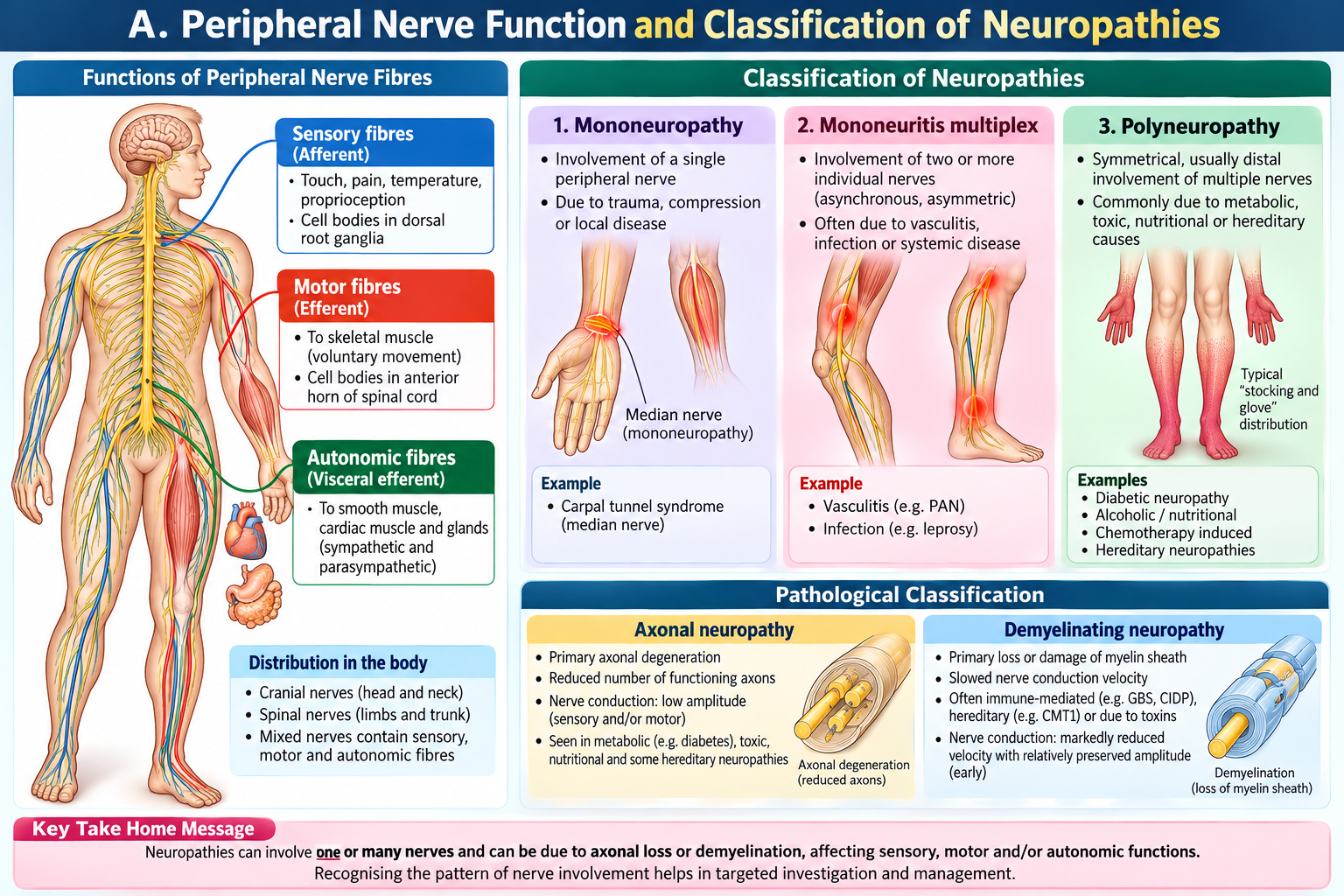
Peripheral neuropathy means damage to nerves outside the brain and spinal cord. Sensory fibres carry touch, pain, temperature, vibration and joint position; motor fibres activate muscles; autonomic fibres regulate sweating, blood pressure and visceral functions. Damage therefore produces a recognisable combination of sensory, motor and autonomic complaints.
Classify before naming the disease
- Distribution: mononeuropathy affects one nerve (for example median nerve entrapment); mononeuritis multiplex affects several separate nerves asymmetrically; polyneuropathy affects many nerves, typically symmetrically and distally.
- Fibre involved: sensory, motor, autonomic or mixed. Small-fibre injury causes burning pain and altered temperature sensation; large-fibre injury causes loss of vibration and position sense with sensory ataxia.
- Time course: acute (days to four weeks), subacute (weeks), or chronic (months to years). Rapid progression with weakness requires urgent assessment.
- Pathophysiology: axonal loss reduces the number of functioning nerve fibres; demyelination slows or blocks conduction. Either process can be acquired or inherited.
Length-dependent polyneuropathy first affects the longest axons: toes and feet before hands, producing a stocking–glove pattern. Distal reflexes, particularly ankle jerks, may disappear early.
B. Acquired Neuropathies: Causes and Clinical Patterns
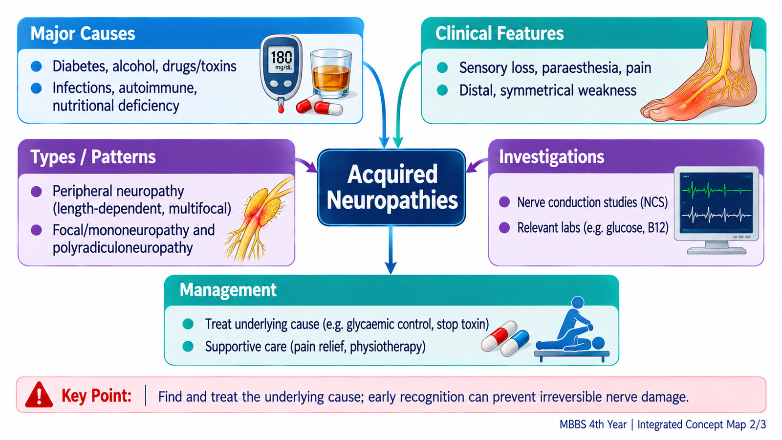
Acquired neuropathies arise from metabolic, nutritional, toxic, inflammatory, infective or compressive processes. A careful history of onset, symmetry, pain, exposures and associated illness narrows the cause before investigations are ordered.
Important causes
- Diabetes mellitus: common cause of chronic distal symmetric sensorimotor polyneuropathy; burning feet, numbness, reduced ankle reflexes and loss of protective sensation predispose to ulcers.
- Nutritional deficiency: vitamin B12 deficiency may produce peripheral neuropathy with impaired vibration/proprioception and sometimes myelopathy; malnutrition and alcohol misuse may cause additional deficiencies.
- Toxins and drugs: alcohol and certain neurotoxic medicines, including selected chemotherapy agents, may damage peripheral axons; review timing and cumulative exposure.
- Renal disease and hypothyroidism: may produce chronic neuropathy; consider systemic symptoms and laboratory abnormalities.
- Immune-mediated disease: Guillain–Barré syndrome (GBS) typically causes rapidly progressive, fairly symmetric weakness with reduced reflexes; chronic inflammatory demyelinating polyradiculoneuropathy (CIDP) causes progression or relapses over more than eight weeks.
- Entrapment: focal numbness or weakness in a specific nerve territory, such as median neuropathy at the wrist.
- Vasculitic neuropathy: painful, asymmetric, stepwise deficits may indicate mononeuritis multiplex.
Infective causes, including leprosy where epidemiologically relevant, may cause focal or multifocal neuropathy with sensory loss and nerve enlargement. These patterns should be interpreted in the patient’s clinical context.

C. Recognising Neuropathy at the Bedside
Patients may describe tingling, burning, electric-shock pain, numbness, weakness, frequent tripping, imbalance or altered sweating. The examination should establish whether the problem is truly peripheral and whether sensory, motor or autonomic fibres predominate.
Focused history
Ask about onset and progression; proximal versus distal weakness; symmetry; falls; pain; diabetes; dietary problems; alcohol and drug exposure; renal or thyroid disease; preceding infection; and family members with similar symptoms. Ask about breathing, swallowing, palpitations and fainting when weakness progresses rapidly.
Focused examination and interpretation
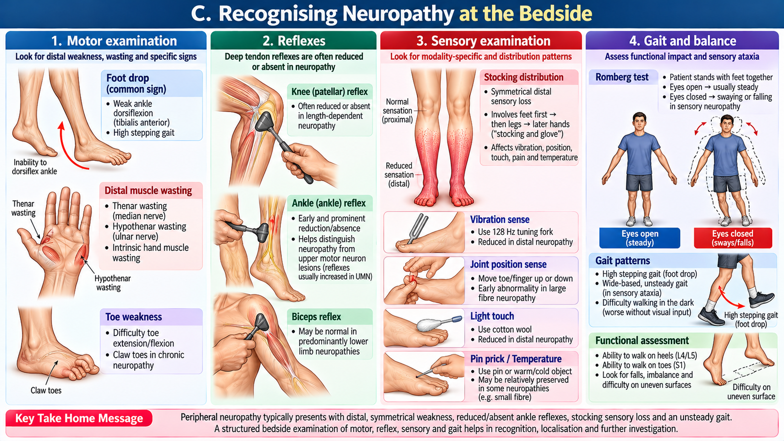
- Motor: assess distal and proximal power, wasting, foot dorsiflexion, hand grip, gait and foot deformity. Distal weakness and muscle wasting support a peripheral motor process.
- Reflexes: reduced or absent tendon reflexes favour peripheral nerve disease; brisk reflexes, spasticity and extensor plantar responses suggest central nervous system involvement instead.
- Sensation: map pinprick/temperature, light touch, vibration and joint position. A distal symmetric gradient suggests polyneuropathy; a single-nerve territory suggests entrapment.
- Gait: high-stepping gait may reflect foot drop; a positive Romberg test with impaired joint position sense suggests large-fibre sensory dysfunction.
- Autonomic features: postural dizziness, sweating changes and gastrointestinal or bladder dysfunction require assessment.
D. Investigating Acquired Neuropathies and Interpreting Results
Investigations should answer two questions: Is there objective peripheral nerve dysfunction? and What treatable cause explains it? Tests are selected according to the pattern rather than ordered indiscriminately.
First-line investigations
- Blood glucose or HbA1c: detect diabetes or poor glycaemic control.
- Full blood count and vitamin B12: assess anaemia and deficiency where clinically indicated.
- Renal function, electrolytes and thyroid function: identify metabolic contributors.
- Additional targeted tests: inflammatory markers, autoimmune tests, infection screening or serum protein studies when clinical clues suggest inflammatory, infective or paraproteinaemic disease.
Nerve conduction studies and electromyography
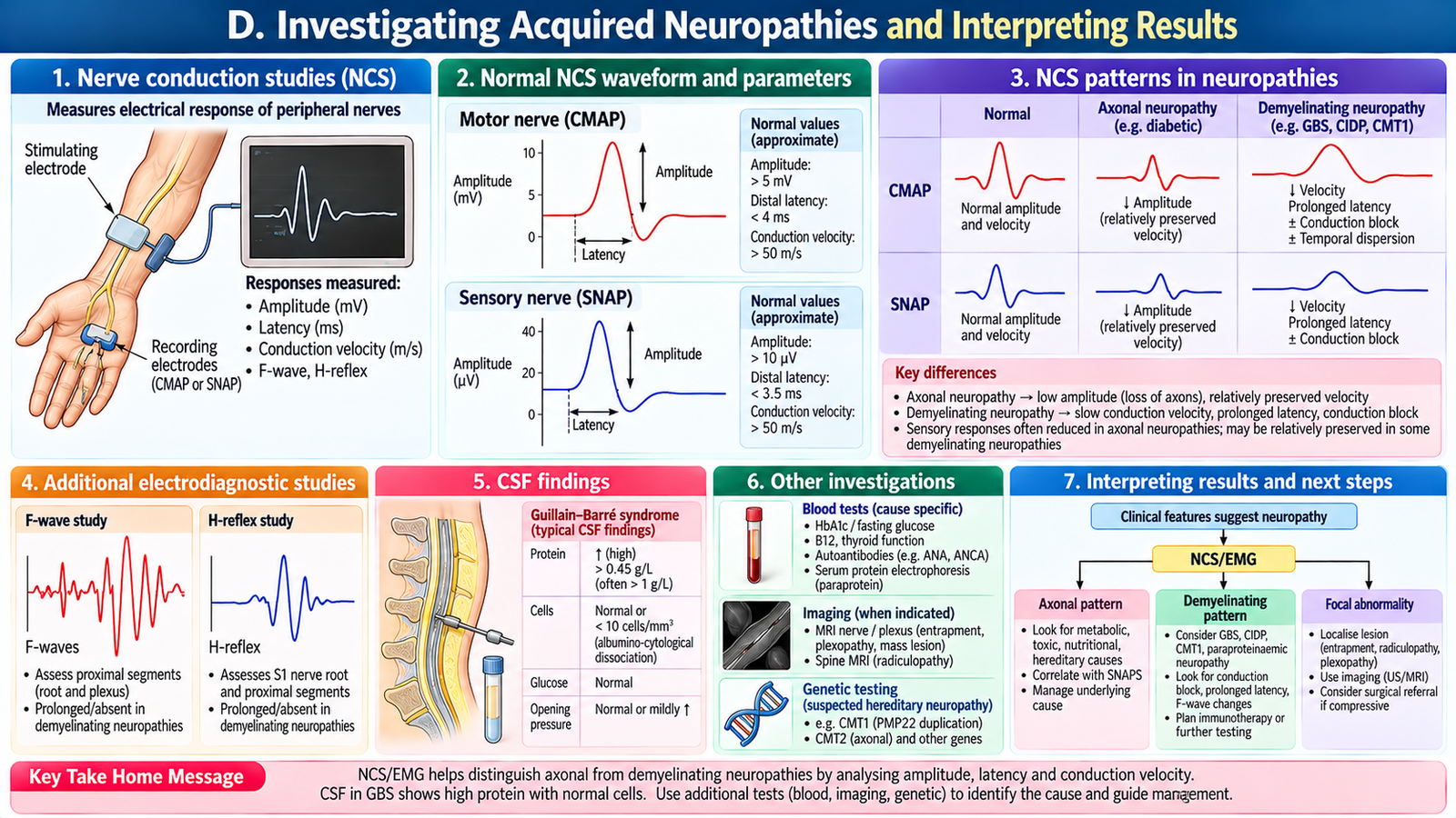
Nerve conduction studies measure electrical responses and conduction velocity. Axonal neuropathy commonly reduces response amplitudes because fewer axons conduct; demyelinating neuropathy produces disproportionate slowing, prolonged distal latencies, conduction block or temporal dispersion. Electromyography can demonstrate denervation and help characterise motor involvement. Very early or pure small-fibre neuropathy may have normal routine conduction studies.
Special situations
In suspected GBS, lumbar puncture may show elevated CSF protein with relatively few cells (albuminocytologic dissociation), but CSF can be normal early. Electrodiagnostic findings also may evolve. Spinal imaging is considered if findings suggest spinal cord compression or another central lesion, rather than routine uncomplicated neuropathy.
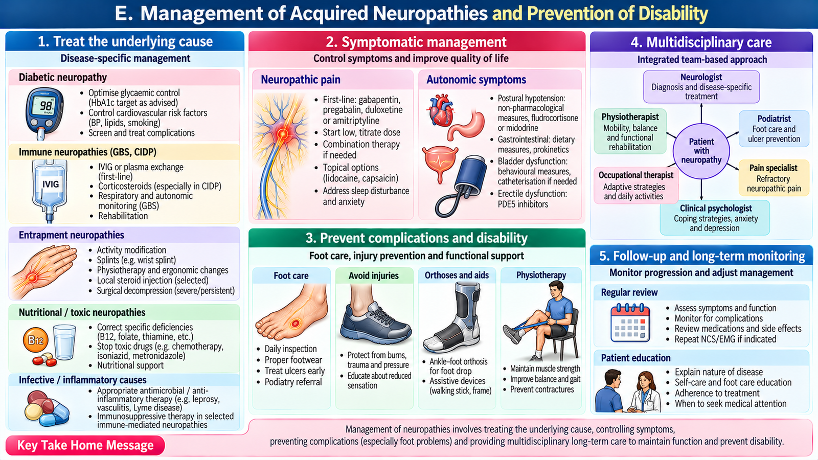
E. Management of Acquired Neuropathies and Prevention of Disability
Treatment has three linked goals: correct the cause, reduce symptoms and preserve function. Improvement depends on the underlying process; long-standing axonal damage may recover slowly or incompletely.
Cause-specific treatment
- Diabetic neuropathy: optimise glycaemic control, cardiovascular risk factors and daily foot protection; address ulcers promptly.
- Deficiency-related neuropathy: replace the deficient nutrient, particularly vitamin B12 when confirmed or strongly suspected, and correct the underlying dietary or absorption problem.
- Toxic neuropathy: stop or modify the responsible exposure with the treating team; manage alcohol-related nutritional deficits.
- Entrapment neuropathy: activity modification and splinting may help; progressive motor deficit or refractory symptoms merit specialist assessment for decompression.
- GBS: admit urgently for respiratory, swallowing and autonomic monitoring; intravenous immunoglobulin or plasma exchange are disease-modifying treatments when indicated. Corticosteroids alone are not effective therapy for typical GBS.
- CIDP: specialist-directed immunotherapy may include corticosteroids, intravenous immunoglobulin or plasma exchange.
Symptom relief and rehabilitation
Neuropathic pain may respond to agents such as gabapentin, pregabalin or duloxetine; selection depends on comorbidity, adverse effects and availability. Simple analgesics often have limited effect on neuropathic burning pain. Physiotherapy, balance training, appropriate ankle-foot orthoses and occupational support reduce falls and maintain independence.
For painful diabetic neuropathy, duloxetine, pregabalin or gabapentin are commonly used first-line options, selected according to comorbidities and tolerability. In suspected Guillain–Barré syndrome, monitor respiratory function using serial forced vital capacity measurements; rapidly declining respiratory function requires urgent intensive-care assessment.
Teach foot inspection, suitable footwear, skin care and avoidance of burns or unnoticed injuries when protective sensation is lost. Review progression, pain, gait, adverse effects and new weakness at follow-up.
F. Hereditary Neuropathies in Children: Classification and Presentation
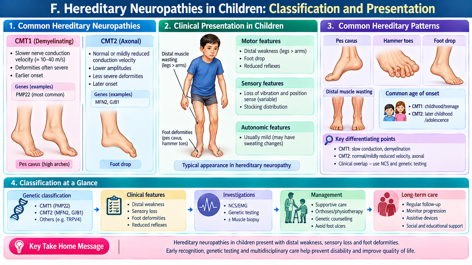
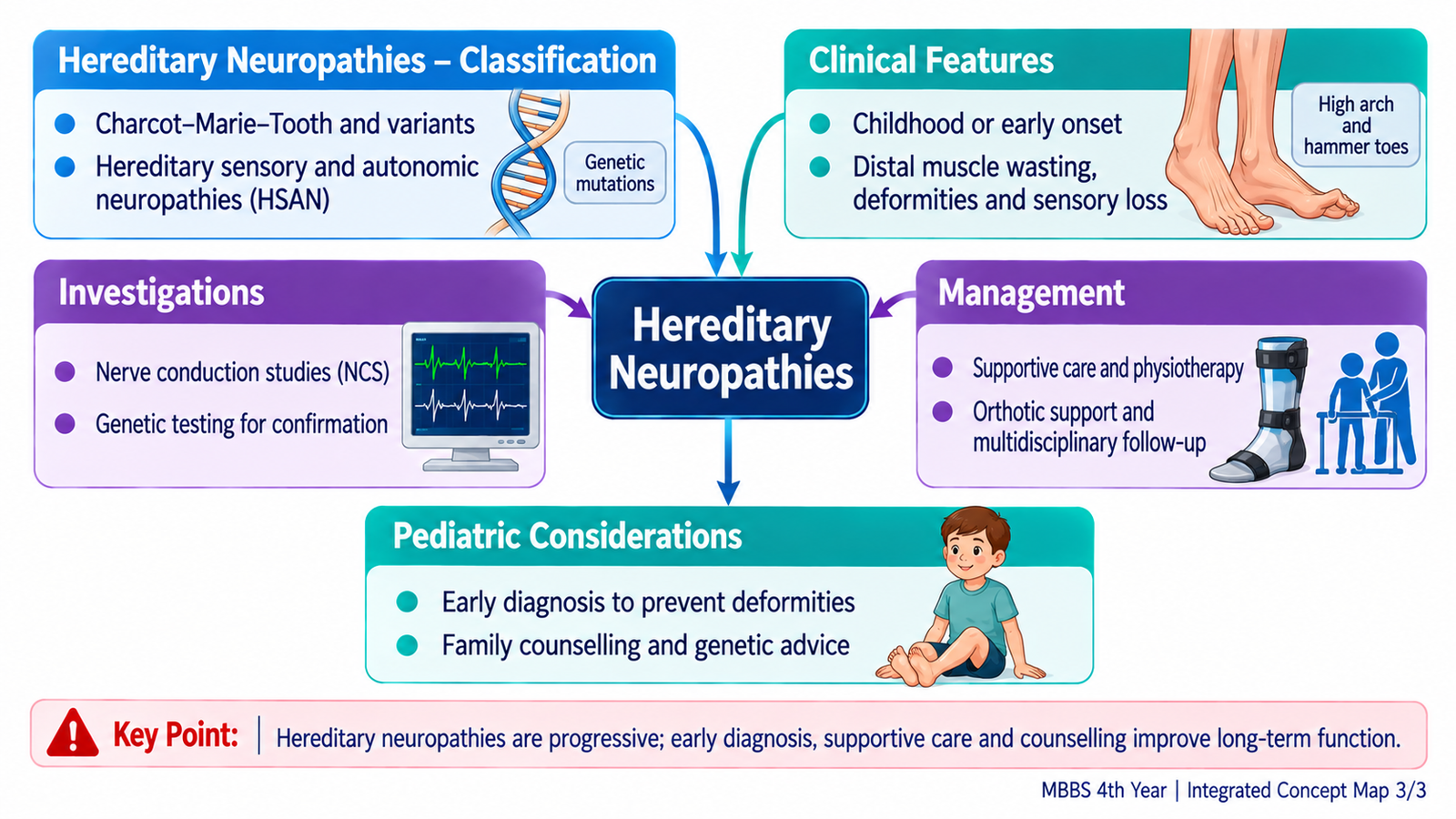
Hereditary neuropathies are genetically determined disorders of peripheral nerves. They commonly develop gradually during childhood or adolescence, but presentation and severity vary widely. A positive family history helps, yet a new genetic variant or recessive inheritance can produce an apparently sporadic case.
Useful clinical classification
- Hereditary motor and sensory neuropathies (HMSN), including Charcot–Marie–Tooth (CMT) disease: predominantly distal weakness, wasting and sensory loss; subtypes may be primarily demyelinating (CMT1) or axonal (CMT2).
- Hereditary sensory and autonomic neuropathies: prominent pain/temperature or autonomic dysfunction; unnoticed injury and ulceration may occur.
- Hereditary neuropathy with liability to pressure palsies (HNPP): recurrent focal palsies following minor compression.
Clinical clues in a child
Clumsiness, repeated ankle sprains, frequent falls, difficulty running, foot drop and slowly progressive distal leg wasting may precede diagnosis. Pes cavus, hammer toes and reduced ankle reflexes support long-standing peripheral neuropathy. Hand weakness can develop later. Some children have relatively mild sensory symptoms despite substantial motor impairment.
Distinguish inherited neuropathy from acquired disease by gradual evolution, skeletal foot changes and similar findings in relatives; however, neither foot deformity nor family history is diagnostic alone.
G. Diagnosis and Long-Term Care of Hereditary Neuropathies
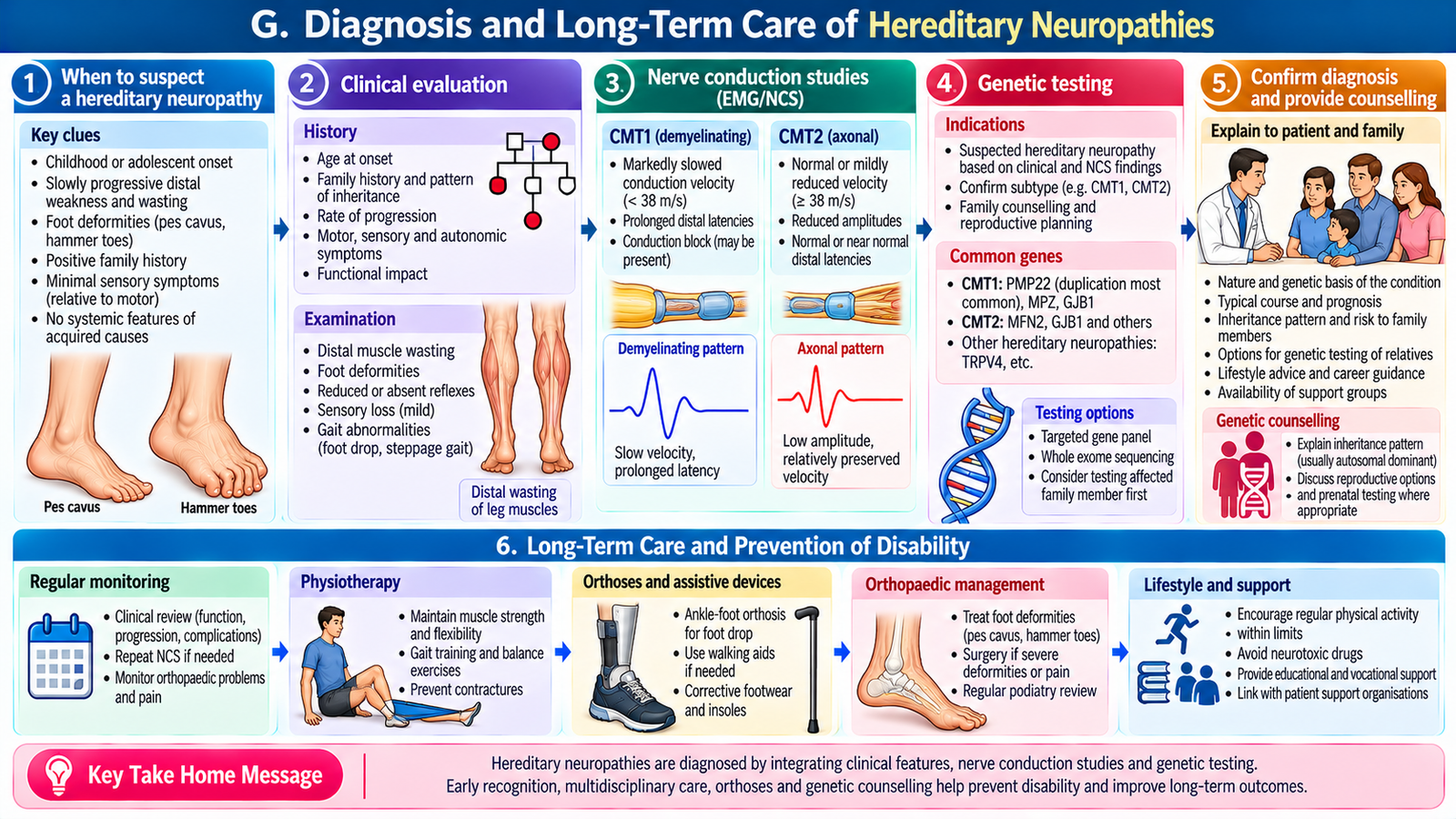
Evaluation begins with a developmental and neurological history, a three-generation family history where feasible, and examination of strength, reflexes, sensation, gait, foot posture and spinal alignment. Assess how symptoms affect school, play, mobility and daily activities.
Diagnostic investigations
- Nerve conduction studies: help confirm peripheral neuropathy and distinguish a predominantly demyelinating from axonal pattern.
- Genetic testing: targeted or panel testing after clinical and electrodiagnostic assessment can establish a molecular diagnosis; for common CMT1A, a PMP22 duplication is a classic cause.
- Screen for treatable mimics: use glucose, B12, thyroid or other targeted investigations when history or examination raises concern; genetic disease does not exclude a superimposed acquired cause.
- Family evaluation: examination and appropriately offered testing of relatives can support diagnosis and counselling.
Management and family-centred care
Many common hereditary neuropathies have no established curative treatment, so care centres on maintaining function. Physiotherapy preserves range of motion and strength; ankle-foot orthoses improve foot clearance and stability; occupational therapy supports hand function. Manage pain, skin breakdown and foot deformities. Orthopaedic referral is appropriate for painful, progressive or function-limiting deformity.
Explain the variable course without predicting an individual child’s future from the label alone. Discuss inheritance and recurrence risk after the specific diagnosis is known, and offer genetic counselling. Encourage suitable physical activity, safe footwear, regular review and attention to school participation and emotional wellbeing.
AIM High-Yield Review
- Distal symmetric stocking–glove sensory loss points to length-dependent polyneuropathy.
- Reduced amplitudes suggest axonal loss; marked conduction slowing suggests demyelination.
- Diabetes is a major acquired cause; inspect feet and assess protective sensation.
- Rapid ascending weakness with areflexia suggests GBS and requires urgent respiratory and autonomic monitoring.
- GBS: intravenous immunoglobulin or plasma exchange when indicated; corticosteroids alone do not treat typical GBS.
- Progression beyond eight weeks or relapsing weakness suggests CIDP rather than typical acute GBS.
- Childhood distal wasting, pes cavus and foot drop suggest hereditary motor and sensory neuropathy such as CMT.
- CMT1 is classically demyelinating; CMT2 is predominantly axonal.
- Electrodiagnostic studies characterise neuropathy; genetic testing can confirm a hereditary subtype.
- Rehabilitation, orthoses, skin protection and family counselling remain central to long-term care.
AIM Video Learning
Topic 10: Acquired & Hereditary Neuropathies
Final Year MBBS | KMU Curriculum | Neuroscience Module
Video 1: Peripheral Neuropathy — Clinical Features
Cleveland Clinic | Sensory, motor and autonomic manifestations of peripheral nerve disorders.
Focus: Clinical recognition and neurological manifestations.
Video 2: Charcot–Marie–Tooth Disease — Hereditary Neuropathy
Charcot-Marie-Tooth Association | Inherited peripheral neuropathy, progressive weakness, sensory loss and functional limitations.
Focus: Hereditary neuropathy and pediatric clinical presentation.