
AIM OSCE / OSPE • FINAL YEAR MBBS
Station 12 — Systemic Lupus Erythematosus: Clinical Assessment & Laboratory Interpretation
MS Module • Medicine • Pediatrics • Nephrology • Rapid Revision • Clinical Skills • Viva
🎯 Candidate Task
A patient presents with recurrent joint pain, skin lesions and fatigue. Examine the supplied clinical images and laboratory results, identify the characteristic findings, apply appropriate SLE classification criteria, and assess for systemic complications, including possible renal involvement.
⚡ Model Station Answer
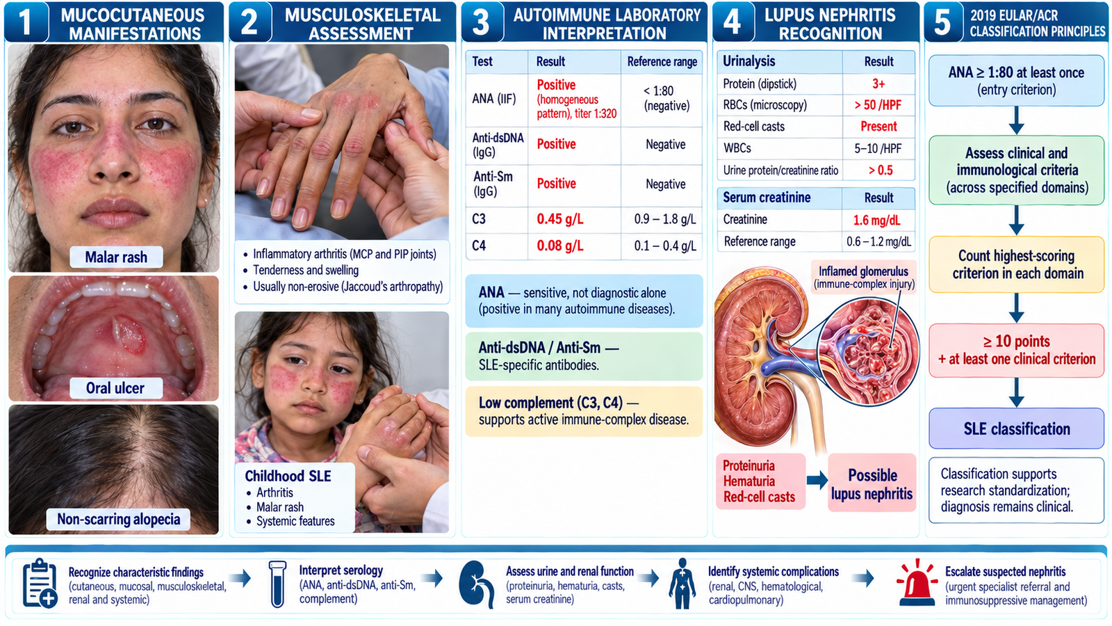
Clinical findings: Malar rash, photosensitivity, oral ulcers, non-scarring alopecia and inflammatory joint involvement support suspected SLE.
Immunology: ANA is a sensitive screening marker; anti-dsDNA and anti-Sm are more specific. Low C3/C4 may support active immune-complex disease.
Classification: The 2019 EULAR/ACR criteria require ANA positivity at least once (≥1:80 on HEp-2 cells or equivalent), followed by weighted clinical and immunological criteria totaling ≥10 points, with at least one clinical criterion.
Renal assessment: Proteinuria, hematuria, red-cell casts, hypertension or rising creatinine suggest possible lupus nephritis and require prompt renal evaluation.
🔎 How to Perform / Approach
- Prepare and examine: Introduce yourself, obtain consent and perform hand hygiene. Inspect the face, exposed skin, scalp and oral mucosa for malar rash, photosensitive lesions, alopecia and ulcers.
- Assess joints and systemic findings: Look for joint tenderness or swelling, edema, pallor and features of serositis. Check blood pressure and assess for clinical evidence of renal involvement.
- Interpret immunological investigations: Review ANA, anti-dsDNA, anti-Sm, C3/C4 and antiphospholipid antibodies. Recognize that ANA positivity alone does not establish SLE.
- Apply classification principles: Confirm the ANA entry criterion, identify qualifying clinical and immunological features and calculate the relevant weighted EULAR/ACR score, excluding findings better explained by another condition.
- Assess renal involvement: Review urinalysis for protein, blood and casts; quantify proteinuria using a urine protein-creatinine ratio and interpret serum creatinine/eGFR. Identify findings suggestive of lupus nephritis.
- Recognize complications: Review the full blood count for cytopenias and consider serosal, neurological and renal manifestations. In children, recognize that SLE may present with rash, arthritis, cytopenias, hypertension or nephritis.
- State the clinical conclusion: Integrate clinical findings with immunology and urine results. Suspected lupus nephritis warrants prompt specialist assessment, with renal biopsy considered to confirm and classify renal pathology when indicated.
🖼️ Visual Learning
Systemic Lupus Erythematosus — Clinical Signs, Autoimmune Serology & Lupus Nephritis
📝 Important Viva Questions
Q1. What are the characteristic mucocutaneous and musculoskeletal features of SLE?
Answer: Malar rash, photosensitivity, oral ulcers, non-scarring alopecia and inflammatory arthritis or arthralgia are characteristic findings.
Q2. Which autoantibodies are particularly useful in SLE assessment?
Answer: ANA is sensitive but nonspecific; anti-dsDNA and anti-Sm are more specific for SLE. Reduced complement may indicate active disease.
Q3. What are the key principles of the 2019 EULAR/ACR SLE classification criteria?
Answer: ANA positivity at least once at ≥1:80 is the entry criterion, followed by a weighted score of at least 10 points including one clinical criterion, with other explanations excluded.
Q4. What urinary findings suggest lupus nephritis?
Answer: Proteinuria, microscopic hematuria and red-cell casts, particularly with reduced renal function or hypertension, suggest renal involvement.
Q5. What important systemic manifestations should be considered in childhood SLE?
Answer: Children may develop rash, arthritis, cytopenias, serositis and renal involvement. Blood pressure, urine examination and renal function assessment are particularly important.