
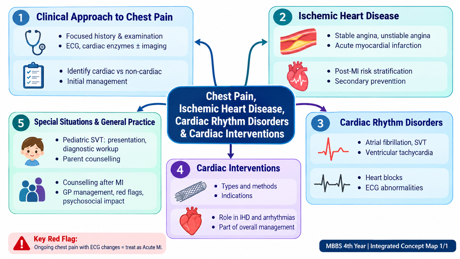
This chapter follows the KMU outcomes for chest pain, ischemic heart disease, common rhythm disorders and cardiac interventions. First understand how symptoms, ECG findings and hemodynamic stability guide clinical decisions; use the AIM High-Yield Review at the end for rapid revision.
Chest Pain, Ischemic Heart Disease, Cardiac Rhythm Disorders & Cardiac Interventions
Cardiorespiratory / Cardiovascular Module — a patient-centred approach from chest pain recognition to ischemia, arrhythmias, intervention, secondary prevention and follow-up.
Topic Introduction
Chest pain is a common presentation, but its importance lies in rapidly separating potentially life-threatening cardiac ischemia from less dangerous causes. Ischemic heart disease occurs when myocardial oxygen supply becomes insufficient for demand, most commonly because coronary blood flow is limited by atherosclerotic disease. The same patient may also develop electrical instability, producing important arrhythmias or conduction blocks. In this chapter, you will learn how to assess chest pain, recognize stable angina and acute coronary syndromes, interpret common rhythm abnormalities, understand their management principles, appreciate the role of cardiac interventions, and plan prevention, counselling and follow-up after myocardial infarction.
A. Clinical Approach to Chest Pain
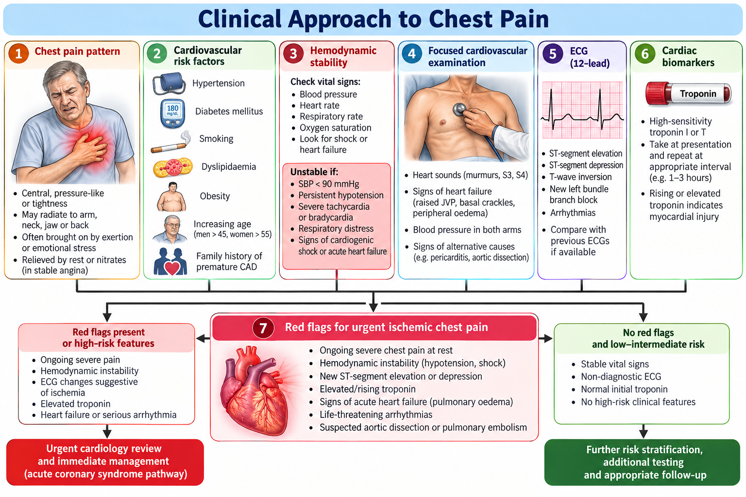
The first task in a patient with chest pain is not to name a disease immediately. It is to determine whether the pain could represent myocardial ischemia or another dangerous cardiopulmonary condition, assess whether the patient is stable, and then use focused history, examination and investigations to narrow the diagnosis.
Understanding ischemic chest pain
Myocardial ischemia develops when oxygen delivery to cardiac muscle cannot meet its metabolic requirement. Ischemic discomfort is therefore commonly provoked by situations that increase cardiac work, while acute reduction of coronary blood flow may produce pain even at rest.
Increased myocardial demand or reduced coronary supply → myocardial ischemia → accumulation of ischemic metabolites and altered cardiac afferent signalling → characteristic chest discomfort.
Focused history
A useful history defines the pain pattern and simultaneously estimates cardiovascular risk. Important features include:
- Site and character: ischemic discomfort is commonly central or retrosternal and may be described as pressure, heaviness, tightness or squeezing.
- Radiation: discomfort may extend to the arm, shoulder, neck, jaw or back.
- Precipitating factors: exertion or emotional stress supports demand-related ischemia.
- Relieving factors: predictable improvement with rest supports stable exertional angina.
- Duration and progression: new, prolonged, worsening or rest pain raises concern for acute coronary syndrome.
- Associated symptoms: dyspnea, sweating, nausea, presyncope or palpitations increase clinical concern.
- Risk context: hypertension, diabetes, dyslipidemia, smoking, obesity, family history and previous vascular disease increase the probability of IHD.
Focused examination
Examination may be normal in uncomplicated ischemia. Its major role is to identify instability, complications and important alternative diagnoses.
- Assess pulse, blood pressure, respiratory rate, oxygenation and overall perfusion.
- Look for pallor, sweating, respiratory distress or altered consciousness.
- Assess rhythm and pulse characteristics.
- Examine the cardiovascular system for abnormal heart sounds, murmurs or signs of heart failure.
- Look for pulmonary congestion and peripheral signs of vascular disease where relevant.
Ongoing severe chest pain, hemodynamic instability, syncope, significant arrhythmia, acute heart failure, marked respiratory distress or evidence of myocardial infarction requires urgent assessment and escalation.
Initial diagnostic approach
The ECG is central because it may demonstrate myocardial ischemia, infarction or an arrhythmia. Cardiac biomarkers help identify myocardial injury. Additional investigations are selected according to the clinical presentation and the possibility of complications or alternative diagnoses.
B. Ischemic Heart Disease: From Stable Angina to Acute Coronary Syndrome
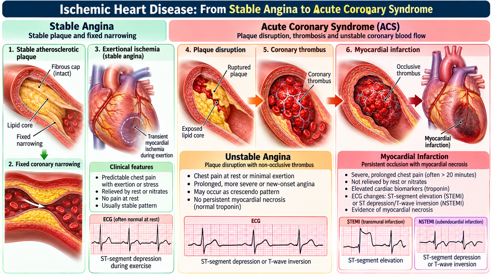
Ischemic heart disease is best understood as a spectrum. The underlying problem is inadequate coronary blood flow relative to myocardial demand, but the clinical presentation depends greatly on whether the coronary lesion is relatively stable or has undergone an acute change such as plaque disruption and thrombosis.
Stable angina
In stable angina, a relatively fixed coronary narrowing limits the ability to increase blood flow during exertion. At rest, supply may remain adequate. During exercise or emotional stress, myocardial oxygen demand rises beyond the available supply, producing predictable transient ischemic discomfort.
Acute coronary syndrome
Acute coronary syndrome represents a sudden deterioration in coronary blood flow. Plaque disruption may trigger platelet activation and thrombus formation. The resulting reduction in perfusion can produce unstable angina or myocardial infarction depending on whether myocardial necrosis occurs.
Unstable angina produces an unstable ischemic pattern but without biomarker evidence of myocardial necrosis. Myocardial infarction involves myocardial injury and necrosis caused by acute ischemia and is supported by the appropriate clinical setting together with cardiac biomarker and ECG findings.
ST-elevation and non-ST-elevation patterns
The ECG helps define the acute ischemic pathway. Persistent ST-segment elevation in a compatible acute presentation suggests an occlusive coronary process requiring an urgent reperfusion strategy. An acute coronary syndrome without persistent ST elevation may still represent myocardial infarction when cardiac biomarkers demonstrate myocardial injury.
| Feature | Stable Angina | Unstable Angina | Myocardial Infarction |
|---|---|---|---|
| Clinical pattern | Predictable with exertion | New, worsening or rest ischemia | Acute ischemia with myocardial injury |
| Underlying process | Relatively fixed flow limitation | Acute coronary instability | Ischemia severe enough to cause necrosis |
| Cardiac biomarkers | No acute myocardial injury | No myocardial necrosis | Evidence of myocardial injury |
| Clinical priority | Symptom control and risk reduction | Urgent ACS assessment | Urgent ACS treatment and reperfusion assessment |
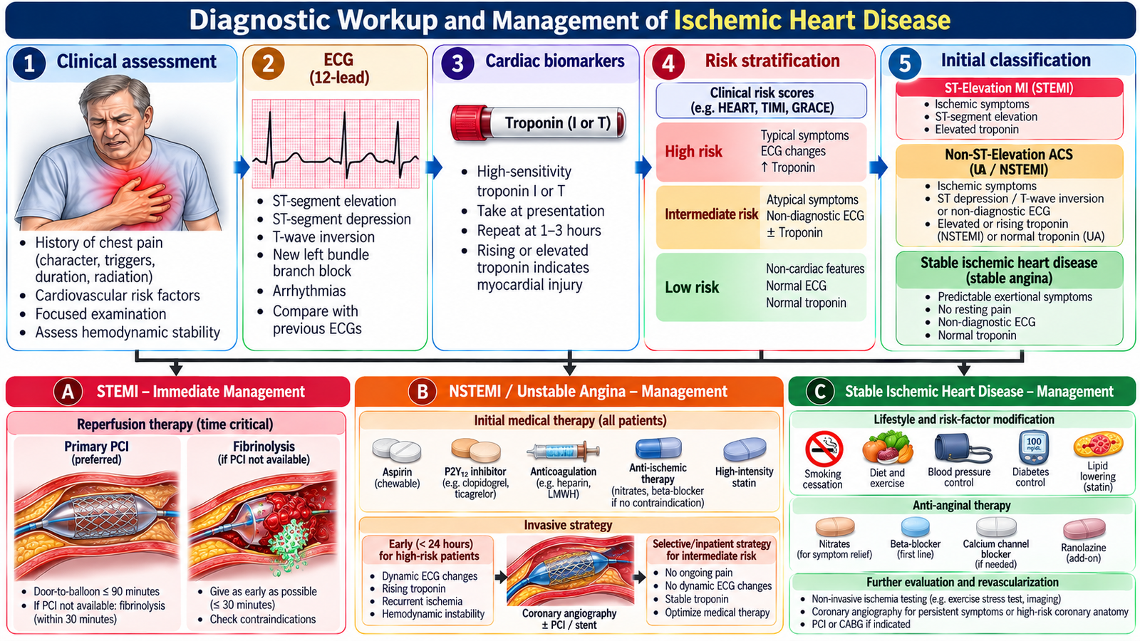
C. Diagnostic Workup and Management of Ischemic Heart Disease
Investigation and management depend on whether the patient has chronic stable ischemia or a possible acute coronary syndrome. The key Final-Year principle is to connect each investigation with a clinical decision rather than simply memorizing a test list.
ECG
An ECG should be interpreted in the clinical context. Ischemic ST-segment or T-wave abnormalities support myocardial ischemia, while an acute ST-elevation pattern may identify a patient who requires an urgent reperfusion pathway. A normal or nondiagnostic initial ECG does not by itself exclude ischemic heart disease when the clinical suspicion remains important.
Cardiac biomarkers
Cardiac troponin is used to detect myocardial injury. In a patient with an appropriate acute ischemic presentation, a characteristic rise or fall in troponin supports acute myocardial infarction. Biomarkers therefore help distinguish myocardial infarction from ischemia without necrosis.
Further assessment
Depending on the presentation, additional assessment may evaluate ventricular function, inducible ischemia or coronary anatomy. Echocardiography can identify ventricular dysfunction and mechanical consequences of infarction. Functional ischemia testing is useful in appropriately selected stable patients, while coronary angiography defines coronary anatomy when invasive assessment or revascularization is required.
ECG change → electrical evidence of ischemia or infarction.
Troponin change → myocardial injury in the correct clinical context.
Echocardiography → ventricular function and complications.
Coronary angiography → coronary anatomy and possible revascularization planning.
Stable ischemic heart disease
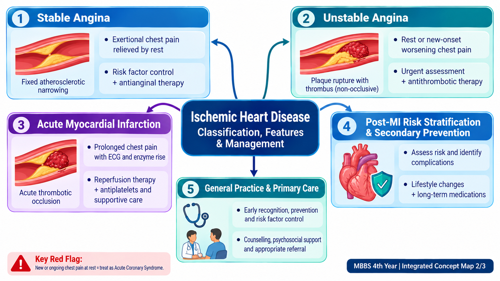
Management aims to reduce ischemic symptoms and future cardiovascular events. Important principles include modification of cardiovascular risk factors, appropriate anti-ischemic therapy, antiplatelet and lipid-lowering strategies where indicated, and assessment for revascularization when symptoms or coronary anatomy justify intervention.
Essential medical treatment: Sublingual glyceryl trinitrate (GTN) provides rapid relief of angina. A beta-blocker (e.g., bisoprolol) or calcium-channel blocker (e.g., amlodipine) is used for long-term symptom control. Aspirin and a high-intensity statin are indicated in appropriate patients with established coronary atherosclerotic disease. Persistent symptoms despite medical therapy warrant cardiology assessment for revascularization.
Acute coronary syndrome
In suspected ACS, management begins with rapid assessment of stability, ECG evaluation, cardiac biomarkers and treatment directed at ongoing ischemia and coronary thrombosis. Patients with an acute occlusive pattern require urgent consideration of reperfusion. Patients without persistent ST elevation are managed according to their clinical risk, evidence of myocardial injury and need for invasive evaluation.
Initial ACS treatment: Give aspirin 162–325 mg chewed unless contraindicated, followed by antiplatelet therapy including a P2Y12 inhibitor (ticagrelor, prasugrel or clopidogrel as appropriate). Initiate anticoagulation (e.g., unfractionated heparin or enoxaparin), high-intensity atorvastatin and sublingual GTN when appropriate; give oxygen only for hypoxemia. STEMI requires urgent primary PCI, or fibrinolysis in eligible patients when timely PCI is unavailable. NSTEMI and unstable angina require risk-based assessment for early invasive management.
Persistent ischemic symptoms, hemodynamic compromise, serious arrhythmia, acute heart failure or an ECG suggesting acute coronary occlusion signals a high-risk situation requiring urgent specialist management.
🩺 AIM Clinical Bridge
A middle-aged patient with cardiovascular risk factors develops central pressure-like chest discomfort while walking. For several months it settled with rest, but today the pain occurred at rest, lasted longer and was accompanied by sweating. The patient appears anxious and the ECG shows new ischemic changes.
Think clinically:
- How has the pattern changed from stable to potentially unstable ischemia?
- Which immediate investigations help distinguish ischemia from myocardial infarction?
- Which findings would make urgent invasive or reperfusion management more likely?
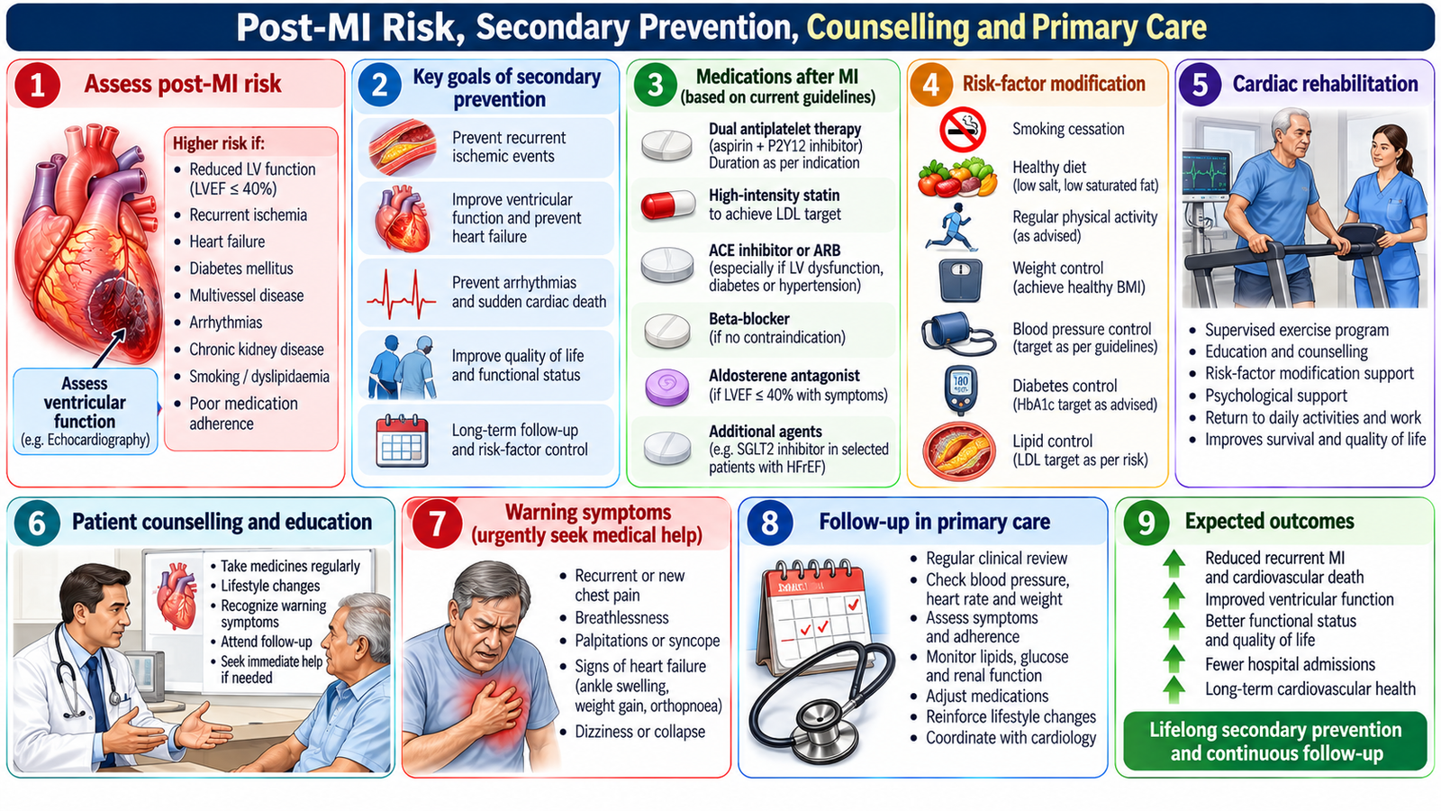
D. Post-MI Risk, Secondary Prevention, Counselling and Primary Care
Survival from an acute myocardial infarction does not end the disease process. The patient remains at risk of recurrent ischemia, ventricular dysfunction, arrhythmia and further cardiovascular events. Post-MI care therefore combines risk assessment, secondary prevention, adherence, rehabilitation, follow-up and patient education.
Post-MI risk assessment
Clinical risk increases when infarction has produced important ventricular dysfunction, persistent ischemia, electrical instability or heart failure. Assessment of symptoms, rhythm, ventricular function and complications helps determine the intensity of follow-up and specialist care.
Secondary prevention
Secondary prevention acts on the mechanisms that caused the first event and on modifiable factors that increase future risk.
- Antiplatelet therapy reduces platelet-mediated thrombotic events where indicated.
- Lipid-lowering therapy reduces atherosclerotic cardiovascular risk.
- Appropriate cardiac medication is selected according to ventricular function, ischemic symptoms, blood pressure and other clinical indications.
- Risk-factor control includes smoking cessation, blood-pressure control, diabetes management, healthy diet, weight management and appropriate physical activity.
- Cardiac rehabilitation supports safe recovery, functional improvement and long-term adherence.
Post-MI medicines: Continue aspirin with a P2Y12 inhibitor (usually for 12 months after ACS unless bleeding risk requires modification), plus high-intensity atorvastatin or rosuvastatin. Use a beta-blocker and ACE inhibitor/ARB when indicated, particularly with reduced left ventricular function, heart failure or hypertension. Reassess lipid levels, ventricular function and medication adherence during follow-up.
Counselling after myocardial infarction
The patient should understand that treatment continues after discharge because coronary disease remains present. Counselling should address medication adherence, modification of cardiovascular risk factors, graded recovery, follow-up and recognition of warning symptoms.
Psychological responses also matter. Fear of another infarction, anxiety about physical activity, reduced confidence, financial concerns and changes in family roles may affect recovery. Primary care can reinforce adherence, monitor risk factors, identify psychosocial difficulties and coordinate referral when symptoms or complications recur.
New or recurrent significant chest pain, syncope, marked breathlessness or sustained palpitations should not be treated as routine follow-up symptoms; they require prompt reassessment.
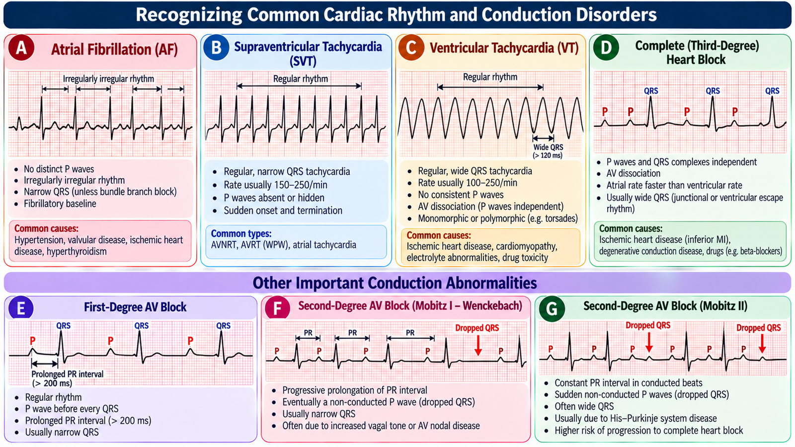
E. Recognizing Common Cardiac Rhythm and Conduction Disorders
An arrhythmia is an abnormality of impulse formation, impulse conduction or both. The clinical importance of an abnormal rhythm depends not only on its ECG appearance but also on its effect on cardiac output and patient stability. A rapid or very slow rhythm can reduce ventricular filling or effective cardiac output, causing palpitations, dizziness, syncope, hypotension, chest pain or heart failure.
A systematic ECG approach
When assessing a rhythm, first determine the rate and regularity, then assess atrial activity, the relationship between atrial and ventricular activity, QRS width and conduction pattern. This prevents a striking ECG feature from distracting from the overall rhythm.
Atrial fibrillation
Atrial fibrillation is characterized by disorganized atrial electrical activity with loss of coordinated atrial contraction. Ventricular conduction varies from beat to beat, producing an irregularly irregular rhythm. Loss of effective atrial contraction also promotes blood stasis in the atria, creating an important thromboembolic risk.
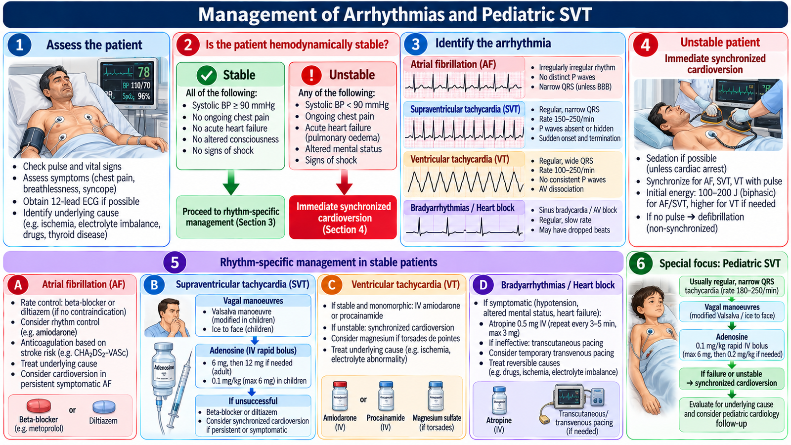
Treatment: Stable AF is commonly managed with rate control using bisoprolol or metoprolol; diltiazem may be used when left ventricular systolic function is preserved. Assess stroke risk using CHA₂DS₂-VASc and prescribe anticoagulation when indicated, preferably apixaban or another DOAC in eligible patients. Warfarin is required for mechanical heart valves or moderate-to-severe rheumatic mitral stenosis. Hemodynamically unstable AF requires urgent synchronized cardioversion.
Supraventricular tachycardia
SVT commonly produces a rapid, regular tachycardia arising above the ventricles. Because the rate is fast, ventricular filling time falls. Patients may develop palpitations, dizziness, chest discomfort or hemodynamic compromise if the rhythm is poorly tolerated.
Treatment: Stable regular narrow-complex SVT is initially managed with modified Valsalva manoeuvre. If unsuccessful, give adenosine 6 mg rapid IV push, followed by 12 mg if necessary, under ECG monitoring. Unstable SVT requires immediate synchronized cardioversion. Avoid AV nodal blockers in pre-excited atrial fibrillation.
Ventricular tachycardia
Ventricular tachycardia originates in the ventricles and commonly produces a rapid broad-complex rhythm. Effective ventricular filling and coordinated contraction may be impaired, so sustained VT can cause hypotension, syncope, myocardial ischemia or progression to cardiac arrest.
Drug treatment: Hemodynamically stable monomorphic VT may be treated with IV amiodarone or procainamide under continuous monitoring and specialist supervision. Unstable VT with a pulse requires synchronized cardioversion; pulseless VT requires CPR and immediate defibrillation.
Heart blocks
Heart block reflects impaired conduction from the atria toward the ventricles. The clinical effect ranges from delayed conduction to intermittent failure of conduction or complete atrioventricular dissociation. More advanced block can produce clinically important bradycardia and reduced cardiac output.
Symptomatic bradycardia: Give atropine 1 mg IV, repeated every 3–5 minutes if required (maximum 3 mg). Persistent instability or high-grade AV block requires urgent pacing assessment; Mobitz II and complete heart block may require a permanent pacemaker after evaluation of reversible causes.
| Rhythm | Key ECG Idea | Main Clinical Concern |
|---|---|---|
| Atrial fibrillation | Irregularly irregular ventricular rhythm without organized P waves | Rate-related symptoms and thromboembolism |
| SVT | Rapid regular supraventricular rhythm, commonly narrow complex | Reduced filling and possible instability |
| Ventricular tachycardia | Rapid broad-complex ventricular rhythm | Hemodynamic collapse and cardiac arrest |
| Heart block | Abnormal atrioventricular conduction | Symptomatic bradycardia and low cardiac output |
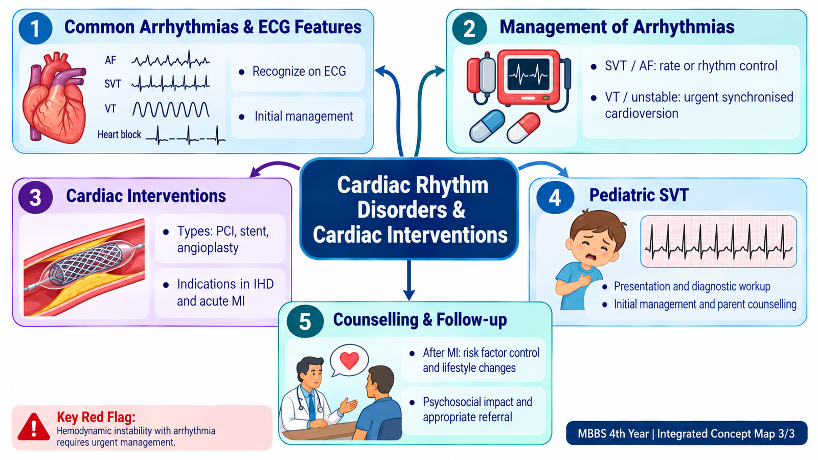
F. Management of Arrhythmias and Pediatric SVT
The first management question in an arrhythmia is whether the rhythm is causing clinical instability. The ECG identifies the electrical disorder, but blood pressure, consciousness, chest pain, heart failure and tissue perfusion determine how urgently the rhythm must be treated.
Stable versus unstable rhythm
An unstable tachyarrhythmia associated with significant hypotension, ischemic chest discomfort, acute heart failure, altered consciousness or other evidence of poor perfusion requires urgent rhythm-directed treatment. A stable patient allows more time to identify the rhythm and choose specific therapy.
Atrial fibrillation
Management has three major clinical questions: control of excessive ventricular rate when required, restoration or maintenance of sinus rhythm in selected patients, and assessment of thromboembolic risk to determine whether anticoagulation is appropriate. Underlying precipitants and associated cardiovascular disease should also be addressed.
Supraventricular tachycardia
In a stable regular SVT, measures that increase vagal influence on AV nodal conduction may terminate an AV node-dependent tachycardia. If appropriate initial measures fail, an AV nodal blocking strategy can be used under clinical supervision. Hemodynamic instability changes the priority toward urgent synchronized electrical cardioversion.
Pediatric SVT treatment: In stable children, attempt age-appropriate vagal manoeuvres under monitoring. If unsuccessful, give adenosine 0.1 mg/kg rapid IV/IO push (maximum 6 mg); a second dose of 0.2 mg/kg (maximum 12 mg) may be given. Hemodynamic compromise requires synchronized cardioversion, initially 0.5–1 J/kg, increasing to 2 J/kg if unsuccessful.
Ventricular tachycardia
VT must be taken seriously because it can rapidly compromise circulation. Management depends on whether a pulse is present and whether the patient is stable. Unstable VT with a pulse requires urgent synchronized cardioversion, while a pulseless ventricular rhythm is managed as cardiac arrest with immediate resuscitation and defibrillation as appropriate.
Heart block
Management depends on the degree of conduction disturbance and its clinical effect. Mild conduction delay may require observation and treatment of reversible causes, whereas clinically important bradycardia or advanced conduction block may require urgent support and pacing assessment.
SVT in children
Children, particularly infants, may not describe palpitations. Pediatric SVT may therefore present with persistent tachycardia, irritability, poor feeding, pallor, respiratory difficulty or signs of heart failure when prolonged. Diagnosis is supported by ECG demonstration of a rapid supraventricular rhythm.
Management again begins with stability. A stable child is assessed for an appropriate rhythm-termination strategy under monitored clinical care, while shock, impaired consciousness or significant cardiovascular compromise requires urgent escalation. Parents should be told what the rhythm disturbance means, why ECG assessment and treatment are needed, the importance of follow-up, and which symptoms require urgent reassessment.
Treat the patient, not only the ECG: an identical rhythm can require a very different response when it causes hypotension, ischemia, heart failure or impaired consciousness.
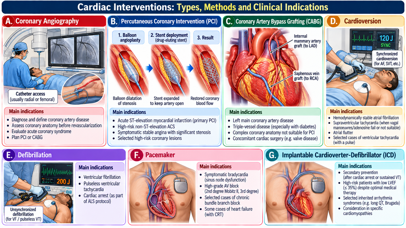
G. Cardiac Interventions: Types, Methods and Clinical Indications
Cardiac interventions are used when diagnosis or treatment requires direct access to the heart or coronary circulation. For Final-Year learning, the important principle is to understand what each intervention is intended to achieve and the clinical situations in which it becomes relevant.
Coronary angiography
Coronary angiography uses intravascular catheter access and contrast imaging to define coronary anatomy. It helps identify the site and severity of coronary obstruction and can guide a decision between medical management and revascularization.
Percutaneous coronary intervention
PCI restores blood flow through a significantly narrowed or acutely occluded coronary artery using catheter-based techniques, commonly balloon dilatation with stent placement. In acute coronary occlusion it can provide reperfusion, while in selected chronic disease it can relieve clinically important ischemia.
Coronary artery bypass grafting
CABG provides an alternative route for blood flow beyond significantly diseased coronary segments. It is considered when the extent or anatomical pattern of coronary disease makes surgical revascularization more appropriate than a catheter-based approach.
Electrical and rhythm interventions
Electrical cardioversion can restore an organized rhythm in selected tachyarrhythmias. Defibrillation delivers an unsynchronized shock for shockable cardiac-arrest rhythms. Permanent pacing provides electrical stimulation when clinically important bradycardia or conduction failure requires ongoing rhythm support. Catheter ablation may be used for selected recurrent arrhythmias by targeting tissue responsible for abnormal electrical circuits.
The choice of intervention therefore follows the clinical problem: coronary obstruction requires a reperfusion or revascularization strategy, while an electrical disorder requires rhythm-specific intervention.
Angiography defines coronary anatomy; PCI treats suitable coronary obstruction. Cardioversion and defibrillation treat electrical instability, while pacing supports clinically important bradyarrhythmia or conduction failure.
Integrated Mechanism Flow
AIM High-Yield Review
- ⭐ Predictable exertional chest discomfort relieved by rest supports stable angina; new, worsening or rest ischemia raises concern for acute coronary syndrome.
- Plaque disruption can trigger platelet activation and coronary thrombosis, converting relatively stable coronary disease into an acute ischemic event.
- ⭐ In suspected ACS, ECG identifies important ischemic patterns, while cardiac troponin identifies myocardial injury in the correct clinical context.
- Persistent ST elevation in a compatible acute presentation suggests an occlusive process requiring urgent reperfusion assessment.
- Post-MI care continues beyond hospital discharge: secondary prevention combines appropriate medication, cardiovascular risk-factor control, rehabilitation, adherence and follow-up.
- ⭐ Atrial fibrillation produces an irregularly irregular rhythm and creates an important thromboembolic risk because coordinated atrial contraction is lost.
- SVT is commonly a rapid regular supraventricular rhythm; pediatric SVT may present with poor feeding, irritability or heart-failure features rather than a verbal complaint of palpitations.
- ⭐ A rapid broad-complex rhythm should raise serious concern for ventricular tachycardia, particularly when accompanied by hypotension, syncope or ischemic symptoms.
- Heart block represents impaired atrioventricular conduction; advanced block can produce significant bradycardia and reduced cardiac output.
- The immediate management of an arrhythmia depends on clinical stability as well as ECG diagnosis.
- Coronary angiography defines coronary anatomy; PCI treats suitable coronary obstruction, while CABG creates a surgical route around important coronary disease.
- Cardioversion, defibrillation, pacing and catheter ablation address electrical problems and should not be confused with coronary revascularization procedures.
- Recurrent chest pain, syncope, acute heart failure, sustained significant arrhythmia or hemodynamic compromise are red flags requiring urgent reassessment or specialist care.
Angina Pectoris — Stable & Unstable Ischemic Chest Pain
Review the mechanism and clinical distinction of ischemic chest pain before moving to acute coronary syndrome and myocardial infarction.
Supraventricular Tachycardia — ECG Recognition & Management
Focus on clinical presentation, ECG recognition, stability assessment and the main management principles of SVT.