
Learn this topic by separating three problems: airflow obstruction, impaired gas exchange, and diffuse lung disease. Then connect symptoms and investigations to the underlying physiology. This makes asthma, COPD, ILD and respiratory failure much easier to distinguish.
Topic 4. Obstructive & Interstitial Lung Disease, Respiratory Failure and Cystic Fibrosis
CVS Module • Respiratory Medicine, Pulmonology & Pediatrics
A clinical journey from airflow obstruction and restrictive lung disease to gas-exchange failure, oxygen therapy and cystic fibrosis.
Topic Introduction
Breathlessness and wheeze may result from narrowed airways, damaged lung parenchyma or failure of the respiratory system to maintain adequate gas exchange. In this topic, you will learn how asthma and COPD produce obstructive physiology, how interstitial lung disease produces restrictive physiology, and how severe respiratory dysfunction can progress to respiratory failure. You will also learn how pulmonary function tests and arterial blood gases translate physiology into clinical information, how oxygen is used safely, and how cystic fibrosis produces important respiratory disease in children. The aim is to connect mechanism, presentation, investigation and management rather than memorize isolated disease lists.
1. Bronchial Asthma & the Chronic Wheezy Chest
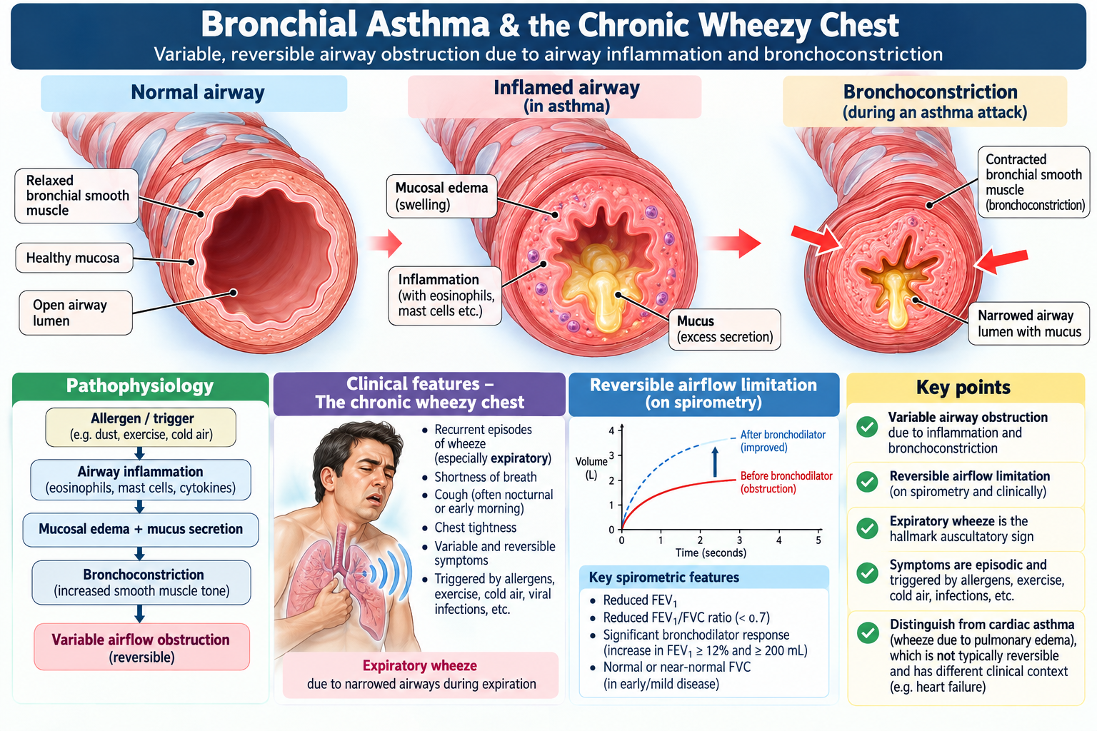
Bronchial asthma is a chronic inflammatory airway disorder characterized clinically by variable respiratory symptoms and physiologically by variable expiratory airflow limitation. Airway narrowing occurs because of bronchial smooth-muscle constriction, mucosal inflammation and edema, together with increased mucus production. Because this obstruction is variable, symptoms can improve spontaneously or after treatment.
Recognizing the pattern
Typical symptoms include episodic wheeze, breathlessness, chest tightness and cough. Variability is important: symptoms may fluctuate over time and may be triggered by exercise, allergens, respiratory infections, smoke, irritants or other recognized triggers.
A focused history should therefore establish the pattern, variability, triggers, nocturnal symptoms, previous exacerbations, treatment response and relevant exposures. Examination may be normal between episodes. During active obstruction, prolonged expiration and widespread expiratory wheeze may be present. Severe disease may produce marked respiratory distress; a very quiet chest in a severely breathless patient is dangerous because it can indicate critically reduced airflow rather than improvement.
How the diagnosis is supported
The clinical pattern suggests asthma, while objective evidence of variable airflow limitation strengthens the diagnosis. Spirometry may demonstrate an obstructive pattern with improvement following bronchodilation. Peak expiratory flow monitoring can also demonstrate clinically meaningful variability when appropriate.
Management logic
Management has two linked goals: relieve bronchoconstriction and control airway inflammation. Bronchodilator therapy improves airflow, while inhaled anti-inflammatory therapy—particularly corticosteroid-based treatment—reduces underlying inflammation and future exacerbation risk. Trigger avoidance, correct inhaler use, adherence and recognition of worsening symptoms are important parts of long-term care.
Practical management: In adults and adolescents, low-dose inhaled corticosteroid (ICS)–formoterol, such as budesonide–formoterol, is the preferred reliever-based treatment; short-acting beta-agonist (SABA) therapy alone is not recommended. Persistent symptoms may require maintenance ICS–formoterol therapy. Acute exacerbations are treated with repeated inhaled salbutamol, oxygen when hypoxemic, and early systemic corticosteroids such as prednisolone (40–50 mg daily for 5–7 days in adults). Severe or life-threatening attacks require emergency referral and may need inhaled ipratropium and intravenous magnesium sulfate.
Bronchial asthma versus cardiac asthma
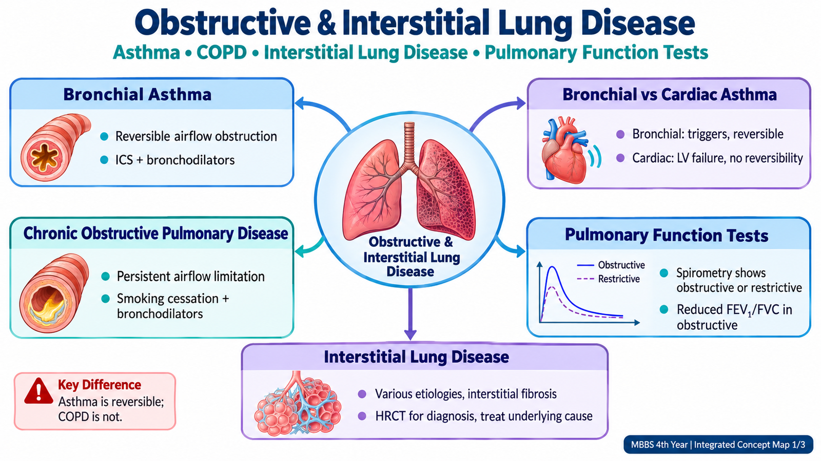
Cardiac asthma is not bronchial asthma. It refers to wheezing associated with pulmonary congestion, usually from left-sided cardiac failure. Orthopnea, paroxysmal nocturnal dyspnea, basal crackles, peripheral edema or other evidence of cardiac disease favour a cardiac cause, whereas variable wheeze with typical triggers and reversible airflow limitation supports bronchial asthma.
2. Chronic Obstructive Pulmonary Disease
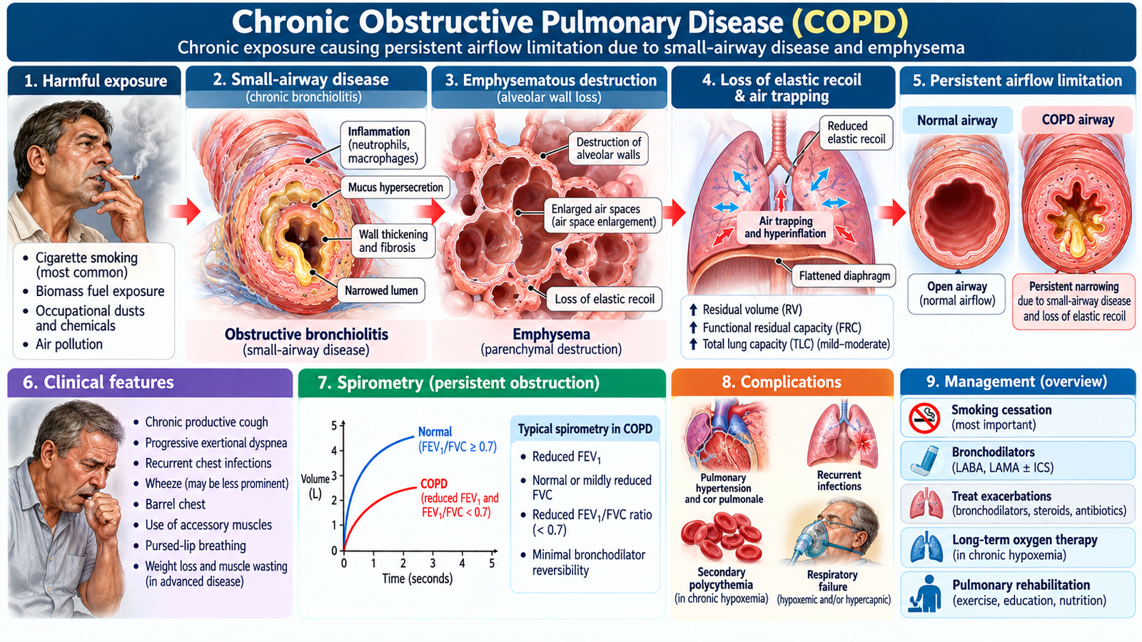
Chronic obstructive pulmonary disease (COPD) is characterized by persistent respiratory symptoms and airflow limitation associated with chronic airway and/or alveolar abnormalities. Long-term exposure to harmful particles or gases—most importantly tobacco smoke in many populations—produces airway inflammation, mucus hypersecretion and structural lung damage.
Two processes help explain the physiology. Small-airway disease narrows the conducting airways, while emphysematous destruction reduces elastic recoil and supporting alveolar attachments. Both make expiration difficult and promote air trapping.
Clinical recognition
The typical adult has persistent or progressive dyspnea, chronic cough and sometimes sputum production, usually in the presence of a relevant exposure history. Examination may show prolonged expiration, wheeze, reduced breath sounds, hyperinflation and increased work of breathing. Advanced disease may be associated with hypoxemia, cyanosis, respiratory failure or features of pulmonary hypertension and right-sided cardiac strain.
Diagnostic approach
The history raises suspicion, but spirometry is central to confirming persistent airflow obstruction. Imaging is useful when assessing structural changes, complications or alternative diagnoses. Arterial blood gases become particularly important when disease is severe, oxygenation is impaired or respiratory failure is suspected.
Management principles
- Remove the harmful exposure: smoking cessation is a major disease-modifying intervention when smoking is the cause.
- Bronchodilation: inhaled bronchodilators reduce symptoms and improve airflow.
- Anti-inflammatory treatment: selected patients benefit from inhaled corticosteroid-containing regimens according to clinical context.
- Prevent exacerbations: vaccination, inhaler technique, adherence and appropriate follow-up are important.
- Rehabilitation: pulmonary rehabilitation can improve exercise tolerance, symptoms and functional status.
- Treat exacerbations and respiratory failure promptly: worsening dyspnea, hypoxemia, hypercapnia or altered mental status requires urgent assessment.
Drug treatment and exacerbations: Stable COPD is managed with long-acting inhaled bronchodilators such as tiotropium (LAMA) or formoterol (LABA); combined LABA/LAMA treatment is commonly preferred in symptomatic patients. Inhaled corticosteroids are added selectively, particularly with frequent exacerbations and raised blood eosinophils. Acute exacerbations require inhaled salbutamol, with or without ipratropium, and a short course of oral prednisolone (40 mg daily for 5 days). Antibiotics are indicated when bacterial infection is suspected, particularly with purulent sputum; acute hypercapnic respiratory acidosis may require non-invasive ventilation.
3. Pulmonary Function Tests: From Numbers to Physiology
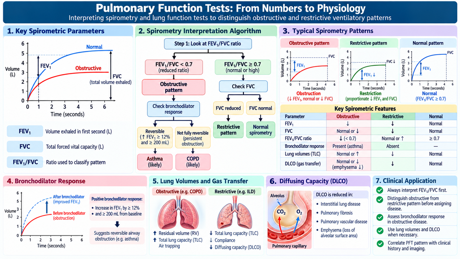
Pulmonary function tests help determine whether respiratory symptoms reflect an obstructive or restrictive ventilatory problem. The most important starting measurements are forced expiratory volume in the first second (FEV1) and forced vital capacity (FVC).
Obstructive pattern
In airflow obstruction, the patient has difficulty expelling air rapidly. FEV1 falls disproportionately compared with FVC, so the FEV1/FVC ratio is reduced. Asthma and COPD are major examples in this topic. Demonstration of significant reversibility after bronchodilation supports variable obstruction in the appropriate clinical context.
Restrictive pattern
In restriction, the total volume of air that can be moved is reduced. FVC falls, while the FEV1/FVC ratio is usually preserved or increased. Spirometry can therefore suggest restriction, but measurement of lung volumes is used to confirm true restriction when required.
Gas transfer
Diffusing capacity provides information about transfer of gas across the alveolar-capillary interface. It can be reduced when the effective gas-exchanging surface is destroyed or when the interstitial barrier is abnormal, helping connect diseases such as emphysema and ILD to their physiological consequences.
| Feature | Obstructive pattern | Restrictive pattern |
|---|---|---|
| Main problem | Difficulty getting air out rapidly | Reduced expandable lung volume |
| FEV1 | Markedly reduced relative to FVC | Reduced with reduced lung volume |
| FEV1/FVC | Reduced | Usually preserved or increased |
| Examples here | Asthma, COPD | Interstitial lung disease |
🩺 AIM Clinical Bridge
An older adult with a long smoking history reports gradually progressive exertional breathlessness and chronic cough. Expiration is prolonged, breath sounds are reduced, and spirometry demonstrates persistent airflow obstruction.
- How does chronic airway and alveolar injury explain the obstructive spirometry?
- Which historical feature helps distinguish this pattern from variable bronchial asthma?
- When would ABG assessment become particularly important?
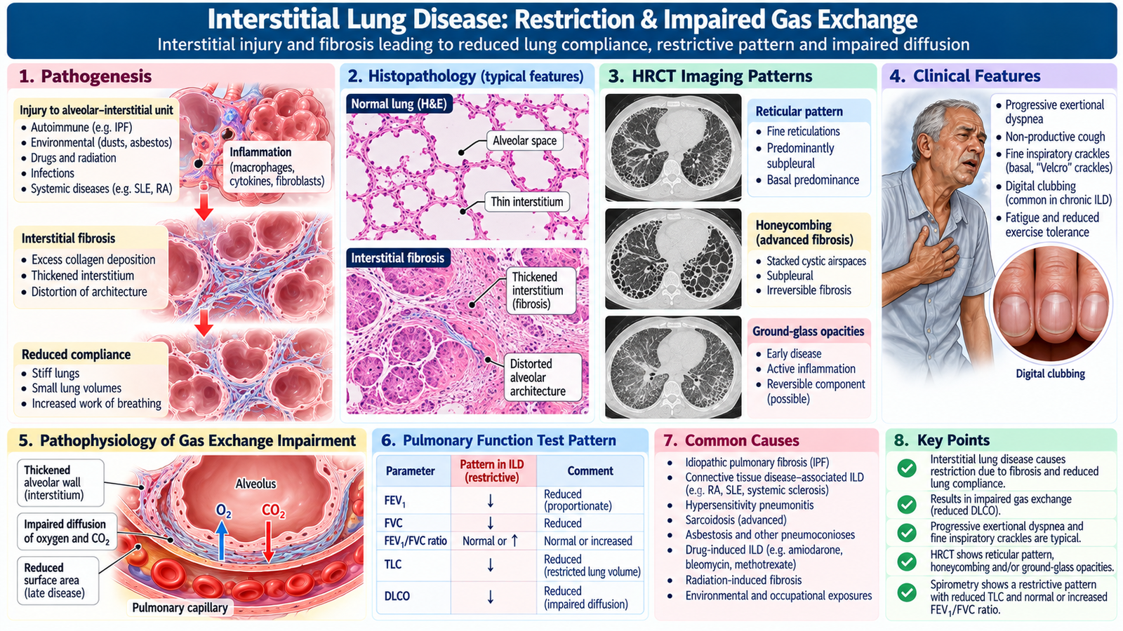
4. Interstitial Lung Disease: Restriction & Impaired Gas Exchange
Interstitial lung disease (ILD) describes a broad group of disorders affecting the pulmonary interstitium and, in many conditions, the alveolar-capillary unit. The key physiological consequence is a lung that becomes less compliant and less efficient at gas exchange. This produces a clinical picture that differs from the predominantly airway-centered obstruction of asthma and COPD.
Types and causes
ILD may be idiopathic or occur in association with recognized causes such as occupational or environmental exposures, systemic connective-tissue disease and certain drugs. The practical undergraduate approach is to search systematically for a cause while recognizing that some patients have no immediately identifiable underlying trigger.
From interstitial injury to symptoms
Persistent injury and inflammation may lead to interstitial thickening and fibrosis. The lungs become stiff, increasing the work of breathing. Thickening or destruction of the alveolar-capillary interface also impairs diffusion. Patients therefore commonly develop progressive exertional dyspnea and a dry cough. Examination may reveal fine inspiratory crackles; clubbing can occur in some forms of fibrotic disease.
Investigations and radiology
Pulmonary function testing commonly suggests a restrictive ventilatory defect and may show impaired gas transfer. Chest imaging may demonstrate bilateral interstitial abnormalities. High-resolution CT provides much greater definition of the distribution and pattern of parenchymal disease and helps characterize ILD in the appropriate clinical setting. Additional investigations are directed by the suspected underlying cause.
Management principles
Management begins by identifying and treating a reversible or avoidable cause when possible. Relevant harmful exposure should be removed. Depending on the ILD, treatment may target inflammation or fibrotic progression. Supportive care includes appropriate oxygen when hypoxemia is present, rehabilitation and management of complications. Progressive or uncertain disease requires specialist assessment.
Disease-specific treatment: In inflammatory ILDs, such as selected connective-tissue-disease-associated ILDs, corticosteroids or other immunosuppressants may be indicated. Idiopathic pulmonary fibrosis is treated with antifibrotic medicines such as nintedanib or pirfenidone under specialist supervision; routine corticosteroids are not recommended for IPF.
5. Respiratory Failure & Arterial Blood Gas Interpretation
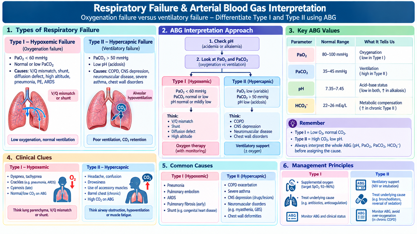
Respiratory failure occurs when the respiratory system can no longer maintain adequate gas exchange. The failure may primarily involve oxygenation, ventilation, or both. Understanding this distinction makes ABG interpretation clinically meaningful.
Type I: hypoxemic respiratory failure
The dominant problem is inadequate arterial oxygenation without primary carbon-dioxide retention. Important mechanisms include ventilation-perfusion mismatch, diffusion impairment and shunt. Severe parenchymal lung disease can produce this pattern.
Type II: hypercapnic respiratory failure
Here, alveolar ventilation is inadequate, causing carbon dioxide retention, commonly accompanied by hypoxemia. Severe obstructive lung disease is an important clinical setting. Other processes that markedly reduce effective ventilation can produce the same physiological endpoint.
Acute versus chronic failure
The distinction matters because the body needs time to compensate for sustained carbon-dioxide retention. In acute hypercapnia, pH may fall substantially because renal compensation has not yet developed. With chronic hypercapnia, renal bicarbonate retention provides partial compensation. ABGs must therefore be interpreted as a pattern rather than by looking at a single value.
A simple ABG approach
- Assess the pH: is there acidemia or alkalemia?
- Assess PaCO2: is ventilation contributing to the disturbance?
- Assess bicarbonate: is there a metabolic component or compensation?
- Assess PaO2: is oxygenation impaired?
- Integrate the pattern with the patient’s clinical condition.
Severe respiratory distress, worsening hypoxemia, cyanosis, exhaustion, reduced consciousness, inability to maintain ventilation or progressive carbon-dioxide retention indicate serious physiological deterioration and require urgent management.
Management principle
Management combines stabilization with treatment of the underlying cause. Airway patency, breathing and circulation are assessed first. Hypoxemia is corrected appropriately, while ventilation is supported when the patient cannot maintain adequate alveolar ventilation. The underlying disorder—such as severe obstructive or parenchymal lung disease—must be treated simultaneously.
Respiratory support: Hypoxemic respiratory failure requires controlled oxygen therapy, with escalation to high-flow nasal oxygen or ventilatory support when necessary. Acute hypercapnic respiratory failure with persistent respiratory acidosis (pH below 7.35 and elevated PaCO₂) commonly requires non-invasive ventilation (BiPAP), particularly in COPD exacerbations. Endotracheal intubation and invasive mechanical ventilation are indicated when ventilatory support fails or the patient cannot protect the airway.

6. Oxygen Therapy: Indications, Delivery & Safe Use
Oxygen is a treatment for hypoxemia. It should be prescribed and monitored according to the patient’s physiological need rather than simply because the patient feels breathless. The key principle is to provide sufficient oxygen while continuing to identify and treat the underlying cause of respiratory compromise.
When oxygen is used
Oxygen is indicated when clinically important hypoxemia is present or strongly suspected in an acutely unwell patient while assessment proceeds. It may also be required in selected chronic hypoxemic respiratory disorders. The required concentration and delivery method depend on severity, clinical context and the need for controlled versus higher-concentration oxygen.
Approaches to delivery
- Nasal cannulae: comfortable for patients who require relatively modest supplemental oxygen.
- Simple face-mask systems: provide supplemental oxygen when a greater inspired concentration is needed.
- Controlled-performance masks: useful when a more predictable inspired oxygen concentration is required.
- Higher-concentration systems: may be required in severe acute hypoxemia while definitive treatment is arranged.
Why COPD requires thoughtful oxygen therapy
Some patients with severe COPD are vulnerable to worsening hypercapnia during excessive oxygen administration. This does not mean that a hypoxemic patient with COPD should be denied oxygen. It means oxygen should be controlled, monitored and adjusted, with clinical assessment and ABG reassessment when indicated.
Target oxygen saturation: Aim for SpO₂ 94–98% in most acutely ill adults without risk of hypercapnic respiratory failure. In patients with COPD or other risk factors for CO₂ retention, initially target 88–92% using controlled oxygen, such as a Venturi mask, pending arterial blood gas results. Adjust oxygen according to clinical response and ABG findings.

7. Cystic Fibrosis in Children
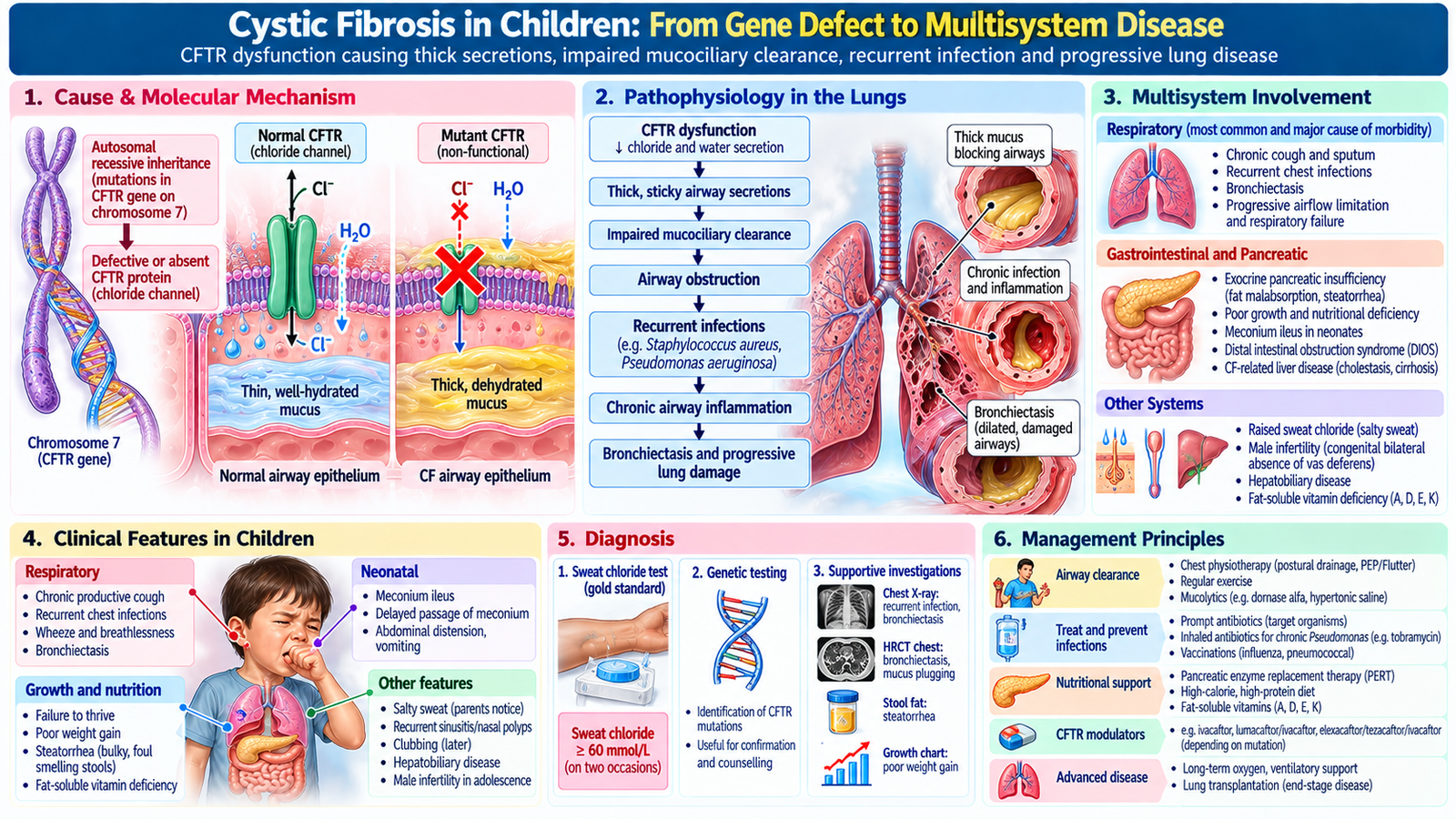
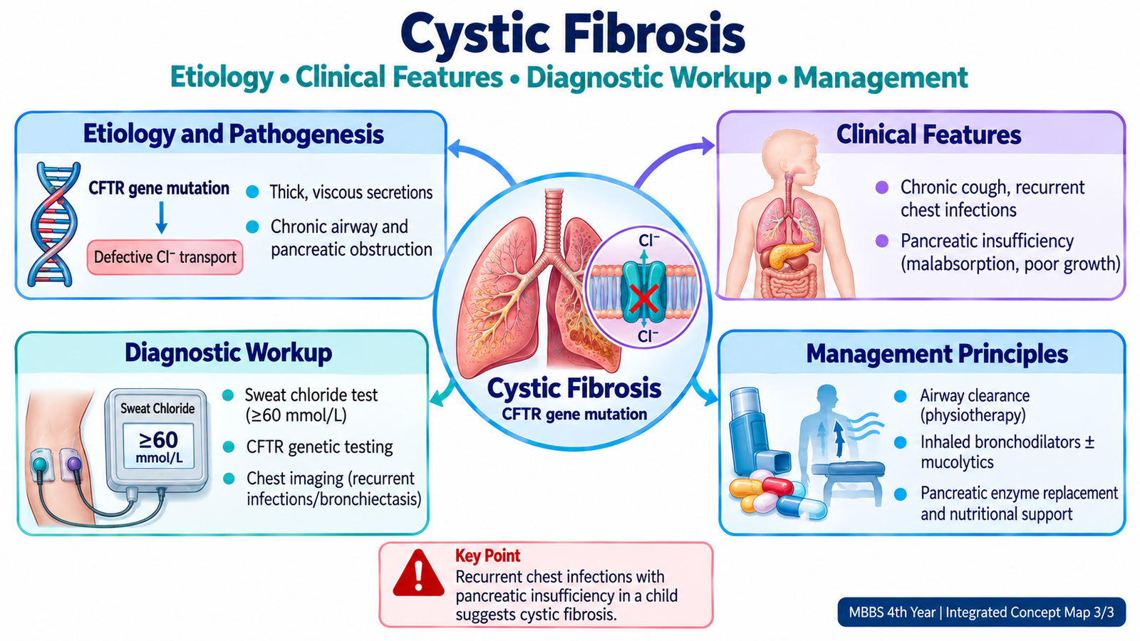
Cystic fibrosis is an inherited disorder caused by abnormal function of the CFTR chloride channel. Abnormal epithelial ion and water transport produces dehydrated, unusually viscous secretions. The disease affects several organs, but chronic respiratory disease is a major source of morbidity.
How the respiratory disease develops
Thick airway secretions impair mucociliary clearance. Mucus retention then promotes airway obstruction and recurrent infection, which drives persistent inflammation and progressive structural airway damage. Over time this cycle can lead to bronchiectatic change and declining pulmonary function.
Clinical presentation
Children may present with recurrent or persistent respiratory symptoms, chronic cough, recurrent chest infections, wheeze or progressive lung disease. Because CF is multisystem, poor weight gain or nutritional difficulty and other manifestations related to thick secretions can provide additional diagnostic clues.
Diagnostic workup
Diagnosis combines the clinical context with evidence of CFTR dysfunction. Sweat chloride testing is an important diagnostic test, while genetic testing can support or clarify the diagnosis where appropriate. Respiratory assessment includes evaluation of infection and pulmonary function according to the child’s age and clinical status.
Management
Management is multidisciplinary and aims to interrupt the cycle of mucus retention, infection, inflammation and progressive lung injury. Important principles include airway-clearance measures, treatment and prevention of respiratory infection, nutritional support, management of pancreatic insufficiency where present, and selected CFTR-directed therapy when appropriate for the underlying defect. Long-term monitoring is essential because pulmonary disease may progressively impair respiratory function.
Specific treatment: Regular chest physiotherapy and airway-clearance techniques are essential. Respiratory infections require culture-guided antibiotics, while pancreatic insufficiency is treated with pancreatic enzyme replacement (pancrelipase), nutritional supplementation and fat-soluble vitamins A, D, E and K when indicated. Eligible children may receive mutation-specific CFTR modulators under specialist care.
Important Comparison: Asthma, COPD & Interstitial Lung Disease
| Feature | Asthma | COPD | ILD |
|---|---|---|---|
| Main site/problem | Variable airway narrowing | Persistent airway/alveolar disease | Lung interstitium/parenchyma |
| Typical symptom pattern | Variable wheeze and dyspnea | Progressive dyspnea ± chronic cough/sputum | Progressive dyspnea and dry cough |
| PFT pattern | Obstructive, often variable/reversible | Persistent obstructive pattern | Restrictive pattern |
| Important examination clue | Expiratory wheeze | Prolonged expiration / reduced breath sounds | Fine inspiratory crackles |
| Core management logic | Control inflammation + bronchodilation | Exposure reduction + bronchodilation + supportive care | Identify cause + disease-specific/supportive treatment |
AIM High-Yield Review
- ⭐ Asthma: variable symptoms + variable expiratory airflow obstruction are the central clinical and physiological concepts.
- Bronchial asthma results from airway inflammation and bronchoconstriction; cardiac asthma reflects pulmonary congestion from cardiac dysfunction.
- ⭐ COPD: persistent airflow limitation in an appropriate exposure context should be confirmed physiologically with spirometry.
- Obstruction produces a disproportionately reduced FEV1 and therefore a reduced FEV1/FVC ratio.
- Restriction produces reduced lung volumes with a usually preserved or increased FEV1/FVC ratio; spirometry suggests restriction and lung-volume measurement confirms it when required.
- ⭐ ILD: interstitial injury/fibrosis → reduced compliance + impaired diffusion → progressive dyspnea, dry cough and restrictive physiology.
- Fine inspiratory crackles and compatible interstitial imaging support ILD in the appropriate clinical setting.
- ⭐ Respiratory failure: Type I is predominantly an oxygenation problem; Type II includes inadequate ventilation with carbon-dioxide retention.
- ABGs should be interpreted systematically using pH, PaCO2, bicarbonate and PaO2 together with the clinical picture.
- Acute hypercapnia causes greater acidemia because renal compensation has had little time to develop; chronic hypercapnia may show compensatory bicarbonate retention.
- Oxygen treats hypoxemia; in patients at risk of hypercapnia it should be controlled and monitored rather than withheld.
- ⭐ Cystic fibrosis: CFTR dysfunction → dehydrated secretions → impaired mucus clearance → recurrent infection/inflammation → progressive airway damage.
- Sweat chloride testing is an important diagnostic test for cystic fibrosis in the appropriate clinical context.
Obstructive & Interstitial Lung Disease, Respiratory Failure and Cystic Fibrosis
Use these selected videos to reinforce the mechanisms, clinical presentation, investigation and management concepts covered in the AIM Learning Material.
1. Obstructive Lung Disease — Asthma & COPD
Focus on airflow obstruction, asthma, COPD, clinical features, pulmonary function testing and management principles.
2. Restrictive & Interstitial Lung Disease
Focus on restrictive physiology, interstitial lung disease, clinical presentation, PFT interpretation, imaging and management.
While watching, concentrate on: obstructive versus restrictive PFT patterns → clinical presentation → gas-exchange abnormalities → respiratory failure → management principles.