
This chapter follows the supplied KMU outcomes for Topic 9, covering lung malignancy and the cardiovascular and pulmonary effects of systemic disease. First understand the clinical patterns and diagnostic logic; use the AIM High-Yield Review at the end for rapid revision. :chatgpt-content-reference{index=”0″}
TOPIC 9. Lung Malignancy & Cardiorespiratory Manifestations of Systemic Disease
CVS Module • A clinically integrated approach to lung tumors, bronchogenic carcinoma, pleural mesothelioma and systemic diseases that involve the heart, circulation and lungs.
Topic Introduction
This topic brings together two important clinical ideas. First, primary lung and pleural malignancies may present with respiratory symptoms, local invasion, metastatic disease or systemic effects. Second, diseases that begin outside the heart and lungs can still produce major cardiorespiratory problems through inflammation, vascular injury, immune mechanisms, abnormal tissue deposition or treatment-related effects. The key is not to memorize long lists. Instead, learn to move from the presenting pattern to the affected structure, select investigations that answer a clinical question, and then connect the diagnosis with prognosis and appropriate management.
A. Lung Tumors: Classification & Clinical Framework
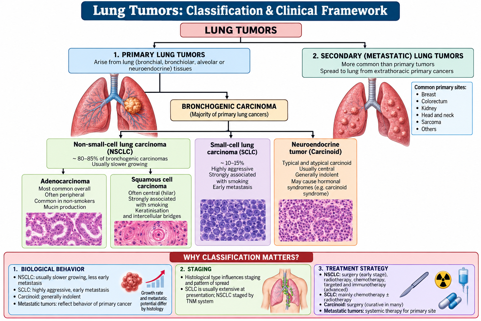
A lung mass is not a diagnosis by itself. Lung tumors are first understood by identifying whether the lesion is primary or metastatic and, for primary bronchogenic carcinoma, by recognizing the major histological grouping because tumor type influences biological behavior, staging and treatment.
Classification
Primary malignant epithelial lung tumors are broadly divided into:
- Non-small-cell lung carcinoma (NSCLC) — principally adenocarcinoma, squamous cell carcinoma and large-cell carcinoma.
- Small-cell lung carcinoma (SCLC) — a highly aggressive neuroendocrine carcinoma with early metastatic potential.
Other pulmonary tumors include neuroendocrine tumors such as carcinoid tumors and less common primary neoplasms. The lungs are also a frequent site of secondary metastatic deposits from malignancies elsewhere in the body.
Why histological type matters
Histology is not merely a pathology label. It helps determine the expected behavior of the tumor and the treatment pathway. NSCLC is staged primarily according to anatomical extent and may be amenable to local treatment when sufficiently localized. SCLC tends to disseminate early and is therefore approached mainly as a systemic malignancy.

B. Bronchogenic Carcinoma: Presentation & Clinical Assessment
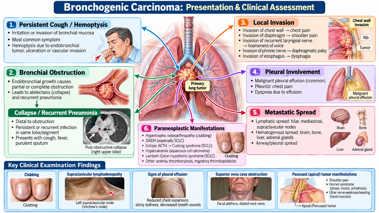
Bronchogenic carcinoma may become apparent because of the primary lung lesion, obstruction of an airway, invasion of neighboring structures, metastatic spread or systemic effects. Understanding these mechanisms makes the apparently diverse clinical features easier to remember.
Clinical presentation
- Local pulmonary disease: persistent or changing cough, hemoptysis, dyspnea and chest discomfort.
- Bronchial obstruction: impaired drainage may cause recurrent or non-resolving pneumonia and distal lung collapse.
- Pleural involvement: pleuritic discomfort, dyspnea and pleural effusion may occur.
- Local invasion: hoarseness may reflect recurrent laryngeal nerve involvement; diaphragmatic dysfunction may follow phrenic nerve involvement.
- Apical disease: an apical tumor may invade nearby structures, producing shoulder/arm pain and neurological or sympathetic-chain manifestations.
- Central venous obstruction: compression or invasion of the superior vena cava can cause facial and neck swelling, venous congestion and breathlessness.
- Metastatic disease: symptoms depend on the involved site and may include bone pain, neurological manifestations or features of hepatic involvement.
- General systemic effects: anorexia, weight loss, fatigue and declining functional status suggest significant malignant disease.
Paraneoplastic manifestations
A tumor can produce symptoms without direct invasion or metastasis by releasing biologically active substances or triggering immune effects. Clinically important associations include:
- Small-cell carcinoma → ectopic hormone production, particularly SIADH and ectopic ACTH-related manifestations.
- Squamous cell carcinoma → parathyroid hormone-related peptide production → hypercalcemia.
- Some lung cancers may be associated with neurological paraneoplastic syndromes or hypertrophic pulmonary osteoarthropathy.
Focused clinical assessment
History should define the respiratory complaint, progression, constitutional symptoms, smoking and occupational exposure, previous malignancy and clues to metastatic or paraneoplastic disease. Examination should look beyond the chest.
Important findings include cachexia, clubbing, lymphadenopathy, signs of collapse or pleural effusion, superior vena cava obstruction, neurological abnormalities and evidence of distant metastatic disease.
C. Bronchogenic Carcinoma: Diagnostic Workup, Management & Complications
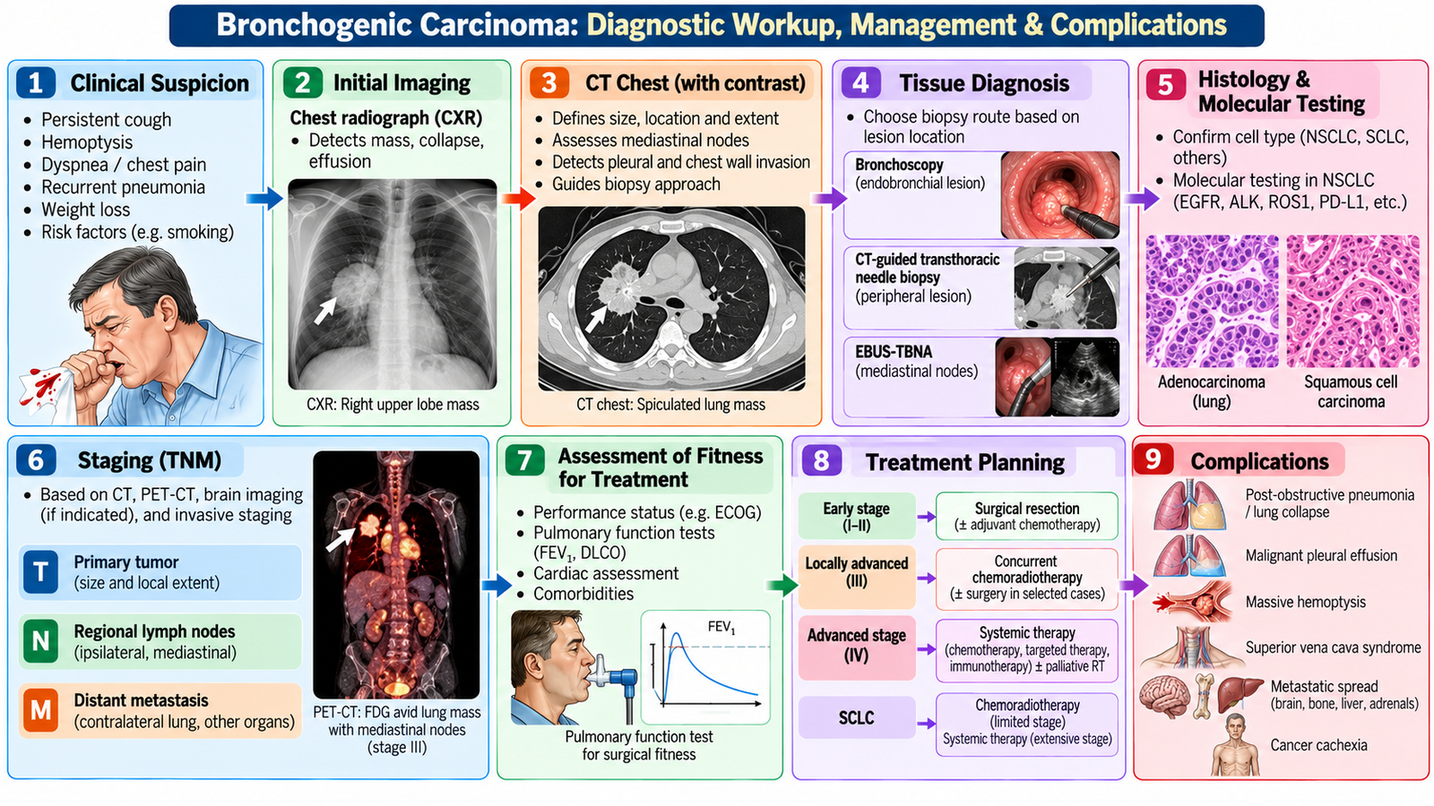
The diagnostic pathway answers three different questions: Is there a suspicious lesion? What is it histologically? How far has it spread? These correspond broadly to detection, tissue diagnosis and staging. Management then depends on tumor type, extent of disease, patient fitness and treatment goals.
Diagnostic workup
- Initial imaging: a chest radiograph may identify a mass, collapse, consolidation, hilar enlargement or pleural effusion.
- Cross-sectional imaging: contrast-enhanced CT of the chest better defines the primary lesion, lymph nodes, pleura and adjacent structures and helps guide further investigation.
- Tissue diagnosis: obtain material by the safest high-yield route. A central endobronchial lesion may be approached bronchoscopically; a peripheral lesion may require image-guided biopsy. Accessible lymph nodes or metastatic sites may provide diagnostic tissue.
- Staging: assess local extension, nodal involvement and distant metastasis using appropriate imaging and targeted tests according to the clinical situation.
- Fitness assessment: performance status and cardiopulmonary reserve influence whether surgery or other therapies can be tolerated.
Imaging can strongly suggest malignancy, but definitive tumor classification generally requires tissue. The biopsy method should be chosen according to lesion location and accessibility rather than using the same procedure for every patient.
Management principles
NSCLC: localized, operable disease may be treated surgically, while radiotherapy and systemic anticancer therapy have important roles according to stage, tumor characteristics and patient fitness.
SCLC: because early dissemination is common, systemic therapy is central, with radiotherapy used in selected settings.
Specific treatment options:
-
NSCLC: Early-stage operable disease is usually treated with surgical resection (commonly lobectomy). Unresectable or advanced disease may require radiotherapy, platinum-based chemotherapy, immunotherapy (e.g., pembrolizumab) or targeted therapy (e.g., osimertinib for suitable EGFR-mutated tumors), depending on staging and molecular testing.
-
SCLC: Standard systemic chemotherapy includes cisplatin or carboplatin with etoposide. Limited-stage disease usually receives combined chemotherapy and thoracic radiotherapy; extensive-stage disease commonly receives chemotherapy plus immunotherapy (atezolizumab or durvalumab).
In advanced disease, symptom control and palliative care remain active treatment. Breathlessness, pain, cough, hemoptysis, pleural effusion and psychosocial distress should be addressed alongside tumor-directed therapy.
Important complications
- Airway obstruction → atelectasis or recurrent/post-obstructive infection.
- Pleural involvement → malignant pleural effusion and progressive dyspnea.
- Local invasion → superior vena cava obstruction, nerve involvement or chest-wall pain.
- Metastatic spread → organ-specific complications, including skeletal and neurological disease.
- Paraneoplastic syndromes → metabolic, endocrine or neurological manifestations.
Major hemoptysis, severe airway compromise, superior vena cava obstruction, acute neurological deterioration or other evidence of major tumor-related organ compromise requires urgent assessment.
🩺 AIM Clinical Bridge
An older adult with progressive cough, weight loss and intermittent hemoptysis has a persistent unilateral hilar opacity on chest imaging. Examination also reveals supraclavicular lymphadenopathy.
- The combination of persistent respiratory symptoms, systemic features and abnormal imaging raises suspicion of lung malignancy.
- An accessible abnormal lymph node may provide diagnostic tissue while also giving information about disease spread.
- The next steps must establish histology and extent before definitive treatment is planned.
D. Pleural Mesothelioma: Recognition, Diagnosis & Management
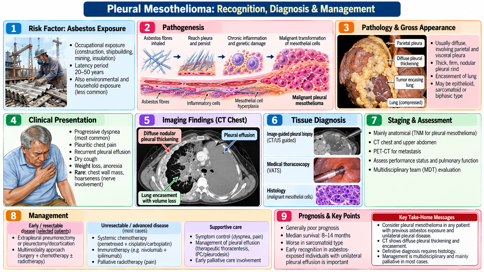
Pleural mesothelioma is a malignant tumor arising from pleural mesothelial cells. A previous history of asbestos exposure is an important clinical clue because disease may become apparent long after the exposure. Unlike a tumor confined to the lung parenchyma, mesothelioma characteristically involves the pleural surface and may progressively encase and restrict the lung.
Clinical features
Common manifestations include progressive dyspnea, persistent chest pain, cough, weight loss and recurrent pleural effusion. Examination may show reduced chest expansion, stony dull percussion and reduced breath sounds when a substantial pleural effusion is present.
Diagnostic workup
- Chest imaging may demonstrate pleural effusion, pleural thickening or a pleural-based abnormality.
- CT defines the distribution and extent of pleural disease and its relationship to adjacent thoracic structures.
- Pleural fluid assessment is useful when an effusion is present, but fluid cytology alone may not establish the diagnosis.
- Pleural tissue biopsy is important for definitive histological diagnosis when mesothelioma is suspected.
- Staging evaluates the extent of local and metastatic disease and helps guide treatment planning.
Management and prognosis
Management requires multidisciplinary assessment. Depending on disease extent and patient fitness, treatment may include systemic anticancer therapy, radiotherapy and selected surgical approaches. Symptom control is particularly important because recurrent pleural fluid, pain and progressive respiratory restriction can markedly affect quality of life.
Specific treatment: Systemic therapy for unresectable pleural mesothelioma may include platinum-based chemotherapy (cisplatin or carboplatin with pemetrexed) or immunotherapy (nivolumab with ipilimumab) in suitable patients. Symptomatic recurrent pleural effusions may require therapeutic drainage, an indwelling pleural catheter or pleurodesis. Palliative radiotherapy may relieve localized chest-wall pain.
Prognosis is influenced by stage, tumor biology, functional status and treatment options. Mesothelioma is frequently difficult to treat curatively because disease may already be extensive when recognized.
Complications
Progressive pleural encasement can restrict lung expansion; recurrent effusion worsens breathlessness; local invasion can produce severe chest-wall pain; and advanced disease may spread beyond the hemithorax.
E. Cardiovascular Manifestations of Systemic Disease
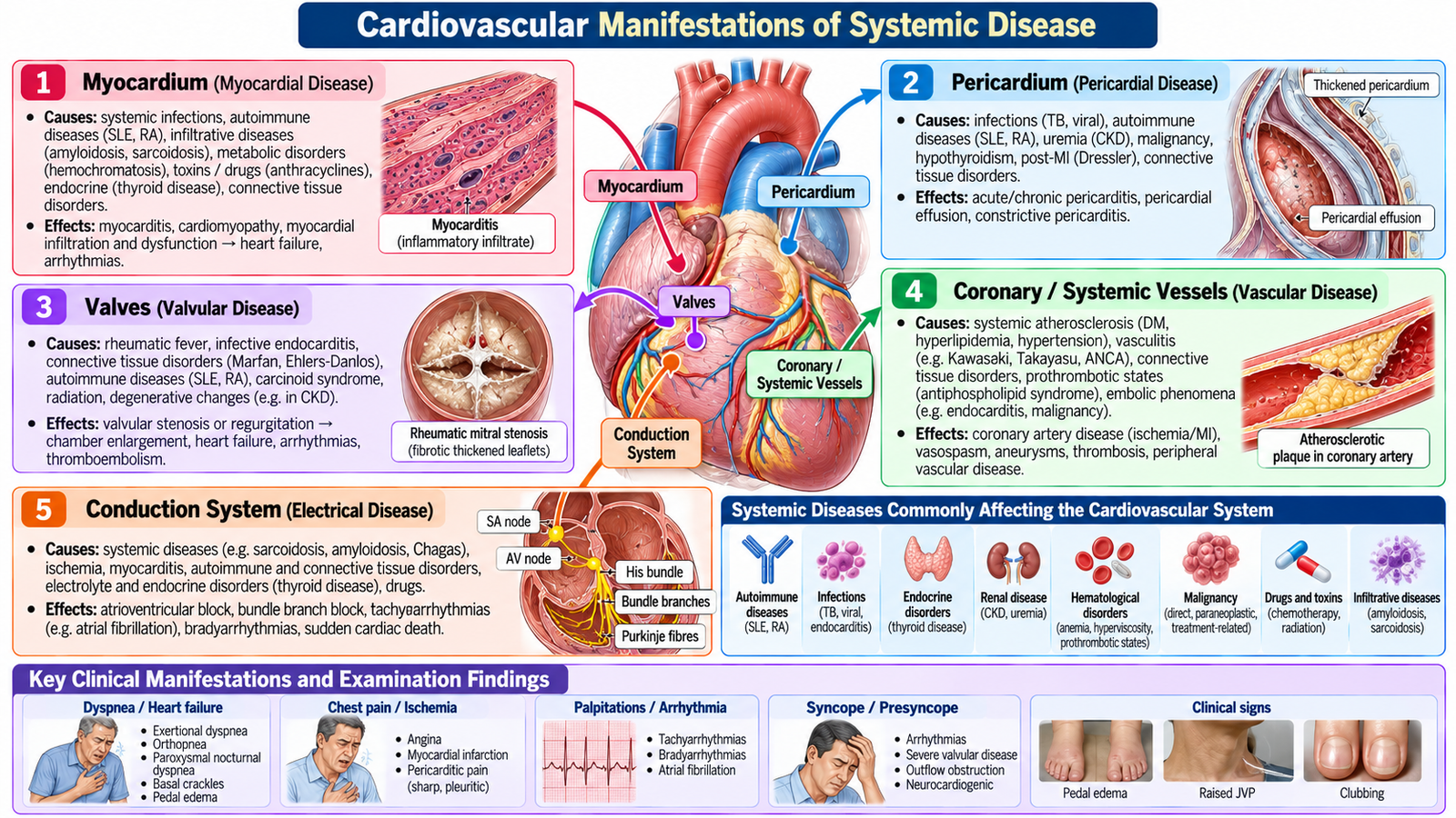
A systemic disease may affect almost any cardiovascular structure: myocardium, pericardium, valves, conduction system or blood vessels. The useful clinical approach is therefore systemic disease → cardiovascular structure affected → functional disturbance → clinical manifestation.
Major patterns of cardiovascular involvement
| Affected component | Functional consequence | Possible clinical expression |
|---|---|---|
| Myocardium | Inflammation, infiltration or impaired myocardial function | Dyspnea, exercise intolerance, heart failure or arrhythmia |
| Pericardium | Inflammation or fluid accumulation | Pericarditic pain, effusion or hemodynamic compromise |
| Valves / endocardial structures | Structural or inflammatory dysfunction | Murmur, regurgitation or secondary heart failure |
| Coronary / systemic vessels | Inflammation, thrombosis or accelerated vascular disease | Ischemia, infarction or vascular complications |
| Conduction system | Electrical instability or conduction disturbance | Palpitations, arrhythmias, syncope or conduction block |
Systemic disease examples
Autoimmune and connective-tissue diseases may cause pericarditis, myocarditis, vascular inflammation, accelerated atherosclerotic disease or pulmonary vascular involvement. Systemic inflammatory disorders may similarly involve the myocardium, pericardium or vessels.
Endocrine and metabolic disorders can alter heart rate, vascular resistance, blood pressure, lipid metabolism and myocardial function. Infiltrative disorders may impair myocardial relaxation, conduction or pump function through abnormal tissue deposition.
The clinical lesson is to avoid assuming that chest pain, dyspnea, edema, palpitations or syncope in a patient with systemic disease must represent an unrelated primary cardiac disorder.
Clinical assessment
Look for symptoms of heart failure, ischemia, pericardial disease and arrhythmia while also assessing the activity and complications of the underlying systemic condition. Examination should assess pulse, blood pressure, volume status, jugular venous pressure, precordium, heart sounds, murmurs, peripheral edema and systemic clues relevant to the underlying disorder.
F. Pulmonary Manifestations of Systemic Disease
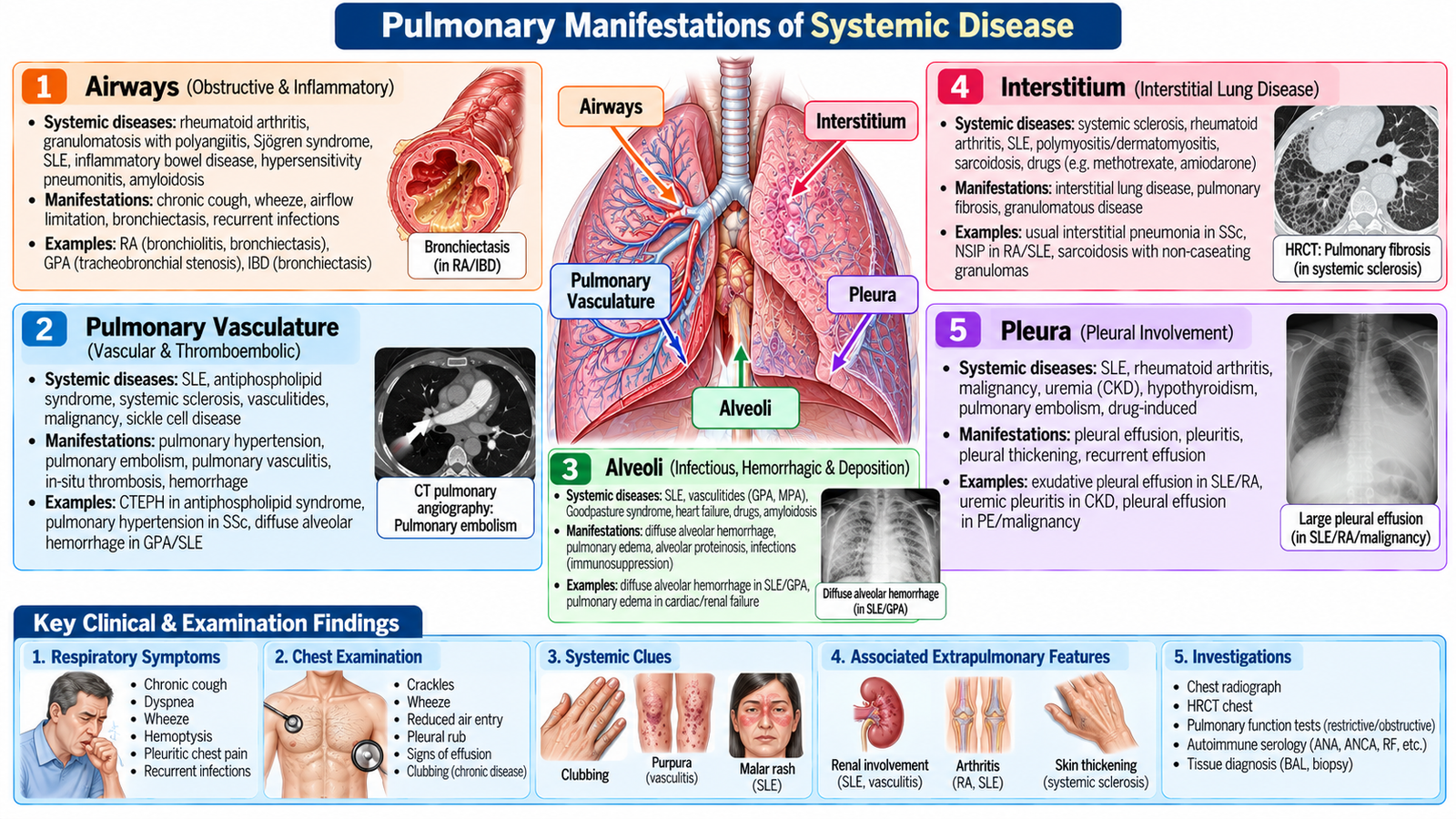
Systemic diseases can involve the pulmonary interstitium, pleura, pulmonary vessels, airways, respiratory muscles or alveoli. The same symptom—particularly dyspnea—can therefore arise through very different mechanisms. Identifying the anatomical compartment involved is the key to organized reasoning.
Major patterns
- Interstitial involvement → impaired gas transfer and reduced lung compliance → progressive exertional dyspnea and dry cough.
- Pleural involvement → pleuritis or pleural effusion → pleuritic pain and breathlessness.
- Pulmonary vascular involvement → pulmonary hypertension or vascular obstruction → exertional dyspnea, syncope or right-heart strain.
- Airway involvement → airflow limitation → cough, wheeze and breathlessness.
- Alveolar or capillary injury → impaired oxygenation and, in severe disease, acute respiratory compromise.
- Neuromuscular involvement → respiratory muscle weakness → reduced ventilation and possible ventilatory failure.
Systemic disorders and the lungs
Connective-tissue and autoimmune diseases are important causes of interstitial, pleural and pulmonary vascular abnormalities. Systemic inflammatory and vasculitic disorders may affect pulmonary vessels or produce alveolar injury. Neuromuscular systemic disease may compromise ventilation because the lungs themselves can be structurally normal while the respiratory pump becomes weak.
Treatment of systemic disease can itself contribute to pulmonary abnormalities, so medication and treatment history should be reviewed whenever a new respiratory problem develops.
Clinical clues
Important examination findings include tachypnea, hypoxemia, fine inspiratory crackles in interstitial disease, signs of pleural effusion, wheeze when airways are affected, and evidence of pulmonary hypertension or right-heart strain when the pulmonary circulation is involved.

G. Investigation, Prognosis & Management of Cardiorespiratory Involvement in Systemic Disease
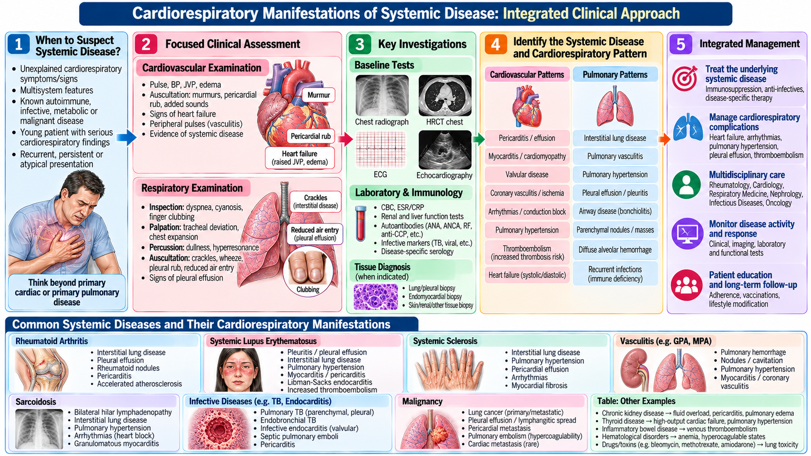
Investigation should be directed by the suspected organ compartment rather than by ordering every cardiac and respiratory test. The aim is to establish whether involvement is present, define its severity, identify complications and distinguish active systemic disease from another common cardiorespiratory condition.
Cardiovascular investigation
- ECG assesses rhythm, conduction and electrical evidence of cardiac involvement.
- Echocardiography evaluates ventricular function, valves, pericardial fluid and important hemodynamic consequences.
- Cardiac biomarkers may support evaluation of myocardial injury or cardiac stress when clinically indicated.
- Further imaging or specialist investigation is selected when initial assessment suggests a specific myocardial, vascular or structural problem.
Pulmonary investigation
- Chest radiography provides an initial structural overview and may reveal interstitial change, pleural disease or other abnormalities.
- CT chest provides greater anatomical definition when interstitial, pleural or other structural pulmonary disease is suspected.
- Pulmonary function testing helps define restrictive or obstructive physiological impairment and can support monitoring.
- Oxygenation assessment helps determine physiological severity when significant respiratory involvement is suspected.
- Additional investigations should target the suspected systemic disease and exclude important alternative explanations.
Management principles
Management has two linked targets: control the underlying systemic disease and treat the cardiorespiratory consequence. Depending on the condition, this may involve appropriate disease-modifying or immunomodulatory treatment under specialist guidance together with standard management of heart failure, pericardial disease, arrhythmia, pulmonary hypertension, interstitial lung disease, pleural disease or respiratory impairment.
Examples of disease-specific treatment: Autoimmune cardiac inflammation may require corticosteroids or other immunosuppressive therapy when indicated, alongside treatment of heart failure or pericarditis. Connective-tissue disease–associated interstitial lung disease may require mycophenolate mofetil, while progressive fibrotic disease may benefit from nintedanib under specialist care. Treatment should address the underlying systemic disorder and the affected organ.
Treatment-related toxicity must also be considered. A new cardiac or pulmonary abnormality during therapy should not automatically be attributed to progression of the systemic disease.
Prognosis and follow-up
Prognosis depends on the underlying disease, the organ involved, severity of functional impairment, reversibility, response to treatment and development of complications. Significant myocardial dysfunction, severe pulmonary vascular disease, progressive interstitial lung disease or respiratory failure generally indicates more serious organ involvement and requires close specialist care.
Systemic disease + organ injury means treatment must address both levels. Treating only breathlessness or edema without controlling the responsible systemic process leaves the driver of organ damage active.
Integrated Mechanism Flow
immune, inflammatory, metabolic, vascular or infiltrative process
heart, vessels, pleura, interstitium, airways or pulmonary circulation
pump failure, inflammation, impaired gas transfer or vascular dysfunction
dyspnea, chest pain, edema, cough, hypoxemia or arrhythmia
define compartment, mechanism and severity
control systemic disease + manage organ consequence
Important Comparison
| Feature | NSCLC | SCLC | Pleural Mesothelioma |
|---|---|---|---|
| Main origin | Bronchopulmonary epithelium | Pulmonary neuroendocrine lineage | Pleural mesothelium |
| Clinical behavior | Variable; localized disease may be surgically treatable | Aggressive with early dissemination | Diffuse pleural growth and local encasement |
| Important association | Smoking strongly associated with several subtypes | Strong smoking association | Previous asbestos exposure |
| Diagnostic emphasis | Tissue diagnosis + anatomical staging | Tissue diagnosis + assessment of extent | Pleural imaging + pleural tissue diagnosis |
| General treatment logic | Local and/or systemic therapy according to stage | Systemic therapy central; radiotherapy in selected settings | Multidisciplinary treatment + symptom control |
AIM High-Yield Review
- ⭐ Bronchogenic carcinoma is broadly divided into NSCLC and SCLC; this distinction influences staging and treatment strategy.
- Persistent cough, hemoptysis, unexplained weight loss, recurrent focal pneumonia or a persistent radiological abnormality should raise suspicion of lung malignancy.
- ⭐ Lung-cancer workup follows the logic detection → tissue diagnosis → staging → fitness assessment → treatment planning.
- Central lesions are commonly accessible bronchoscopically, whereas peripheral lesions may require image-guided sampling; an accessible metastatic site may sometimes provide the safest diagnostic tissue.
- Small-cell carcinoma is associated with important paraneoplastic endocrine manifestations such as SIADH and ectopic ACTH effects; squamous carcinoma may cause hypercalcemia through PTH-related peptide.
- Superior vena cava obstruction, major hemoptysis and acute neurological deterioration are serious complications of thoracic malignancy requiring urgent assessment.
- ⭐ Pleural mesothelioma should be considered when previous asbestos exposure is combined with progressive pleural disease, recurrent effusion or pleural thickening.
- Pleural fluid analysis can contribute to assessment of mesothelioma, but definitive diagnosis commonly requires adequate pleural tissue.
- Systemic disease can affect the myocardium, pericardium, valves, vessels or conduction system; the resulting symptom depends on which structure and function are disturbed.
- Pulmonary systemic-disease involvement may affect the interstitium, pleura, vessels, airways, alveoli or respiratory muscles; identifying the compartment narrows the investigation pathway.
- ⭐ Cardiorespiratory manifestations of systemic disease require dual management: treat the underlying systemic process and treat the resulting cardiac or pulmonary dysfunction.
- ECG and echocardiography are key initial tools for many suspected cardiac manifestations, while chest imaging and pulmonary function testing help characterize many pulmonary manifestations.
- Always consider treatment-related toxicity when a patient with systemic disease develops a new cardiac or respiratory abnormality.
- Prognosis is determined not simply by the diagnostic label but by the underlying systemic disease, organ involved, severity, reversibility, functional impairment and response to treatment.
Lung Malignancy & Cardiorespiratory Manifestations of Systemic Disease
Final Year MBBS • CVS Module