
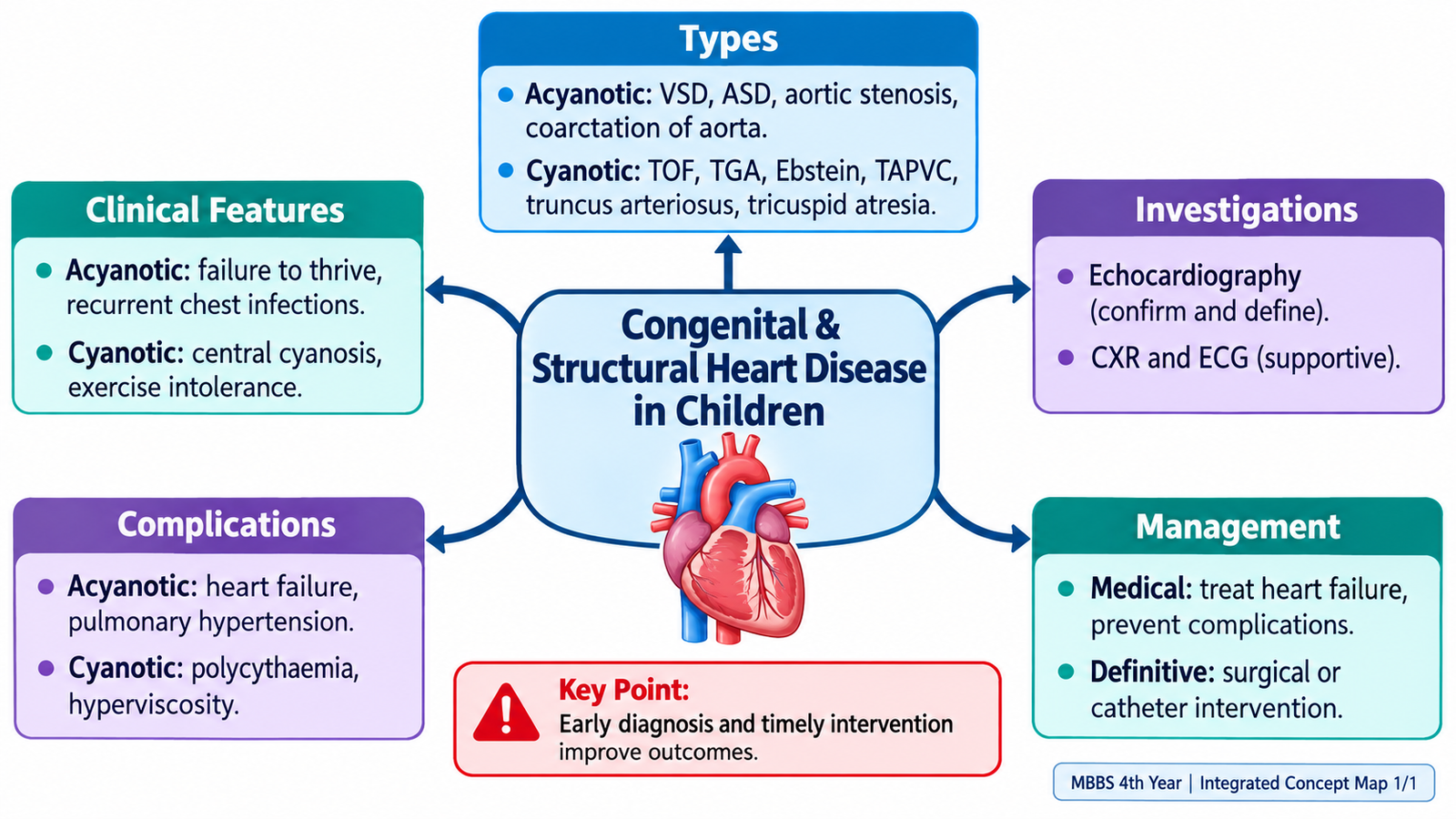
Do not memorize congenital heart diseases as separate lists. First decide whether the child is acyanotic or cyanotic, then connect the anatomical defect to the direction of blood flow, clinical signs, investigations and management.
Topic 5. Congenital & Structural Heart Disease in Children
CVS Module • Pediatrics & Medicine
Understand congenital heart disease by following the abnormal circulation: structural defect → altered blood flow → clinical presentation → diagnostic findings → management.
Topic Introduction
Congenital heart disease (CHD) is a structural abnormality of the heart or great vessels present from birth. For clinical learning, the most useful first division is into acyanotic and cyanotic disease. The key is not simply to remember the name of each defect, but to understand how the abnormal anatomy changes blood flow. Once that relationship is clear, findings such as murmurs, heart failure, cyanosis, pulse abnormalities and characteristic imaging become much easier to interpret.
1. Understanding Congenital Heart Disease Through Blood Flow
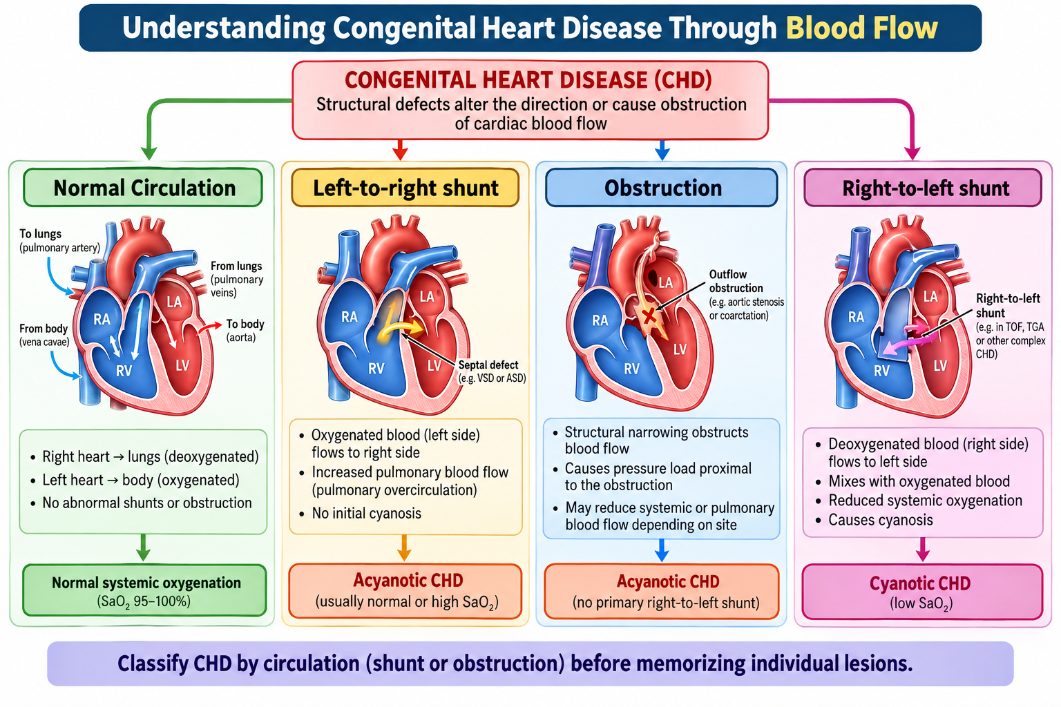
In the normal circulation, systemic venous blood reaches the right heart, passes through the lungs for oxygenation and returns through the left heart to the systemic circulation. A congenital structural defect may create an abnormal communication, obstruct blood flow or alter the normal connections between chambers and great vessels.
Acyanotic lesions in this topic include ventricular septal defect (VSD), atrial septal defect (ASD), aortic stenosis and coarctation of the aorta. VSD and ASD usually produce a left-to-right shunt, whereas aortic stenosis and coarctation are primarily obstructive lesions.
Cyanotic lesions occur when systemic arterial oxygenation is reduced because deoxygenated blood reaches the systemic circulation or pulmonary and systemic blood streams do not mix adequately. The major lesions required here are Tetralogy of Fallot, transposition of the great arteries, Ebstein anomaly, total anomalous pulmonary venous connection, truncus arteriosus and tricuspid atresia.
2. Acyanotic Congenital Heart Disease: VSD & ASD
Ventricular Septal Defect
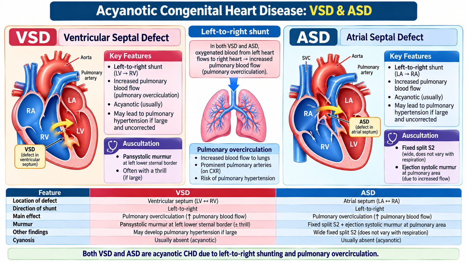
A VSD is an abnormal communication between the right and left ventricles. After birth, left ventricular pressure is normally higher than right ventricular pressure, so blood generally moves:
The clinical effect depends greatly on the size of the defect and pulmonary vascular resistance. A small VSD may cause little hemodynamic disturbance but can produce a prominent murmur because blood moves rapidly through the small opening. A large VSD permits substantial left-to-right flow, increasing pulmonary blood flow and eventually producing volume overload of the heart.
How does a child with VSD present?
A small defect may be discovered because of a murmur in an otherwise well child. A significant defect may present in infancy with poor feeding, sweating during feeds, tachypnea, poor weight gain and recurrent respiratory symptoms. These findings reflect the increased workload of the heart and pulmonary overcirculation.
A characteristic finding is a pansystolic murmur, classically prominent at the lower left sternal border. Large shunts may produce signs of heart failure.
Diagnostic approach
Echocardiography with Doppler defines the defect, demonstrates the direction and importance of shunting and assesses cardiac chamber effects. Chest radiography and ECG can provide supportive evidence of pulmonary overcirculation or chamber enlargement when the defect is significant.
Management logic
Management depends on hemodynamic significance. Small uncomplicated defects may require observation because some close spontaneously. When a large VSD causes heart failure, medical therapy controls symptoms while the child is assessed for definitive closure. Persistent important shunting, failure to thrive or complications may require catheter-based or surgical intervention according to the anatomy.
Symptomatic VSD: Infants with heart failure may require furosemide and nutritional support while awaiting definitive closure. Large defects causing persistent heart failure, failure to thrive or significant pulmonary overcirculation require early pediatric cardiology assessment for surgical repair.
Atrial Septal Defect
An ASD creates communication between the atria. Because left atrial pressure is normally greater than right atrial pressure, blood usually flows from the left atrium to the right atrium. This produces chronic volume loading of the right heart and increased pulmonary blood flow.
Many children remain asymptomatic for years. On examination, a classic clue is a wide, fixed splitting of the second heart sound. A systolic flow murmur may be heard because of increased flow across the pulmonary valve rather than because blood passing through the ASD itself produces a loud murmur.
Echocardiography establishes the diagnosis, defines the type and size of the defect and assesses right-heart volume loading. Significant suitable defects may be closed by a catheter device; other anatomies require surgical closure.

3. Obstructive Acyanotic Lesions: Aortic Stenosis & Coarctation
Aortic Stenosis
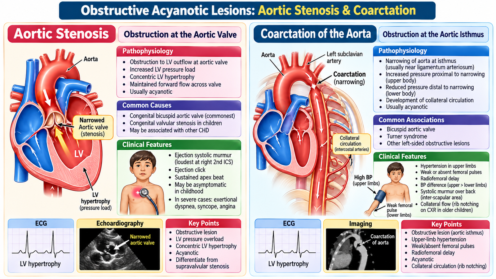
Congenital aortic stenosis obstructs left ventricular outflow. The left ventricle must generate higher pressure to eject blood across the narrowed valve or outflow region. Persistent pressure overload produces left ventricular hypertrophy.
Mild disease may be asymptomatic. More significant obstruction can cause exertional symptoms, reduced exercise tolerance, chest discomfort or syncope in older children. Severe disease in infancy can produce poor systemic perfusion and heart failure.
A typical examination finding is an ejection systolic murmur over the aortic area with radiation toward the neck. Echocardiography defines the anatomy and assesses severity. Management depends on severity and may include catheter-based or surgical relief of the obstruction.
Coarctation of the Aorta
Coarctation is a localized narrowing of the aorta, commonly involving the region near the ductus arteriosus. The narrowing creates increased resistance to blood leaving the left ventricle and reduces flow to the lower part of the body.
Older children or adults may be detected because of hypertension. Important examination clues include upper-limb hypertension, weak femoral pulses and radiofemoral delay. Blood pressure should therefore be assessed in the upper and lower limbs when coarctation is suspected.
Severe coarctation can present in early infancy when ductal flow falls, causing poor feeding, tachypnea, weak peripheral perfusion, metabolic deterioration or heart failure. This is a potentially critical presentation requiring urgent specialist management.
Investigations and management
Echocardiography is central in children. ECG may demonstrate effects of ventricular pressure load, while chest imaging and advanced anatomical imaging may be used when needed to define the lesion and plan intervention. Definitive treatment relieves the obstruction using an appropriate catheter-based or surgical approach. Long-term follow-up remains important because hypertension can persist or recur even after anatomical correction.
Critical neonatal coarctation: A neonate presenting with shock or poor systemic perfusion may require IV prostaglandin E1 (alprostadil) to reopen or maintain the ductus arteriosus until urgent surgical treatment. Older children with significant coarctation may undergo surgical repair or balloon angioplasty/stenting according to age and anatomy. Persistent hypertension requires monitoring and treatment.

4. Recognizing the Cyanotic Infant or Child
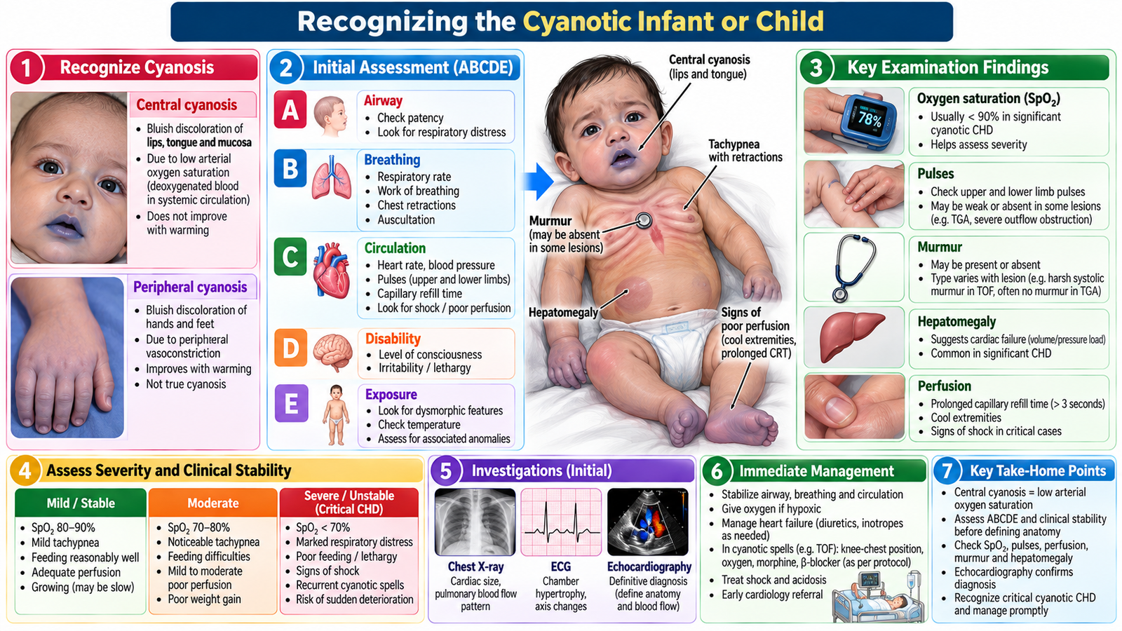
Cyanotic congenital heart disease should be considered when a neonate or child has persistent central cyanosis that cannot be explained by a simple respiratory problem. The underlying cardiovascular problem is inadequate delivery of oxygenated blood to the systemic circulation.
Clinical presentation
The age and severity of presentation vary with the lesion. Important presentations include cyanosis from birth, increasing cyanosis, poor feeding, tachypnea, poor weight gain, exercise limitation, episodic worsening of cyanosis or signs of heart failure. Long-standing cyanosis may eventually produce digital clubbing.
Focused physical examination
In a neonate or infant, examination aims to establish both the severity of illness and clues to the cardiac anatomy. Important components include:
- General appearance, feeding ability and respiratory effort.
- Central cyanosis and oxygen saturation.
- Pulse rate, character and symmetry.
- Peripheral perfusion and capillary refill.
- Precordial activity and cardiac impulse.
- Heart sounds and murmurs.
- Signs of heart failure such as tachypnea and hepatomegaly.
A murmur may provide an important anatomical clue, but absence of a loud murmur does not exclude severe congenital heart disease. Some critical lesions present predominantly with profound cyanosis or circulatory instability.
Diagnostic workup
Pulse oximetry identifies hypoxemia but does not define the lesion. Chest radiography can show pulmonary vascularity, heart size and characteristic cardiac contours. ECG may demonstrate chamber hypertrophy or conduction patterns. Echocardiography with Doppler is the central diagnostic investigation because it demonstrates cardiac anatomy, connections and blood flow.
Emergency stabilization: In a critically cyanosed or poorly perfused neonate with suspected duct-dependent congenital heart disease, start IV prostaglandin E1 (alprostadil) under neonatal intensive-care supervision while arranging urgent echocardiography and pediatric cardiology referral. Monitor oxygenation, circulation and respiration because prostaglandin infusion may cause apnea.
Parent counselling
Parents need a simple explanation of the structural problem, why cyanosis occurs, what investigations are required and whether catheter or surgical treatment may be necessary. They should understand feeding and growth concerns, medication adherence where prescribed, the need for follow-up and the warning signs requiring urgent medical review. Communication should remain clear and supportive without giving unrealistic reassurance or unnecessarily alarming the family.
An infant has poor feeding, sweating and tachypnea. Examination shows a prominent pansystolic murmur and signs of heart failure. Another child has central cyanosis with a systolic murmur and becomes more cyanosed during crying.
- The first pattern suggests significant pulmonary overcirculation from an acyanotic shunt such as VSD.
- The second pattern directs attention toward cyanotic CHD, particularly a lesion with reduced pulmonary flow such as Tetralogy of Fallot.
- In both children, echocardiography connects the clinical pattern to the underlying anatomy.
5. Tetralogy of Fallot & Transposition of the Great Arteries
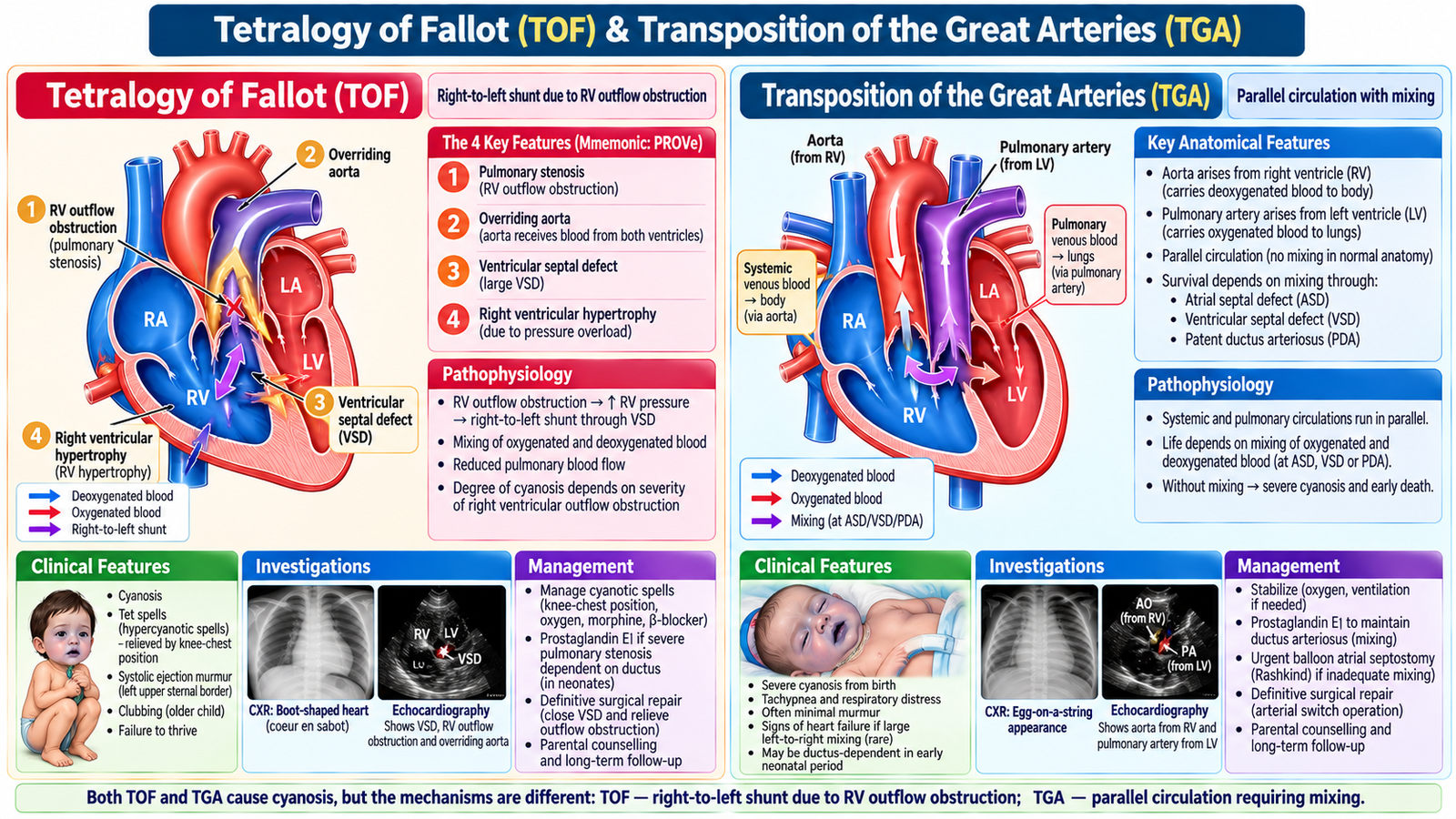
Tetralogy of Fallot
Tetralogy of Fallot (TOF) combines four anatomical abnormalities:
The central physiological problem is right ventricular outflow obstruction. When resistance to pulmonary flow is high, blood crosses the VSD from right to left and enters the overriding aorta. More deoxygenated blood therefore reaches the systemic circulation, causing cyanosis.
Children may have cyanosis, a systolic murmur and reduced exercise tolerance. Some develop episodes of sudden worsening cyanosis, known as hypercyanotic or “tet” spells. Crying, feeding or exertion may precipitate these episodes because right-to-left shunting increases.
Older children may instinctively squat after exertion. Increased systemic vascular resistance reduces right-to-left shunting and can improve pulmonary blood flow and systemic oxygenation.
Chest radiography may show the classic boot-shaped cardiac silhouette in established TOF, while echocardiography demonstrates the defining anatomy. Definitive management is surgical repair; unstable hypercyanotic episodes require urgent supportive management while specialist treatment is arranged.
Management of a hypercyanotic (tet) spell: Place the child in the knee-chest position, keep the child calm, administer oxygen and establish IV access. Correct hypovolemia with cautious IV fluids. Persistent spells may require morphine, propranolol or phenylephrine under monitored specialist care. Arrange urgent pediatric cardiology assessment and definitive surgical repair.
Transposition of the Great Arteries
In transposition of the great arteries (TGA), the great arteries arise from the wrong ventricles: the aorta arises from the right ventricle and the pulmonary artery from the left ventricle. This creates two circulations running in parallel rather than the normal series circulation.
Survival therefore depends on mixing between the two circulations through an atrial communication, ventricular communication or patent ductus arteriosus. TGA typically presents with marked cyanosis early in life. Echocardiography confirms the abnormal ventricular–arterial connections.
Initial management aims to maintain adequate oxygen delivery and sufficient mixing while definitive repair is arranged. In a critically cyanosed neonate, maintaining ductal patency may be essential temporarily. Definitive treatment restores the appropriate relationship between systemic and pulmonary circulation
Specific treatment of TGA: IV prostaglandin E1 helps maintain ductal patency and improve mixing in selected neonates. If interatrial mixing is inadequate, urgent balloon atrial septostomy (Rashkind procedure) may be lifesaving. Definitive treatment for uncomplicated d-TGA is the arterial switch operation, usually performed in the neonatal period.
6. Other Major Cyanotic Congenital Heart Lesions
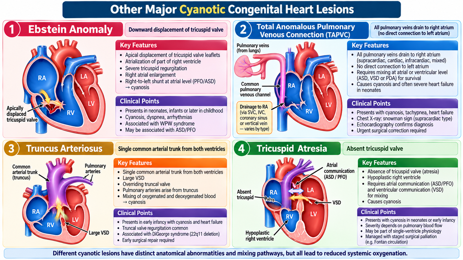
Ebstein Anomaly
Ebstein anomaly involves abnormal downward displacement of the tricuspid valve into the right ventricle. Part of the right ventricle becomes functionally incorporated into the atrium, producing a large right atrium and significant tricuspid regurgitation. When right-sided pressure promotes right-to-left flow across an atrial communication, cyanosis can occur.
Presentation ranges from severe neonatal cyanosis to later symptoms such as exercise limitation, palpitations or signs of right-sided volume overload. Echocardiography demonstrates the abnormal tricuspid valve position and associated hemodynamic effects.
Total Anomalous Pulmonary Venous Connection
In total anomalous pulmonary venous connection (TAPVC), the pulmonary veins do not connect normally to the left atrium. Pulmonary venous blood instead returns to the systemic venous circulation or right side of the heart.
An interatrial communication is therefore necessary for oxygenated blood to reach the left heart and systemic circulation. The child has mixing of oxygenated and deoxygenated blood, producing cyanosis. If the anomalous pulmonary venous pathway is obstructed, severe respiratory distress and cyanosis can develop rapidly. Echocardiography identifies the abnormal venous connection, and definitive treatment is surgical correction.
Truncus Arteriosus
In truncus arteriosus, a single arterial trunk leaves the heart and supplies the systemic, pulmonary and coronary circulations. A VSD is present beneath the common trunk, allowing ventricular blood to mix.
Because both pulmonary and systemic circulations receive mixed blood, cyanosis occurs. Pulmonary blood flow may be excessive, so infants can also develop heart failure and poor growth. Echocardiography demonstrates the common arterial trunk and associated anatomy. Definitive management requires surgical repair.
Tricuspid Atresia
Tricuspid atresia means there is no effective direct connection between the right atrium and right ventricle through the tricuspid valve. Systemic venous blood must therefore cross an atrial communication to reach the left side of the heart.
Additional communication is required to permit pulmonary blood flow. The physiological result is mixing and cyanosis, with severity influenced by the amount of pulmonary blood flow. Echocardiography confirms the absent tricuspid connection and associated defects. Management requires specialist staged intervention appropriate to the anatomy and circulation.
7. From Presentation to Diagnosis, Management & Family Care
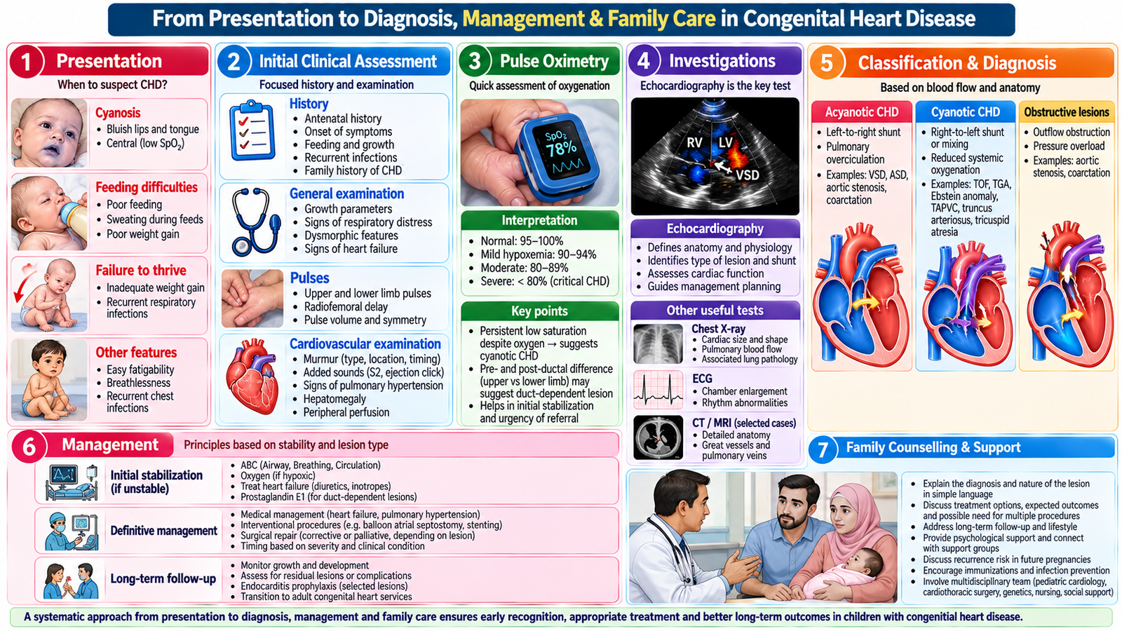
Congenital heart disease becomes easier clinically when assessment follows a consistent reasoning pathway rather than beginning with a long list of named defects.
History: identify the physiological problem
In infants, ask about feeding difficulty, sweating during feeds, rapid breathing, poor weight gain, cyanosis and recurrent respiratory symptoms. In older children, ask about exercise tolerance, breathlessness, chest symptoms, syncope, palpitations and cyanosis. The timing of symptoms is important because severe structural lesions often present early.
Examination: connect signs to circulation
Assess growth, respiratory effort, central cyanosis, pulses, blood pressure where relevant, precordial activity, heart sounds and murmurs. Look for evidence of heart failure or poor systemic perfusion. Pulse differences are particularly important in coarctation, while persistent cyanosis directs attention toward cyanotic CHD.
Investigations: answer specific questions
- Pulse oximetry: Is significant hypoxemia present?
- ECG: Is there chamber hypertrophy, strain or an associated rhythm abnormality?
- Chest radiograph: Is the heart enlarged? Is pulmonary blood flow increased or reduced? Is there a characteristic contour?
- Echocardiography with Doppler: What is the structural defect, direction of blood flow and hemodynamic effect?
- Further anatomical assessment: Selected when detailed anatomy is required for intervention planning.
Management: treat physiology and correct anatomy when needed
Management depends on the lesion and its physiological consequences. The immediate priority may be treatment of heart failure, hypoxemia or poor perfusion. Some critical neonatal lesions require temporary maintenance of ductal flow until definitive intervention. Significant structural abnormalities may require catheter-based procedures or surgery. The aim is not simply to “close a hole” or “repair a valve”; it is to restore a circulation that provides adequate systemic oxygen delivery without harmful pressure or volume loading.
Follow-up and parent counselling
Families should understand the child’s diagnosis in simple terms, the reason for symptoms, planned investigations and treatment, and the importance of follow-up. Discuss feeding and growth where relevant, medication adherence, activity advice appropriate to the child’s condition and warning signs such as worsening cyanosis, breathing difficulty, poor feeding, syncope or marked deterioration. Children who have undergone intervention may still require long-term cardiovascular follow-up.
Integrated Mechanism Flow
Important Comparison
| Lesion | Main physiological problem | Key clinical clue | Core diagnostic idea |
|---|---|---|---|
| VSD | Left-to-right ventricular shunt | Pansystolic murmur; heart failure if large | Echo shows defect and shunt |
| ASD | Left-to-right atrial shunt | Fixed wide split S2 | Echo shows ASD and right-heart loading |
| Aortic stenosis | LV outflow obstruction | Ejection systolic murmur | Echo assesses obstruction |
| Coarctation | Aortic obstruction | Arm hypertension + weak femoral pulses | Echo/anatomical imaging defines narrowing |
| TOF | Reduced pulmonary flow + right-to-left shunt | Cyanosis ± hypercyanotic spells | Echo demonstrates four-part anatomy |
| TGA | Parallel circulations | Severe early cyanosis | Echo shows ventriculoarterial discordance |
AIM High-Yield Review
- ⭐ VSD and ASD usually begin as left-to-right shunts; cyanosis is not expected initially.
- A large VSD → pulmonary overcirculation → infant heart-failure symptoms such as tachypnea, sweating during feeding and poor weight gain.
- VSD → pansystolic murmur; ASD → fixed wide splitting of S2.
- Long-standing major left-to-right shunting can produce pulmonary vascular disease and eventual Eisenmenger physiology.
- ⭐ Arm hypertension + weak/delayed femoral pulses → think coarctation of the aorta.
- Aortic stenosis produces LV pressure overload → LV hypertrophy and an ejection systolic murmur.
- ⭐ TOF: VSD + RV outflow obstruction + overriding aorta + RV hypertrophy; increasing right-to-left shunting produces cyanosis.
- A hypercyanotic spell in TOF represents an acute increase in right-to-left shunting and reduced pulmonary blood flow.
- ⭐ TGA creates parallel circulations; survival initially depends on communication and mixing between them.
- TAPVC requires an atrial communication for systemic output; obstruction of pulmonary venous drainage can cause severe early deterioration.
- Truncus arteriosus → one common arterial trunk + mixing, often with increased pulmonary flow and heart failure.
- Tricuspid atresia → absent effective right atrioventricular connection; blood must cross an atrial communication.
- Ebstein anomaly primarily affects the tricuspid valve and may cause marked tricuspid regurgitation with cyanosis when right-to-left atrial shunting occurs.
- Echocardiography with Doppler is central to defining anatomy, blood-flow direction and hemodynamic consequences in congenital heart disease.
- A severely cyanosed or poorly perfused neonate with suspected CHD requires stabilization and urgent specialist assessment rather than waiting for a precise bedside anatomical diagnosis.
Congenital & Structural Heart Disease in Children
Review the major acyanotic and cyanotic congenital heart lesions, their altered circulation, clinical presentation and diagnostic approach.