
Learn this topic as a connected clinical pathway: pump dysfunction → reduced cardiac output/congestion → symptoms and signs → investigations → cause-directed and syndrome-directed management. Then compare heart failure with cardiomyopathy, myocarditis and pericardial disease.
Topic 3. Heart Failure, Cardiomyopathy, Myocarditis & Pericardial Disease Across the Lifespan
CVS Module • A clinically integrated approach to myocardial dysfunction, congestion, cardiomyopathy, inflammation and pericardial disease in adults and children.
Topic Introduction
Heart failure is a clinical syndrome in which the heart cannot provide adequate circulation without abnormal filling pressures. It may result from impaired contraction, impaired relaxation, structural heart disease or an important rhythm or loading abnormality. Cardiomyopathies and myocarditis can directly damage myocardial function, while pericardial disease affects the membrane surrounding the heart and may impair cardiac filling when fluid accumulates under pressure. In children, the underlying causes and presentation may differ from adults, so feeding, growth, respiratory symptoms and age-appropriate examination become especially important. This chapter connects these conditions through their mechanisms, clinical recognition, investigation and management.
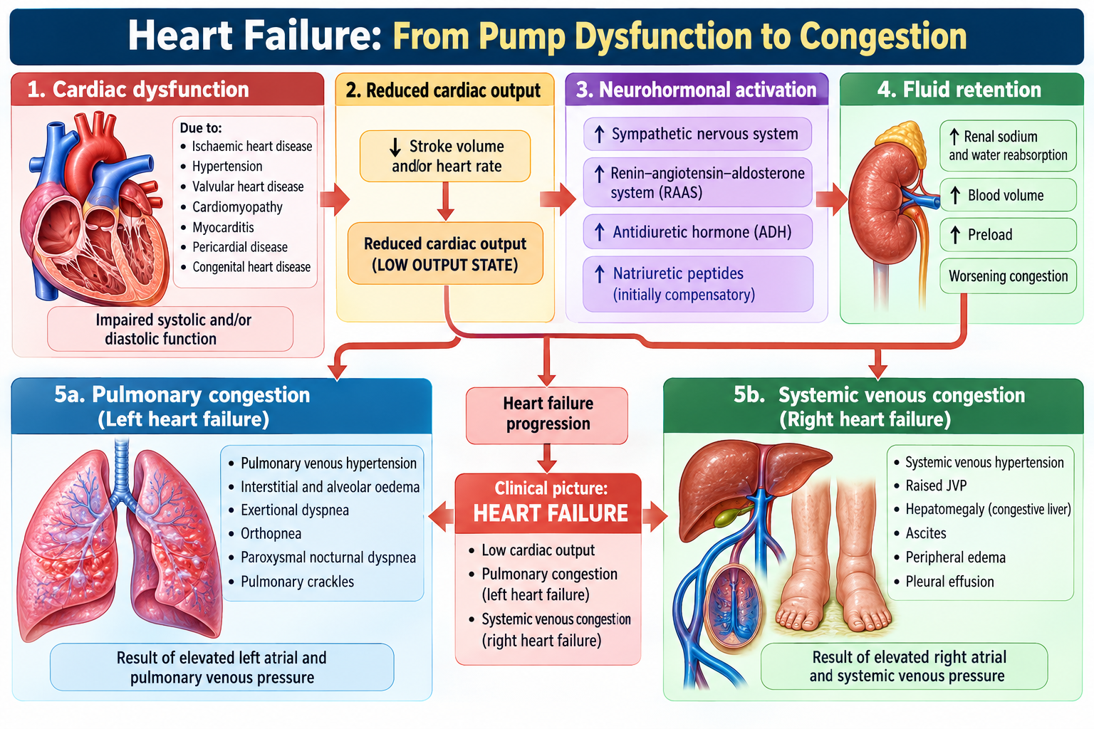
1. Heart Failure: From Pump Dysfunction to Congestion
Heart failure is not a single disease. It is the final clinical syndrome produced when cardiac structure or function becomes abnormal enough to impair effective circulation. Understanding the relationship between cardiac output, ventricular filling and congestion makes the symptoms, signs and treatment much easier to understand.
Types and functional patterns
Heart failure may involve predominantly the left ventricle, right ventricle or both sides. It may present acutely or develop chronically. Another clinically useful distinction is between failure associated with reduced ventricular systolic function and failure in which ventricular contraction is relatively preserved but filling and relaxation are abnormal.
| Pattern | Main disturbance | Clinical consequence |
|---|---|---|
| Left-sided failure | Raised left-sided filling pressure | Pulmonary congestion → dyspnea, orthopnea, crackles |
| Right-sided failure | Raised systemic venous pressure | Peripheral edema, raised JVP, hepatic congestion |
| Reduced systolic function | Impaired ventricular contraction | Reduced forward output with secondary congestion |
| Preserved systolic function | Impaired relaxation/compliance | Raised filling pressure despite relatively preserved contraction |
Why heart failure progresses
A fall in effective cardiac output activates compensatory responses. Sympathetic activation initially supports heart rate and contractility, while renal sodium and water retention attempts to preserve circulating volume. These responses may help temporarily, but persistent vasoconstriction, fluid retention and ventricular stress increase cardiac workload and promote further congestion and remodeling.
Common etiological groups include ischemic heart disease, long-standing hypertension, valvular disease, cardiomyopathy, myocarditis and rhythm disorders. Identifying the underlying cause matters because management must address both the heart-failure syndrome and the disease producing it.
2. Clinical Recognition, Assessment & Investigation of Heart Failure
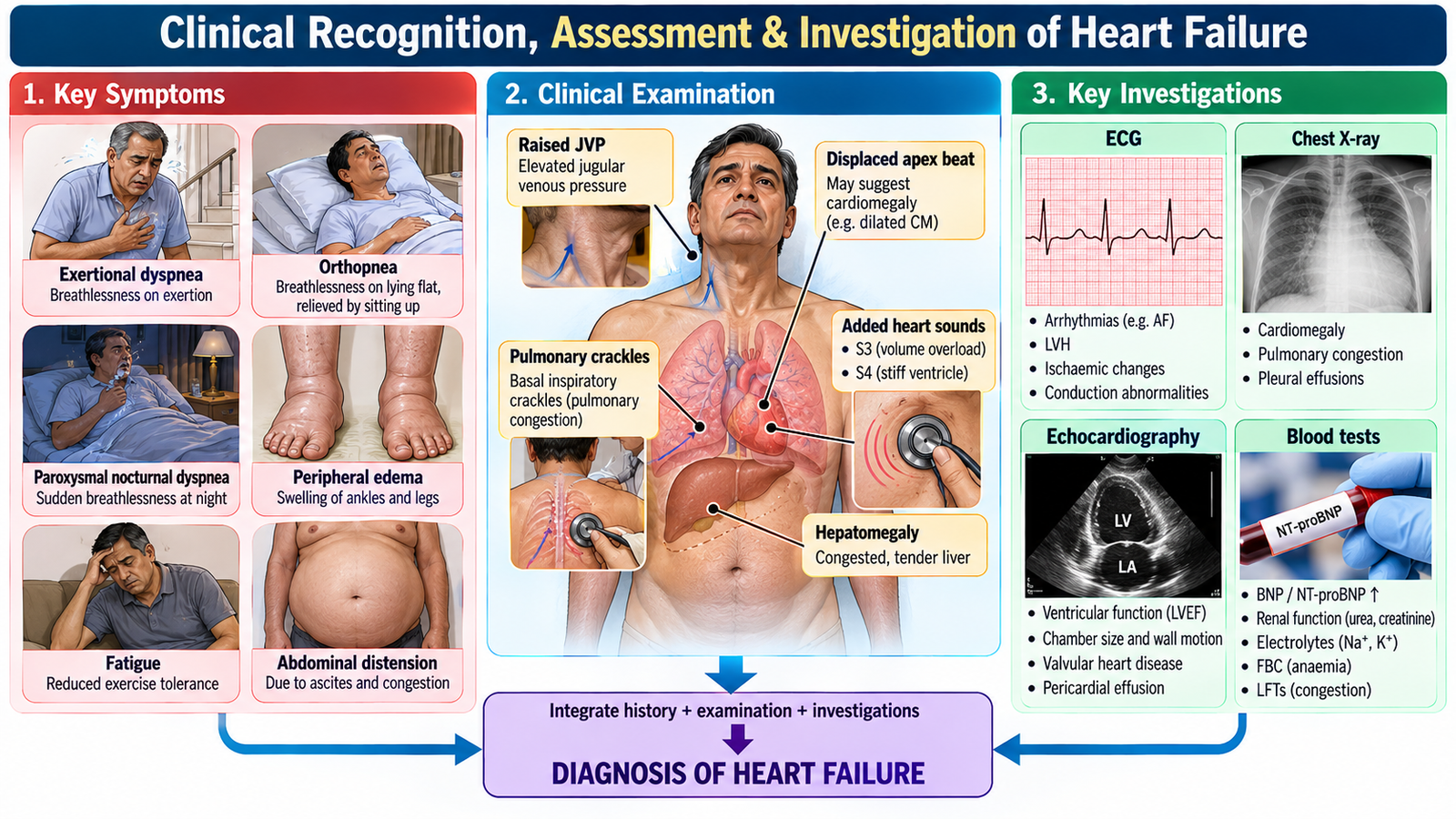
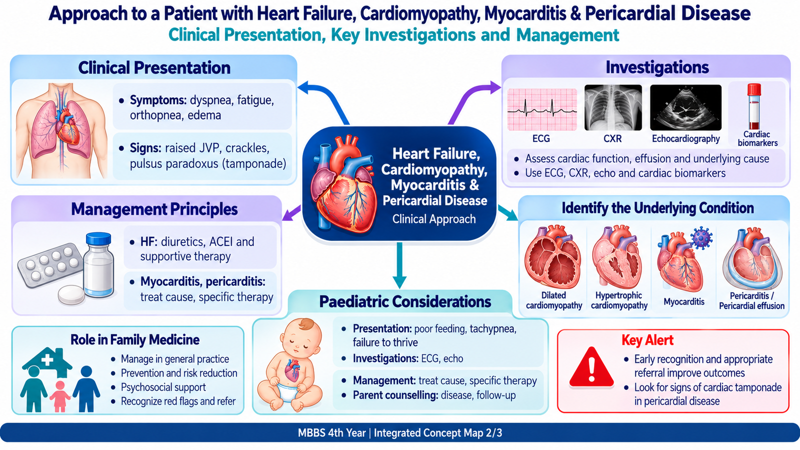
Heart failure is recognized by combining the symptom pattern, evidence of congestion or impaired perfusion, and objective assessment of cardiac structure and function. No single symptom should be interpreted in isolation.
Symptoms and focused history
- Exertional dyspnea develops as pulmonary venous pressure rises and exercise reserve falls.
- Orthopnea and paroxysmal nocturnal dyspnea suggest pulmonary congestion.
- Fatigue and reduced exercise tolerance reflect impaired forward cardiac output.
- Ankle swelling and abdominal fullness suggest systemic venous congestion.
- Ask about ischemic symptoms, hypertension, valvular disease, arrhythmia, previous cardiac disease and factors that may precipitate deterioration.
Focused examination
Assess pulse, blood pressure, oxygenation and general perfusion first. Look for raised jugular venous pressure, peripheral edema and other evidence of systemic congestion. Respiratory crackles support pulmonary congestion. Cardiac examination may reveal tachycardia, displaced apex beat, additional heart sounds or a murmur pointing toward an underlying structural lesion.
Investigations: ask what each test contributes
- ECG: looks for ischemia, previous infarction, rhythm disturbance and conduction abnormalities.
- Chest radiograph: may demonstrate cardiomegaly, pulmonary vascular congestion or pulmonary edema and may suggest an alternative pulmonary cause of dyspnea.
- Natriuretic peptides: support assessment when heart failure is suspected; elevation reflects myocardial wall stress but must be interpreted in clinical context.
- Echocardiography: is central for ventricular function, chamber size, wall motion, valve disease and pericardial assessment.
- Basic laboratory evaluation: helps identify anemia, renal dysfunction, electrolyte disturbance and other clinically relevant contributors or consequences.
Prognosis varies with underlying cause, ventricular dysfunction, symptom burden, recurrent decompensation, comorbidity and response to treatment. Repeated hospitalization or progressive functional limitation suggests more advanced disease.
3. Management, Prevention & Long-Term Care of Heart Failure
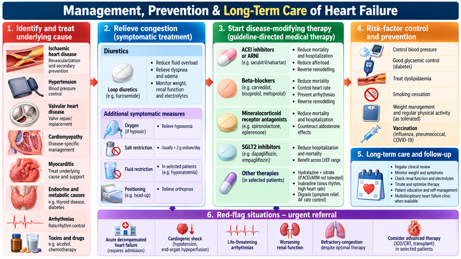
Management has three connected goals: relieve congestion and symptoms, improve cardiac outcomes, and treat the underlying or precipitating cause. The exact plan depends on the type and severity of heart failure and the patient’s associated disease.
Treat congestion and stabilize deterioration
Diuretics reduce excess sodium and water and therefore relieve pulmonary and peripheral congestion. In acute deterioration, management also requires assessment of oxygenation, circulation, precipitating factors and the need for urgent hospital-level care.
Acute decompensated heart failure: Give IV furosemide for significant fluid overload and oxygen if hypoxemic; use non-invasive ventilation for severe pulmonary edema when indicated. IV nitrates may help hypertensive pulmonary edema if blood pressure permits. Cardiogenic shock requires urgent intensive-care management and possible inotropic support.
Modify the disease process
In appropriate patients with chronic heart failure and reduced systolic function, evidence-based therapy targets maladaptive neurohormonal pathways and reduces disease progression. Treatment is individualized according to ventricular function, blood pressure, renal function, electrolytes and comorbidity. Ischemia, hypertension, valve disease and important arrhythmias should be addressed when present.
Chronic heart failure treatment: HFrEF (LVEF ≤40%) requires four disease-modifying drug classes: sacubitril/valsartan (or an ACE inhibitor/ARB), an evidence-based beta-blocker (bisoprolol, carvedilol or metoprolol succinate), spironolactone/eplerenone, and dapagliflozin/empagliflozin. Furosemide relieves congestion when needed. HFpEF is managed with diuretics for congestion, an SGLT2 inhibitor and treatment of associated conditions. Monitor blood pressure, renal function and potassium during therapy.
Patient-centred self-care
Students should understand the purpose of counselling rather than memorize a script. Patients need to understand their diagnosis, medication adherence, lifestyle and cardiovascular risk reduction, follow-up, and symptoms suggesting deterioration. Prevention of ischemic heart disease and good control of cardiovascular risk factors also reduce the development and progression of heart failure.
Family Medicine perspective
Primary care supports long-term medication adherence, risk-factor management, recognition of deterioration and timely referral. Chronic cardiac disease can reduce work capacity, independence and quality of life and can create financial and emotional strain for both patients and families. These psychosocial effects should be actively recognized rather than treated as separate from medical care.
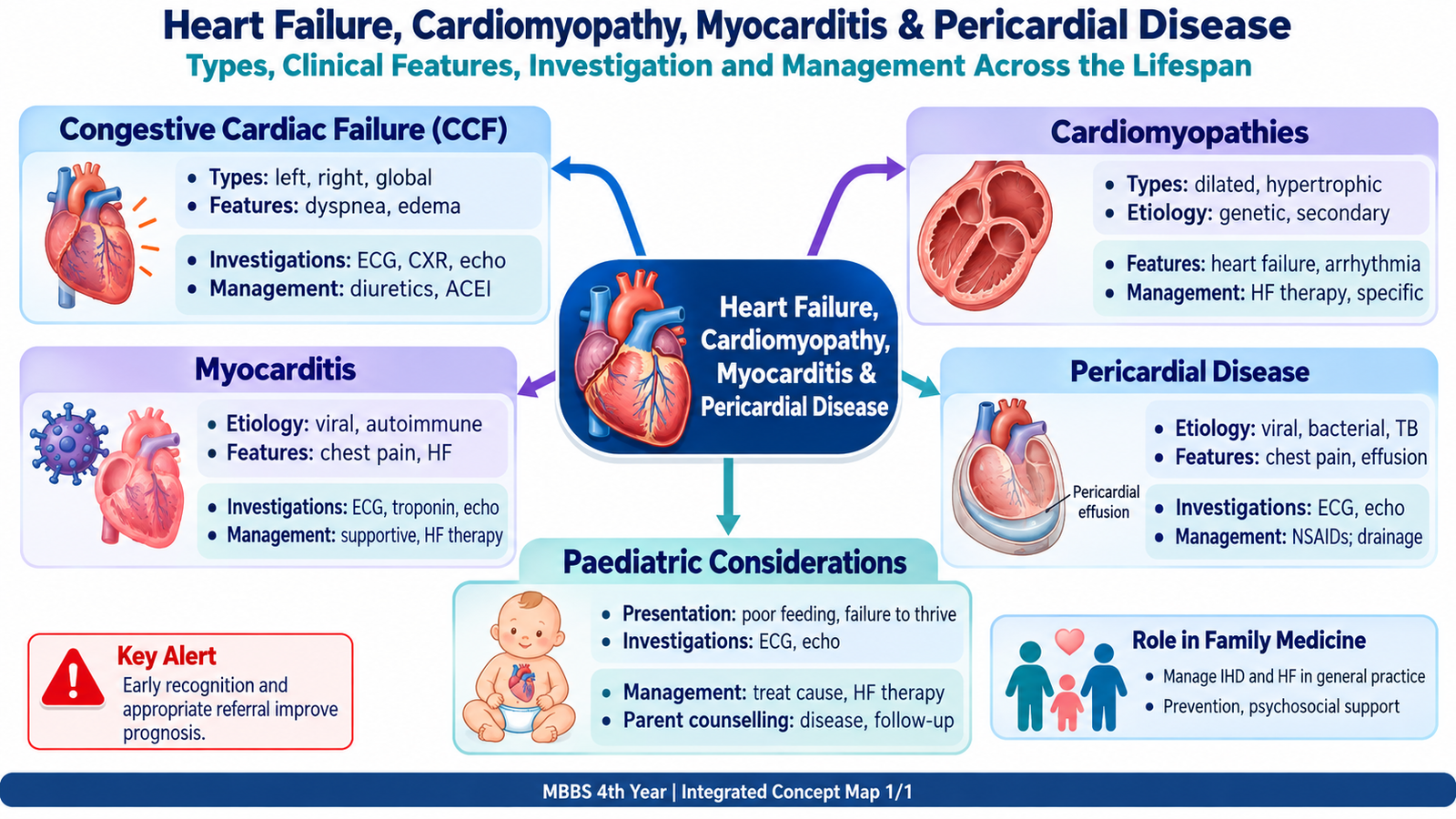
An older adult with known ischemic heart disease develops progressive exertional dyspnea, orthopnea and ankle swelling. Examination shows elevated JVP, bilateral basal crackles and pitting edema. The findings should be connected rather than memorized separately: impaired cardiac function has produced both pulmonary and systemic congestion.
- Which findings indicate pulmonary versus systemic venous congestion?
- Which investigation best defines cardiac structure and ventricular function?
- Which underlying and precipitating causes should be sought?
4. Cardiomyopathies: Recognizing the Major Patterns
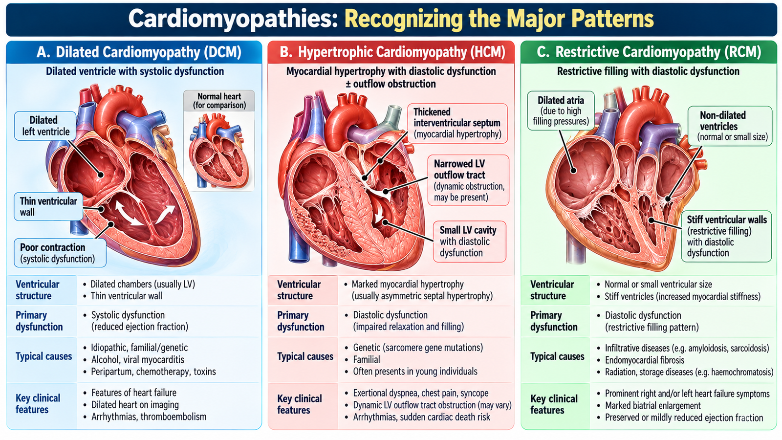
Cardiomyopathies are disorders in which abnormal myocardial structure or function cannot be explained simply by another obvious loading condition. For undergraduate clinical learning, the important task is to recognize the major patterns and understand how each alters ventricular function.
| Type | Structural/functional pattern | Important clinical consequence |
|---|---|---|
| Dilated | Ventricular dilatation with impaired systolic contraction | Heart failure, functional valve regurgitation and arrhythmia may occur |
| Hypertrophic | Abnormal myocardial hypertrophy with impaired relaxation; outflow obstruction may occur | Exertional symptoms, arrhythmia and risk of serious cardiac events |
| Restrictive | Stiff ventricles with impaired diastolic filling | Raised filling pressures and congestive features |
Evaluation is guided by history, family history, physical findings, ECG and cardiac imaging, especially echocardiography. Management depends on the cardiomyopathy phenotype and its consequences, including heart failure, arrhythmia and obstruction where relevant.
5. Myocarditis: Inflammation of the Myocardium
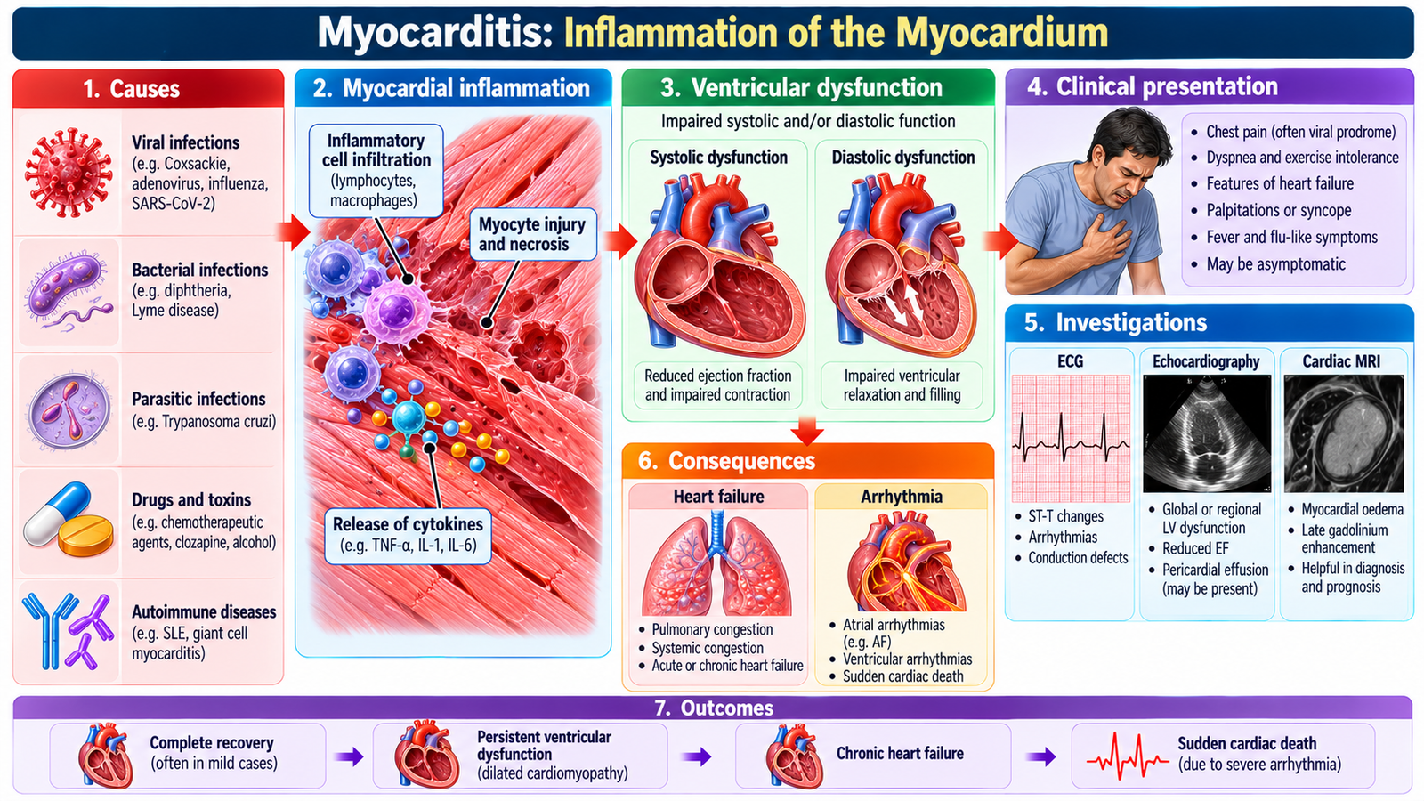
Myocarditis is inflammation of the heart muscle. The clinically important link is myocardial inflammation → myocyte dysfunction → impaired contraction and/or electrical instability. The presentation therefore ranges from mild symptoms to heart failure, arrhythmia or severe circulatory compromise.
Etiology and presentation
Causes include infections and non-infectious inflammatory or toxic processes. A preceding infective-type illness may be present, but it is not essential. Patients may develop chest discomfort, dyspnea, fatigue, palpitations or features of heart failure. Tachycardia or rhythm disturbance may accompany myocardial dysfunction.
Diagnostic reasoning
Suspect myocarditis when new cardiac symptoms or unexplained ventricular dysfunction occur in a compatible clinical setting. ECG abnormalities may be nonspecific or show rhythm disturbance. Cardiac biomarkers may rise when myocardial injury is present, while echocardiography evaluates ventricular function and complications. Other investigations are selected according to severity and suspected cause.
Management principle
Treatment is mainly directed toward the clinical consequences and the identified cause: manage heart failure when present, recognize and treat important arrhythmias, avoid unnecessary cardiac stress during active illness and urgently escalate patients with hemodynamic instability or severe ventricular dysfunction.
Treatment: Most uncomplicated viral myocarditis is managed supportively; antibiotics or immunosuppression are reserved for specific identified causes. Treat associated heart failure with appropriate therapy and monitor significant arrhythmias. Avoid strenuous exercise during active myocarditis and resume activity only after clinical reassessment.
6. Pericarditis & Pericardial Effusion
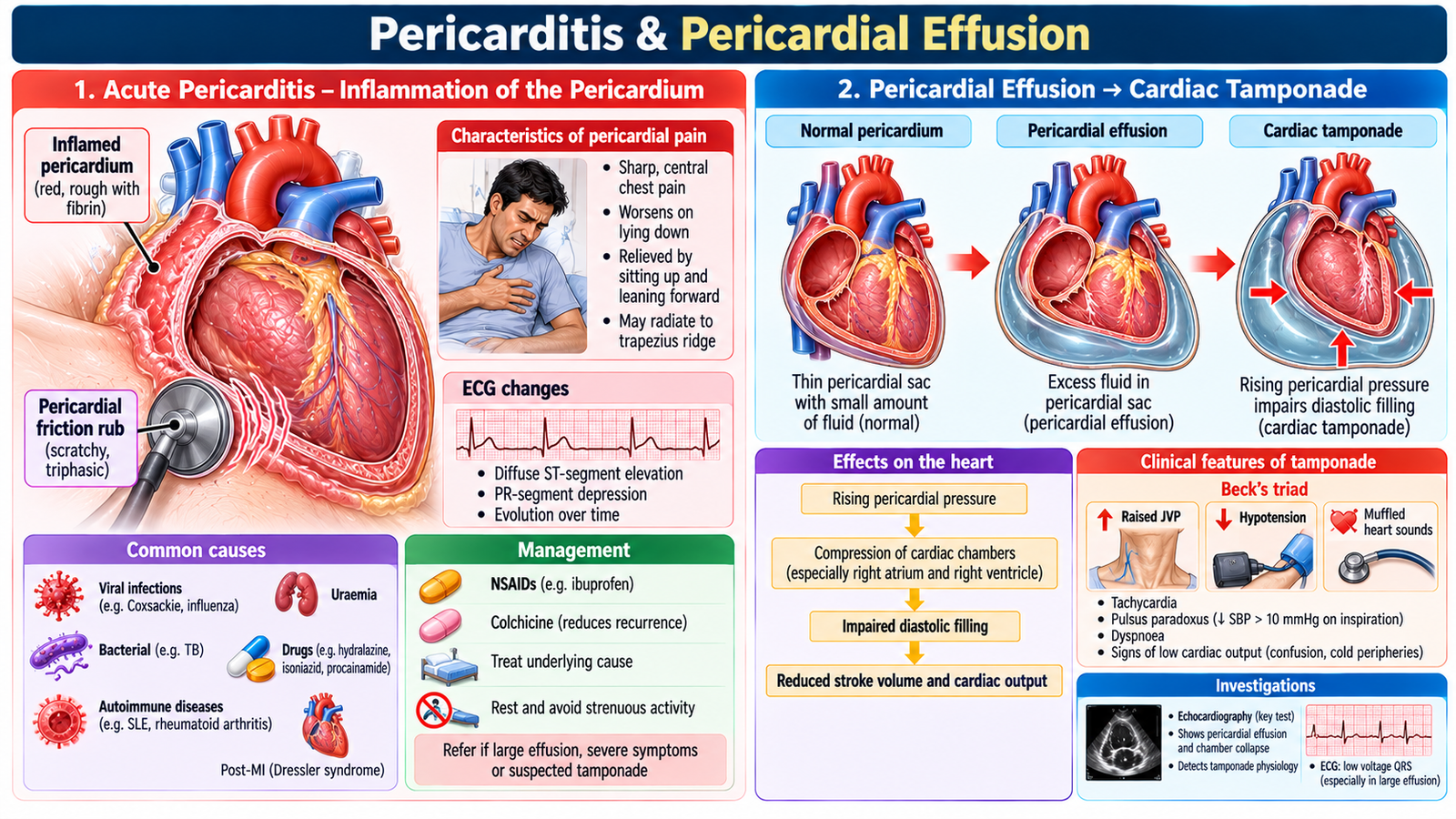
The pericardium normally surrounds the heart with only a small amount of fluid between its layers. Pericarditis produces inflammation of these layers, whereas a pericardial effusion is abnormal accumulation of fluid within the pericardial space. The major danger arises when pressure from the fluid begins to impair cardiac filling.
Acute pericarditis
Typical pain is sharp and pleuritic, may worsen when lying flat and may improve when sitting forward. A pericardial friction rub is an important examination clue when present. ECG may show widespread inflammatory changes rather than a single coronary-territory pattern.
Etiological groups include infectious, inflammatory, post-cardiac injury and other systemic causes. Treatment addresses inflammation and the underlying cause where identified, while monitoring for complications such as increasing effusion.
First-line treatment: Uncomplicated acute pericarditis is treated with ibuprofen or high-dose aspirin plus colchicine to reduce inflammation and recurrence, with proton-pump inhibitor protection when appropriate. Restrict strenuous physical activity until clinical recovery. Corticosteroids are not routine first-line therapy and are reserved for selected indications.
Pericardial effusion and tamponade physiology
The effect of an effusion depends not only on its volume but also on how rapidly it accumulates. Rapid accumulation may sharply increase intrapericardial pressure before the pericardium has time to adapt. Rising pressure restricts diastolic filling, reduces stroke volume and can eventually produce obstructive shock—cardiac tamponade.
Echocardiography is central when a significant effusion or tamponade is suspected because it demonstrates fluid and its hemodynamic effect. A patient with hemodynamic compromise from tamponade requires urgent drainage rather than simple observation.
7. Heart Failure, Cardiomyopathy & Myocarditis in Children
Children may not describe breathlessness, exercise intolerance or palpitations in the way adults do. Clinical recognition therefore depends heavily on age-specific history, feeding, growth, respiratory effort, activity and examination. The underlying disease may be congenital or acquired, and the same cardiac dysfunction may appear differently at different ages.
Recognizing pediatric cardiac failure
In infants, feeding is physiologically demanding. Cardiac failure may therefore present with poor feeding, sweating or breathlessness during feeds, prolonged feeding, poor weight gain and tachypnea. Older children may show reduced exercise tolerance, fatigue, breathlessness, edema or other signs of congestion.
History and physical examination
Ask about feeding, growth, respiratory symptoms, exercise or play tolerance, cyanosis, previous infections and known congenital or acquired cardiac disease. Examination should assess growth, respiratory effort, heart rate and rhythm, perfusion, pulses, cardiac sounds or murmurs, hepatomegaly and evidence of edema or pulmonary congestion.
In infants and young children, hepatomegaly is an especially useful sign of systemic venous congestion. Peripheral edema may be less prominent than in adults.
Diagnostic workup and management
Investigations are selected according to age and suspected cause. ECG, chest radiography and echocardiography commonly contribute to defining rhythm, cardiac size, pulmonary circulation, ventricular function and structural disease. Management treats the heart-failure syndrome while addressing the underlying cause and maintaining appropriate nutritional and developmental support.
Pediatric heart failure treatment: Furosemide is commonly used to relieve pulmonary or systemic congestion. Children with systolic dysfunction may receive an ACE inhibitor (e.g., enalapril) and selected beta-blocker therapy under pediatric cardiology supervision. Acute severe cardiac failure requires hospital admission, respiratory and circulatory support, and treatment of the underlying cause.
Pediatric cardiomyopathy and myocarditis
Cardiomyopathy in a child may lead to ventricular dysfunction, heart failure and rhythm problems. Pediatric myocarditis should be considered when previously unexplained cardiac dysfunction, tachycardia, respiratory distress, poor feeding or exercise intolerance develops in an appropriate clinical context. Management depends on severity and focuses on cardiovascular support, heart-failure treatment and management of important arrhythmias or an identifiable cause.
Specific management: Pediatric dilated cardiomyopathy is treated according to ventricular dysfunction and heart-failure severity, with specialist assessment for advanced treatment when necessary. Hypertrophic cardiomyopathy may require beta-blockers for symptomatic obstruction. Pediatric myocarditis requires supportive care, monitoring for arrhythmias and heart failure, and urgent intensive-care assessment if cardiogenic shock develops.
Parent counselling
Parents should understand the suspected diagnosis, purpose of investigations and treatment, importance of medication adherence and follow-up, and warning symptoms requiring urgent reassessment. Counselling should acknowledge parental anxiety and explain the child’s condition in clear language without giving unrealistic reassurance about prognosis.

Integrated Mechanism Flow
AIM High-Yield Review
- ⭐ Left-sided heart failure raises pulmonary venous pressure → dyspnea, orthopnea and pulmonary congestion.
- Right-sided failure raises systemic venous pressure → raised JVP, hepatic congestion and peripheral edema.
- ⭐ Echocardiography is central to defining ventricular function, structural abnormalities and important pericardial disease.
- Diuretics relieve congestion but management must also address the underlying cardiac disease and long-term disease progression.
- Dilated cardiomyopathy → ventricular dilatation + systolic dysfunction; restrictive cardiomyopathy → stiff ventricle + impaired filling.
- ⭐ Hypertrophic cardiomyopathy prominently impairs diastolic filling and may produce dynamic outflow obstruction.
- Myocarditis may produce heart failure and arrhythmia because myocardial inflammation impairs mechanical and electrical function.
- Pericarditis classically causes pleuritic, positional chest pain; a friction rub is an important clinical clue when present.
- ⭐ Rapidly accumulating pericardial fluid may impair diastolic filling → reduced cardiac output → tamponade and shock.
- Infant heart failure may present as poor feeding, sweating during feeds, tachypnea and poor weight gain rather than a typical adult symptom description.
- Hepatomegaly is an important sign of systemic venous congestion in infants and children.
- Primary care supports prevention, adherence, monitoring, psychosocial care, red-flag recognition and appropriate specialist referral.
Heart Failure, Cardiomyopathy, Myocarditis & Pericardial Disease
Final Year MBBS • CVS Module • Watch alongside the AIM Learning Material