
This chapter follows the KMU curriculum outcomes for chest trauma, pleural disorders and thoracic procedures. First understand the clinical logic linking respiratory compromise to diagnosis and treatment; use the High-Yield Review at the end for rapid revision.
Topic 2. Chest Trauma, Pleural Disorders & Thoracic Procedures
A clinically integrated approach to chest trauma, hemothorax, pneumothorax, pleural effusion, pleural aspiration, thoracostomy and chest-tube care.
Topic Introduction
Chest trauma and pleural disorders can interfere rapidly with ventilation and circulation because the pleural space is essential for normal lung expansion. In trauma, air or blood may enter this space and produce pneumothorax or hemothorax; in medical disease, fluid may accumulate as a pleural effusion. The Final Year student therefore needs to connect initial ABC assessment with recognition of respiratory compromise, appropriate investigations and timely pleural intervention. This chapter also explains the principles of pleural aspiration, thoracostomy and chest intubation without turning procedural learning into an OSCE checklist.
A. Chest Trauma: From Primary Assessment to Clinical Priorities
Chest trauma may be blunt or penetrating. Its immediate importance is not simply the anatomical injury: the key question is whether the injury is interfering with oxygenation, ventilation or circulation. This is why assessment begins with a structured trauma approach rather than with a detailed search for a final diagnosis.
ABC assessment
Airway: determine whether the airway is patent while protecting the cervical spine when trauma circumstances require it. Airway obstruction, altered consciousness or major associated injury can rapidly compromise oxygen delivery.
Breathing: inspect respiratory effort and chest movement, assess oxygenation, palpate the chest, and use percussion and auscultation to identify major asymmetry. Reduced expansion with absent or markedly reduced breath sounds should immediately raise concern for a significant pleural or pulmonary complication.
Circulation: assess pulse, blood pressure, peripheral perfusion and evidence of blood loss. Thoracic bleeding may be concealed within the pleural cavity, so shock after chest trauma can occur without obvious external hemorrhage.
A life-threatening chest injury is treated when recognized during the primary survey. Definitive imaging should not delay immediate treatment when the clinical problem is causing severe respiratory or circulatory compromise.
Focused diagnostic assessment
After immediate threats have been addressed, determine the mechanism of injury, site of impact or penetration, onset of dyspnea or chest pain, associated blood loss and progression of symptoms. Examination should look for chest-wall tenderness, asymmetrical movement, abnormal percussion, altered breath sounds and signs of hemodynamic compromise.
Chest radiography and bedside ultrasonography can help identify pleural air, pleural blood and associated thoracic injury in a sufficiently stable patient. More detailed imaging may be appropriate in stable patients when the nature or extent of injury remains uncertain.

B. Traumatic Pleural Complications: Pneumothorax & Hemothorax
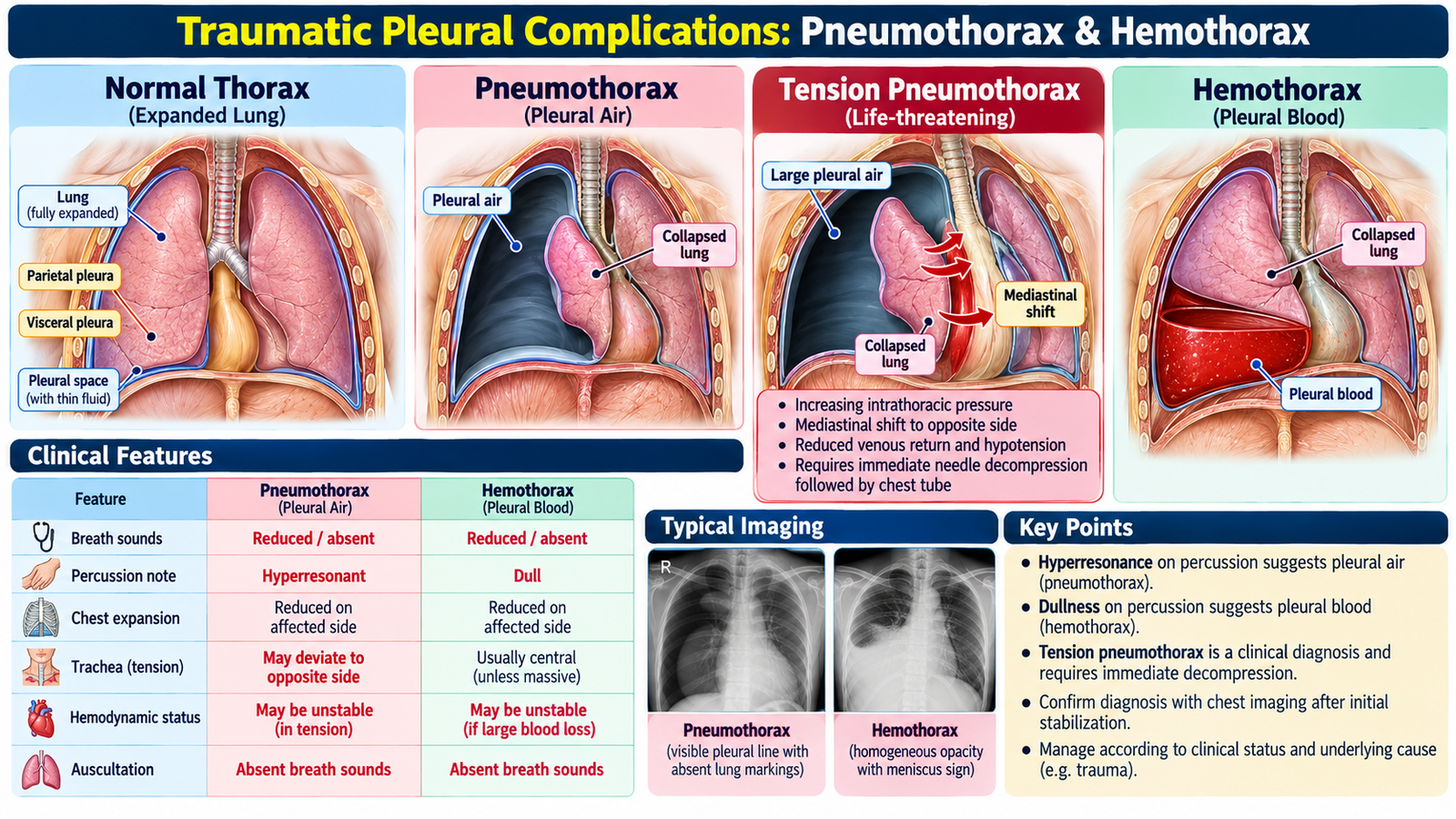
The pleural cavity normally contains only a thin film of fluid and maintains the pressure relationship needed to keep the lung expanded. Trauma can introduce air, blood, or both into this space. The resulting loss of normal pleural mechanics explains many of the important examination findings.
Pneumothorax
A pneumothorax occurs when air enters the pleural space. Loss of the normal negative intrapleural pressure allows the affected lung to recoil inward, reducing effective ventilation.
This explains the combination of dyspnea, reduced movement of the affected hemithorax, reduced breath sounds and a more resonant percussion note.
Tension pneumothorax is particularly dangerous because intrapleural pressure progressively rises. The affected lung becomes compressed and the increasing intrathoracic pressure impairs venous return to the heart. The patient may therefore develop severe respiratory distress together with circulatory compromise.
Severe respiratory distress plus hemodynamic compromise in a patient with suspected pneumothorax is an emergency. Management is based on urgent decompression followed by definitive pleural drainage rather than waiting for routine imaging confirmation.
Management of pneumothorax: A small, stable pneumothorax may be managed with observation, analgesia and follow-up. Symptomatic or clinically significant pneumothorax may require needle aspiration or intercostal chest drainage, depending on its cause and severity. Tension pneumothorax with severe respiratory or circulatory compromise requires immediate chest decompression followed by chest-tube drainage; imaging must not delay treatment.
Hemothorax
Hemothorax is accumulation of blood within the pleural cavity. It creates two simultaneous problems: blood occupying the pleural space compresses the lung and reduces ventilation, while significant intrathoracic blood loss can impair circulation.
Reduced breath sounds with a dull percussion note suggests pleural fluid rather than pleural air. Tachycardia, hypotension and poor perfusion increase concern for clinically important hemorrhage.
Management depends on the patient’s physiological condition and severity of the collection. Initial priorities include resuscitation when required and drainage of significant pleural blood through a chest tube. Persistent or major bleeding requires urgent surgical assessment.
C. Pleural Effusion: Diagnostic & Management Approach
Pleural effusion is an abnormal accumulation of fluid in the pleural space. The important clinical task is not merely to confirm that fluid is present, but to determine why it accumulated and whether the underlying disorder requires specific treatment.
How pleural fluid accumulates
Pleural fluid balance depends on hydrostatic and oncotic pressures, capillary permeability and lymphatic drainage. Disturbance of these mechanisms produces two broad patterns:
Primarily reflects altered systemic hydrostatic or oncotic forces rather than primary pleural inflammation.
Usually reflects local pleural or pulmonary disease causing inflammation, increased permeability or impaired lymphatic drainage.
Clinical recognition
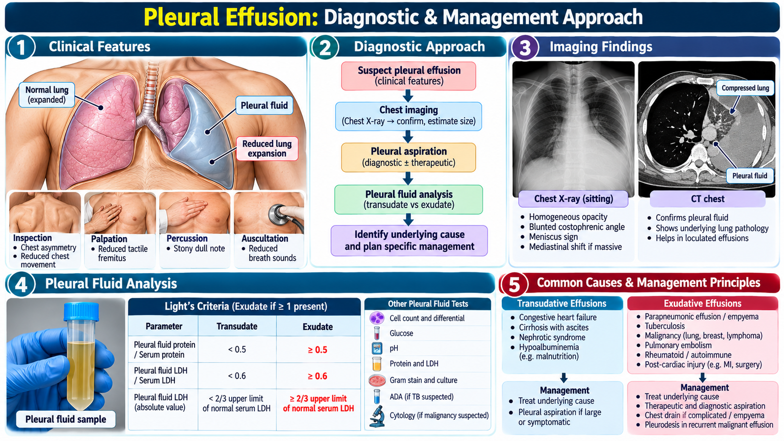
The patient may present with dyspnea, pleuritic chest discomfort or symptoms related to the underlying disease. A sufficiently large effusion reduces expansion of the affected side and produces stony dullness to percussion with reduced breath sounds over the fluid.
Diagnostic approach
Imaging confirms and characterizes pleural fluid. Chest radiography can demonstrate fluid accumulation, while ultrasonography is particularly useful for confirming the presence and location of fluid and for guiding pleural aspiration.
When clinically indicated, pleural fluid is aspirated and assessed alongside the clinical picture. Protein and lactate dehydrogenase relationships between pleural fluid and serum help distinguish an exudative from a transudative process. Further pleural-fluid assessment is selected according to the suspected cause.
Confirm fluid → assess clinical context → classify the fluid → investigate the likely underlying cause → treat the cause and drain when clinically indicated.
Management principle
Treatment is directed at the underlying disease. Pleural drainage may also be required when the collection is causing important symptoms, when diagnostic sampling is necessary, or when the nature of the fluid makes drainage therapeutically important.
Cause-specific management: Heart-failure-related effusions usually respond to diuretics such as furosemide and treatment of the underlying cardiac disease. Parapneumonic effusions require appropriate antibiotics; empyema or complicated infected effusions generally require chest-tube drainage. Tuberculous pleural effusion requires standard anti-tuberculosis therapy, while symptomatic malignant effusion may require therapeutic aspiration, an indwelling pleural catheter or pleurodesis.
D. Pleural Fluid Aspiration: Purpose, Interpretation & Safety
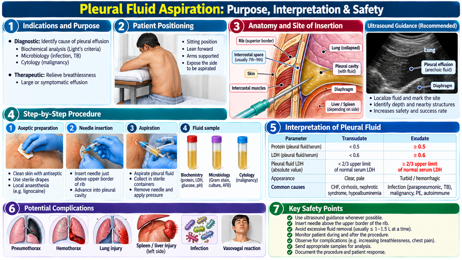
Pleural aspiration, or thoracentesis, involves inserting a needle or catheter into the pleural space to obtain or remove fluid. For Final Year learning, the essential concepts are why it is performed, what information it provides, how imaging improves safety, and what complications must be recognized.
Why aspirate pleural fluid?
- To obtain fluid when the cause of an effusion requires clarification.
- To characterize the fluid and guide further diagnostic reasoning.
- To relieve symptoms in selected patients with a clinically important fluid collection.
Clinical knowledge supporting the procedure
Before aspiration, the clinician confirms the presence and location of pleural fluid and assesses whether the patient can undergo the procedure safely. Ultrasound guidance improves localization and helps avoid inappropriate puncture sites.
The needle is directed into the pleural space while respecting the anatomy of the intercostal neurovascular bundle. The procedural principle is to choose an appropriate image-confirmed site and avoid injury to adjacent structures.
What happens after fluid is obtained?
The appearance and laboratory characteristics of the fluid are interpreted together with the patient’s history, examination and imaging. No single fluid result should be interpreted in isolation from the clinical context.
Potential complications include pneumothorax, bleeding, infection and injury to nearby structures. New respiratory deterioration after pleural aspiration therefore requires prompt reassessment.
E. Thoracostomy & Chest Intubation
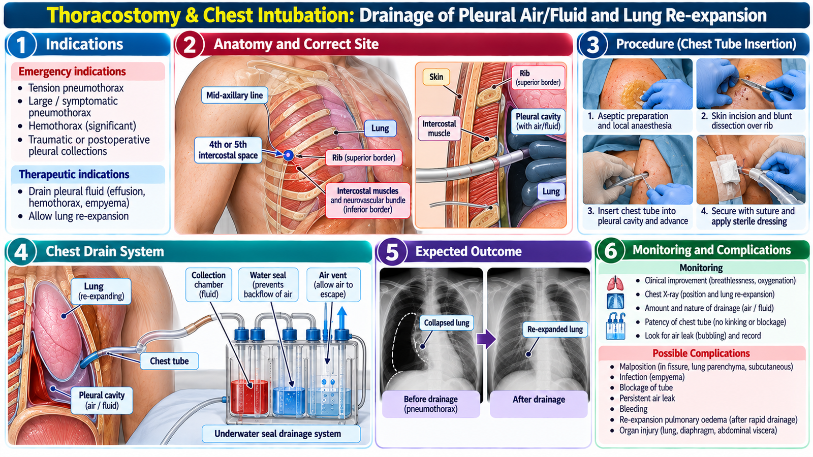
Tube thoracostomy places a drain into the pleural cavity so that accumulated air, blood or other pleural contents can leave the thorax. The purpose is to restore a pleural environment in which the lung can re-expand and respiratory function can improve.
When is pleural drainage required?
Important indications include clinically significant pneumothorax, hemothorax and other pleural collections in which continuous drainage is required. In traumatic disease, the patient’s respiratory and hemodynamic status determines urgency.
Core principle
A chest tube is inserted through an appropriate lateral chest-wall site into the pleural cavity and connected to a drainage system. The system permits removal of pleural air or fluid while helping prevent their return into the chest.
What should a student understand when observing chest-tube insertion?
- The clinical indication must be clear before the procedure.
- Correct patient positioning and anatomical site selection reduce complications.
- Aseptic technique and adequate analgesia are important patient-safety principles.
- The tube must communicate effectively with the pleural cavity and drainage system.
- After insertion, the patient and drainage system require clinical reassessment.
Monitoring after insertion
Reassess respiratory symptoms, oxygenation, chest findings and the patient’s overall clinical condition. The drainage system should be observed for function and the amount and nature of drainage. Unexpected deterioration requires assessment for persistent disease, tube malfunction or a procedure-related complication.
A chest tube is not simply an insertion procedure. Correct positioning, secure connection, ongoing observation and prompt recognition of deterioration are integral to safe pleural drainage.
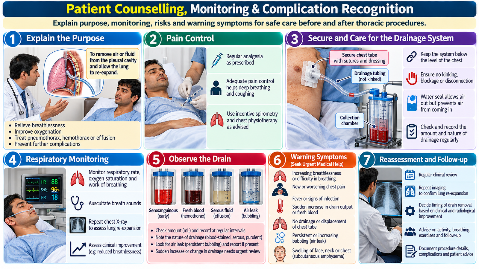
F. Patient Counselling, Monitoring & Complication Recognition
Thoracic procedures can be frightening because patients are already experiencing pain or breathlessness. Good counselling explains the reason for the intervention, what the patient is likely to experience, how the tube or aspiration will help, and which symptoms require immediate attention.
Before the procedure
Explain the clinical problem in understandable language and describe why aspiration or drainage is being recommended. Discuss the general nature of the procedure, expected discomfort, analgesia or local anesthesia as appropriate, important risks and the need for monitoring.
While a chest tube remains in place
The patient should understand that the tube needs to remain connected and secure and that staff will monitor breathing, pain, drainage and recovery. Concerns about movement, discomfort and duration should be addressed honestly without giving unsupported guarantees.
Symptoms requiring reassessment
Increasing breathlessness, worsening chest pain, new hemodynamic instability, unexpected bleeding or clinical deterioration should trigger prompt review. The purpose is to identify recurrence, ongoing disease, tube dysfunction or a procedural complication early.
Patient-centred approach
Use clear language, allow questions, acknowledge anxiety, check understanding and explain the monitoring plan. Counselling should support informed participation without overwhelming the patient with technical procedural detail.
🩺 AIM Clinical Bridge
After blunt chest trauma, a patient becomes breathless. The affected side moves poorly and breath sounds are markedly reduced. Your next interpretation depends on both physiology and percussion: pleural air tends toward hyperresonance, whereas pleural blood produces dullness. If severe respiratory distress is accompanied by circulatory compromise, immediately consider a life-threatening pressure effect rather than waiting for routine investigation.
- Is the dominant problem pleural air, pleural fluid/blood, or another chest injury?
- Is the patient physiologically stable enough for further investigation?
- Does the patient need observation, aspiration, pleural drainage or immediate emergency treatment?
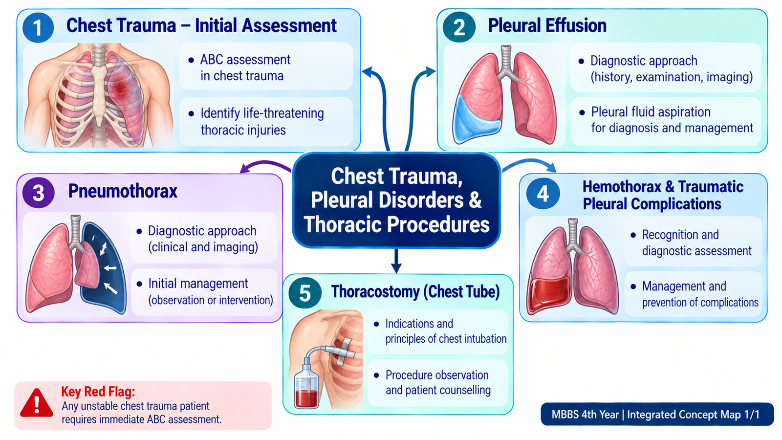

Integrated Mechanism Flow
Important Comparison
| Feature | Pneumothorax | Hemothorax | Pleural Effusion |
|---|---|---|---|
| Pleural content | Air | Blood | Pleural fluid |
| Breath sounds | Reduced | Reduced | Reduced over fluid |
| Percussion | Hyperresonant | Dull | Stony dull |
| Major concern | Loss of lung expansion; tension physiology if pressure rises | Lung compression plus blood loss | Identify underlying cause and effect on breathing |
| Management logic | Observe or evacuate air according to severity | Resuscitate and drain significant blood | Diagnose cause; aspirate or drain when indicated |
AIM High-Yield Review
- Chest trauma begins with physiology: assess airway, breathing and circulation before pursuing detailed anatomical diagnosis.
- Pleural air: pneumothorax reduces lung expansion and typically produces reduced breath sounds with hyperresonance.
- Tension pneumothorax: increasing intrapleural pressure compromises ventilation and venous return; severe respiratory and circulatory compromise demands emergency treatment.
- Pleural blood: hemothorax can cause both respiratory compromise from lung compression and circulatory compromise from blood loss.
- Percussion helps integration: air tends toward hyperresonance, whereas pleural blood or fluid produces dullness.
- Pleural effusion: confirm the fluid, determine its likely mechanism, classify it when appropriate and identify the underlying disease.
- Pleural aspiration: may provide diagnostic fluid or therapeutic relief; ultrasound localization improves procedural safety.
- Thoracostomy: continuous evacuation of pleural air or fluid permits lung re-expansion when drainage is indicated.
- After a pleural procedure: new or worsening respiratory distress requires prompt reassessment for persistent disease, recurrence or a procedural complication.
- Counselling is part of management: explain purpose, expected experience, important risks, monitoring and warning symptoms in clear patient-centred language.
Chest Trauma, Pleural Disorders & Thoracic Procedures
Review the clinical approach to chest trauma, pneumothorax, hemothorax, pleural effusion and thoracic drainage procedures.