
Think of DVT and pulmonary embolism as different points on the same venous thromboembolism pathway. First understand why a thrombus forms, then connect the patient’s risk factors and symptoms to the correct diagnostic test and the urgency of treatment.
Topic 6. Venous Thromboembolism: Deep Vein Thrombosis & Pulmonary Embolism
A clinically connected approach to risk factors, unilateral painful leg swelling, DVT diagnosis and management, pulmonary embolism recognition, diagnostic reasoning, complications and treatment.
Topic Introduction
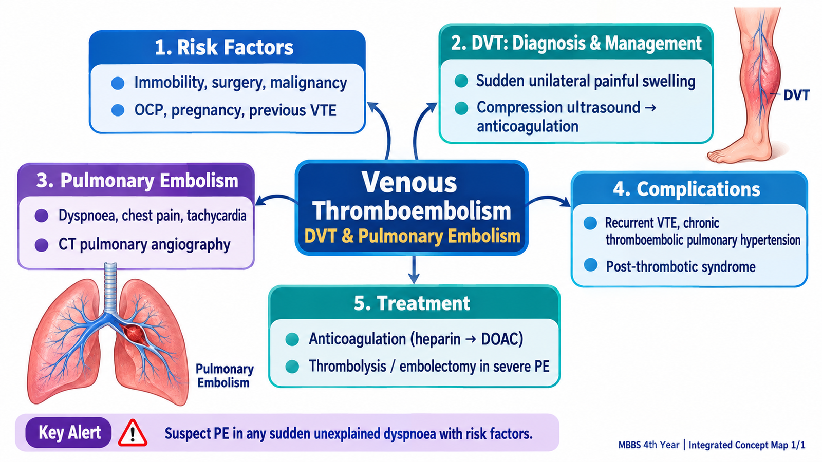
Venous thromboembolism (VTE) includes deep vein thrombosis (DVT) and pulmonary embolism (PE). They are closely linked: a thrombus usually forms in a deep vein, most commonly in the lower limb, and part of it may detach and travel through the venous circulation to the pulmonary arteries. The clinical challenge is therefore not simply to recognize a swollen leg or a breathless patient, but to estimate the probability of VTE, choose appropriate investigations, identify instability, and start treatment before serious complications occur. This chapter develops that reasoning step by step.
A. Venous Thromboembolism: From Risk Factor to Thrombus and Embolus
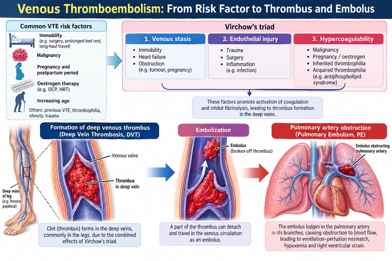
VTE develops when the normal balance that keeps blood flowing without excessive clotting is disturbed. The classical framework is Virchow’s triad: venous stasis, endothelial injury and increased tendency of blood to clot. A patient may have one factor or several acting together. The greater the combination of risk factors, the greater the clinical suspicion when compatible symptoms appear.
Why does venous thrombosis occur?
Venous stasis reduces movement of blood through the veins, allowing activated coagulation factors to accumulate. Endothelial injury exposes procoagulant surfaces. Hypercoagulability means the coagulation system is unusually prone to forming thrombus.
Core mechanism:
Predisposing factor → stasis, endothelial injury or hypercoagulability → fibrin-rich venous thrombus → partial or complete venous obstruction → local leg symptoms ± embolization → pulmonary arterial obstruction.
Important clinical risk contexts
- Immobility: prolonged bed rest, hospitalization or reduced mobility promotes venous stasis.
- Major surgery or trauma: combines tissue injury, immobility and activation of coagulation.
- Malignancy: may create a persistent hypercoagulable state.
- Pregnancy and the postpartum period: increase thrombotic tendency and may be accompanied by venous stasis.
- Estrogen-containing therapy: can increase thrombotic risk.
- Previous VTE: substantially increases suspicion when compatible symptoms recur.
- Inherited or acquired thrombophilia: may predispose to recurrent or apparently unprovoked thrombosis.
Risk factors do not diagnose VTE by themselves. Their importance is that they change the pre-test probability: unilateral leg swelling in a recently immobilized patient is more suspicious for DVT than the same symptom in a patient with a clear traumatic explanation.
B. Sudden Unilateral Painful Leg Swelling: Recognizing Suspected DVT
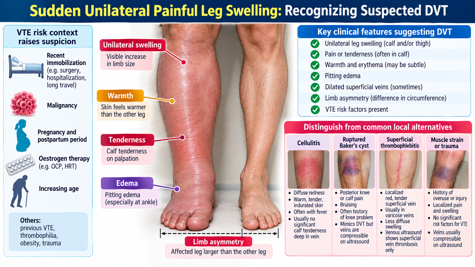
DVT commonly presents as a new unilateral swollen and painful limb, but no single symptom or physical sign is sufficiently reliable to confirm the diagnosis. The clinician first asks whether the pattern is compatible with venous obstruction, whether important VTE risk factors are present, and whether another local condition better explains the swelling.
History clues that increase suspicion
- Recent onset of unilateral swelling or discomfort.
- Previous DVT or pulmonary embolism.
- Recent surgery, hospitalization, trauma or immobility.
- Active malignancy or another strong hypercoagulable context.
- Pregnancy, postpartum state or estrogen exposure where relevant.
- Associated chest pain, breathlessness, syncope or hemoptysis, which may indicate that PE has already occurred.
Examination findings and their meaning
The affected limb may show asymmetrical swelling, edema, warmth and tenderness. These findings result from impaired venous drainage and local inflammatory response around the thrombosed vein. Comparison with the opposite leg helps establish whether swelling is genuinely unilateral.
Clinical examination must also look for an alternative diagnosis. Marked erythema with an obvious skin infection suggests cellulitis, while a history of knee disease with swelling extending into the calf may suggest a ruptured Baker cyst. The purpose is not to create a long differential list, but to avoid assuming that every painful swollen leg is DVT.
| Feature | DVT | Cellulitis | Ruptured Baker Cyst |
|---|---|---|---|
| Main process | Deep venous thrombosis | Skin/subcutaneous infection | Leakage of knee-joint cyst fluid |
| Typical clue | VTE risk context | Prominent erythema or skin source | Associated knee pathology |
| Main danger | Pulmonary embolism | Spreading infection | Usually local morbidity |
Important: clinical findings raise or lower suspicion, but suspected DVT should be assessed using a structured diagnostic pathway rather than clinical impression alone.
C. Diagnosing Deep Vein Thrombosis: Probability, D-Dimer and Ultrasound
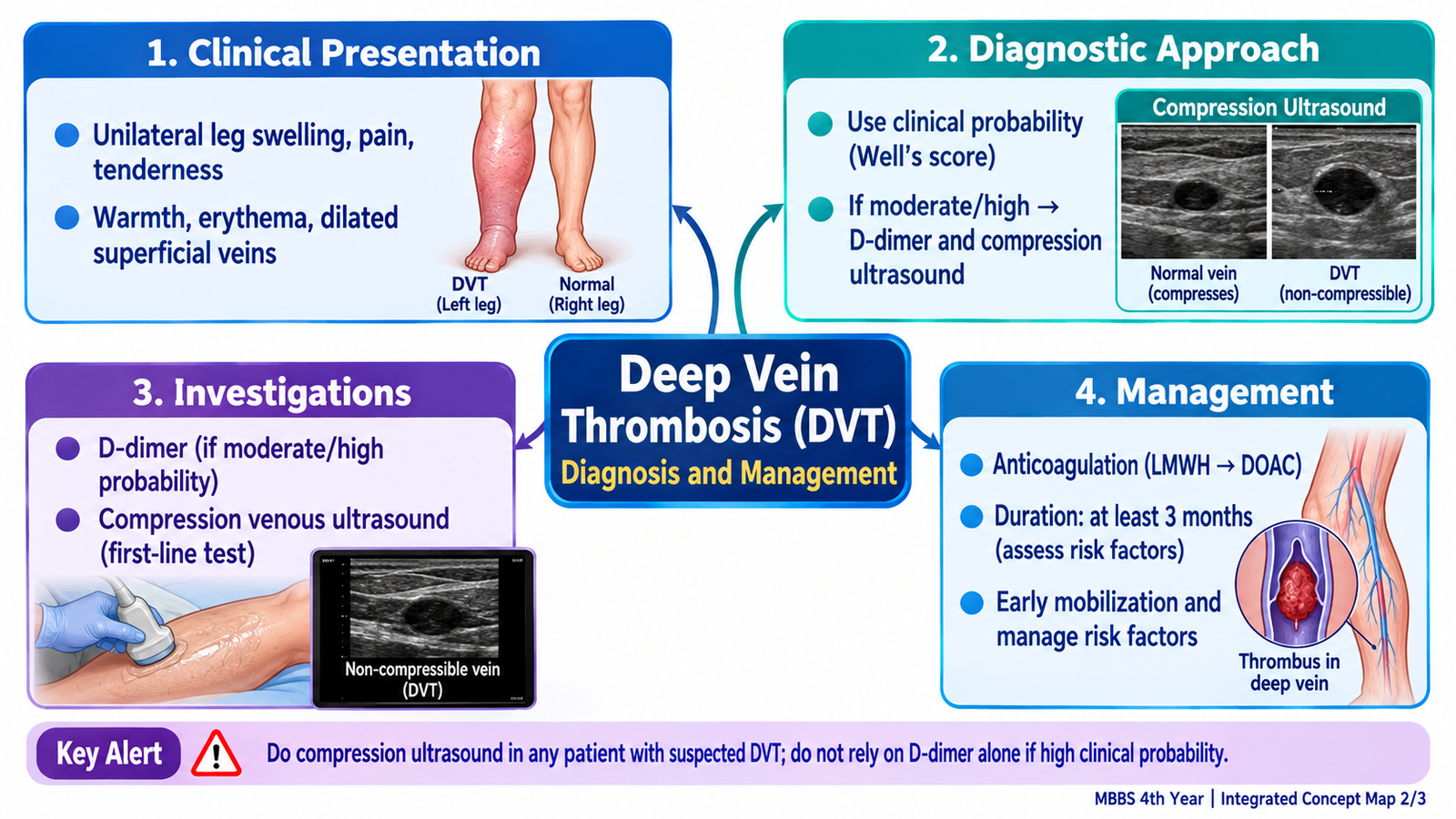
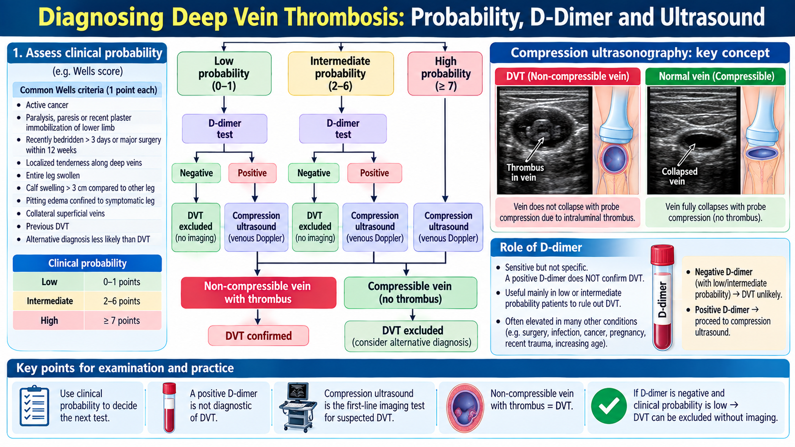
The diagnosis of DVT is based on a combination of clinical probability and objective testing. The important principle is that investigation depends on how likely DVT appears before testing. This prevents unnecessary imaging in very-low-probability patients while ensuring that high-risk patients are investigated promptly.
1. Estimate clinical probability
Structured clinical assessment combines the patient’s symptoms, signs and risk context. A probability score may be used, but the important undergraduate concept is the distinction between a patient in whom DVT is unlikely and one in whom it is clinically likely.
2. Understand the role of D-dimer
D-dimer is produced when cross-linked fibrin is broken down. A raised level therefore indicates increased fibrin formation and degradation, but it is not specific for VTE. It may also rise in infection, inflammation, malignancy, pregnancy, after surgery and in other conditions.
3. Compression ultrasonography
Ultrasound is the key imaging test for suspected lower-limb DVT. A normal vein is compressible with the ultrasound probe. A thrombosed vein loses normal compressibility because thrombus occupies its lumen. Thus:
Non-compressible deep vein → supports the diagnosis of DVT → anticoagulant treatment is considered unless contraindicated.
If the initial assessment does not establish the diagnosis but clinical suspicion remains important, reassessment or repeat imaging may be required according to the clinical pathway being used. The key reasoning principle is to reconcile the test result with pre-test probability.
🩺 AIM Clinical Bridge
A hospitalized patient has been relatively immobile for several days and develops new swelling and pain of one calf. Later, the patient suddenly becomes breathless with pleuritic chest discomfort and tachycardia. The two presentations should not be considered separate problems: the leg symptoms suggest DVT, while the new cardiorespiratory symptoms raise concern that part of the thrombus has embolized to the pulmonary circulation.
- Which features make VTE likely?
- Is the patient hemodynamically stable or unstable?
- Which investigation should confirm the suspected site of thrombosis or embolism?
D. Pulmonary Embolism: Presentation, Severity and Complications
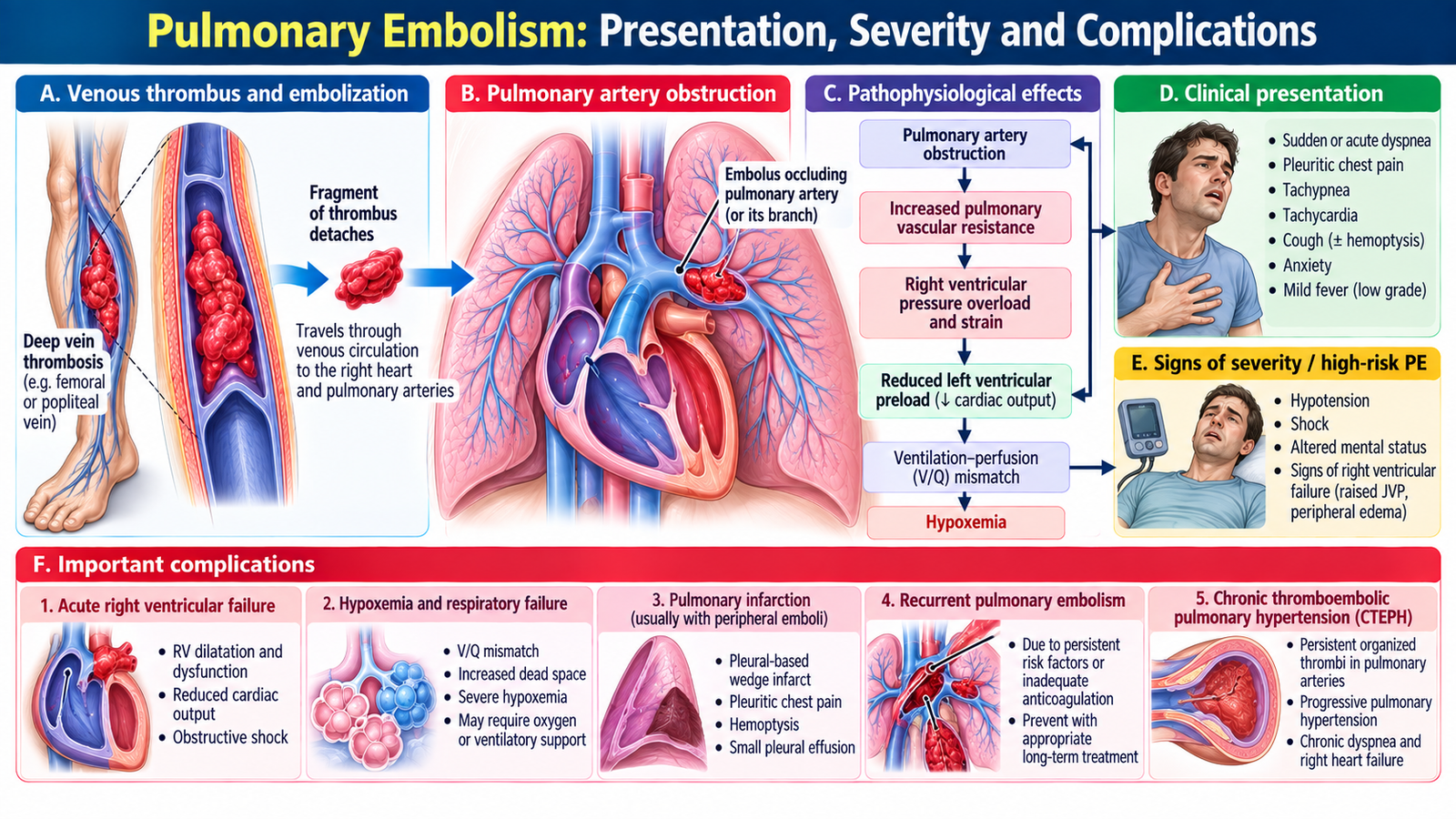
Pulmonary embolism occurs when embolic material, usually thrombus from the deep veins of the lower limb or pelvis, travels through the right side of the heart and obstructs part of the pulmonary arterial circulation. The clinical effect depends on the size of the obstruction and the patient’s cardiopulmonary reserve.
How pulmonary obstruction produces symptoms
Obstruction of pulmonary arteries creates areas of lung that are ventilated but inadequately perfused. This produces ventilation–perfusion mismatch and hypoxemia. Pulmonary vascular resistance also rises. If the embolic burden is large, the right ventricle must suddenly pump against a much higher resistance, which may cause acute right ventricular failure and reduced left ventricular filling.
Pulmonary arterial obstruction → increased pulmonary vascular resistance → right ventricular strain → reduced left-sided filling and cardiac output → hypotension, syncope or shock in severe PE.
Typical clinical presentation
- Sudden or unexplained dyspnea.
- Pleuritic chest pain.
- Tachypnea and tachycardia.
- Reduced oxygen saturation or hypoxemia.
- Hemoptysis may occur when peripheral pulmonary infarction develops.
- Syncope, hypotension or shock suggest a more severe hemodynamic effect.
- Some patients simultaneously have symptoms or signs of DVT.
Stable versus unstable PE
A patient with suspected PE should rapidly be assessed for hemodynamic stability. This distinction matters because it changes the urgency and type of treatment.
No persistent circulatory collapse. Diagnostic confirmation and anticoagulation are the main priorities.
Hypotension, shock or collapse suggests severe obstruction with threatened circulation and requires urgent stabilization and reperfusion consideration.
Important complications
- Acute right ventricular failure.
- Obstructive shock.
- Pulmonary infarction in selected cases.
- Recurrent embolism if the thrombotic source remains untreated.
- Death in severe untreated PE.
- Persistent pulmonary vascular disease may develop after unresolved or recurrent thromboembolism.
E. Diagnosing Pulmonary Embolism: Clinical Probability to Imaging
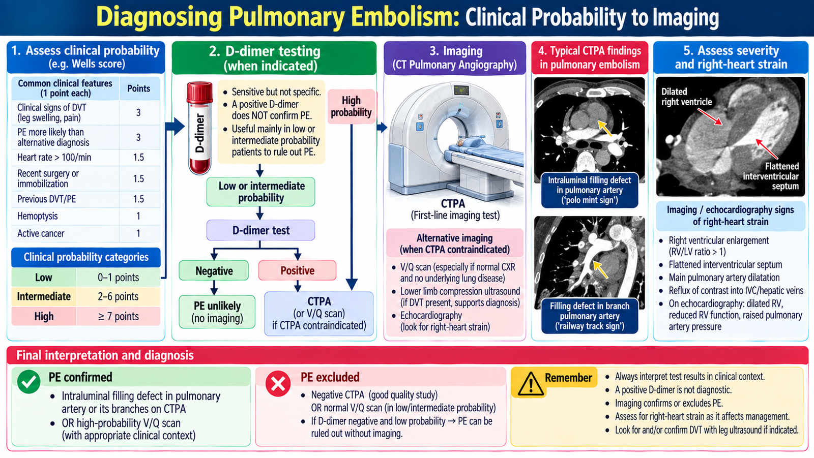
The diagnosis of PE cannot be based on symptoms alone because dyspnea, chest pain and tachycardia occur in many cardiopulmonary disorders. The diagnostic approach therefore starts with clinical probability and then uses laboratory or imaging tests according to that probability.
Clinical probability comes first
A typical presentation, VTE risk factors, signs of DVT and absence of a more convincing alternative increase the probability of PE. Structured clinical probability tools can support this assessment, but the central principle is to decide whether PE is sufficiently likely to justify direct imaging or whether a rule-out strategy is appropriate.
D-dimer
As in suspected DVT, D-dimer is most helpful when the clinical probability is not high. A negative result in the appropriate low-probability setting can make acute VTE very unlikely. A positive result is nonspecific and usually requires further assessment rather than being treated as proof of PE.
CT pulmonary angiography
CT pulmonary angiography (CTPA) is the principal imaging test used to demonstrate thrombus within the pulmonary arterial circulation in most patients being investigated for PE. A visible intraluminal filling defect in a pulmonary artery supports the diagnosis.
Interpretation chain:
Compatible presentation + sufficient clinical probability → appropriate imaging → pulmonary arterial filling defect on CTPA → PE confirmed → determine severity and begin appropriate treatment.
Other investigations support assessment rather than replace confirmation
- Pulse oximetry and blood gases may show hypoxemia but are not diagnostic.
- ECG may show tachycardia or evidence of right-heart strain but cannot exclude PE when normal.
- Chest radiography is often useful for alternative diagnoses but does not reliably confirm or exclude PE.
- Echocardiography can identify right ventricular strain and is particularly useful when severe PE with hemodynamic compromise is suspected.
- Compression ultrasound may demonstrate a lower-limb DVT and thereby support the presence of VTE.
F. Management of DVT and Pulmonary Embolism
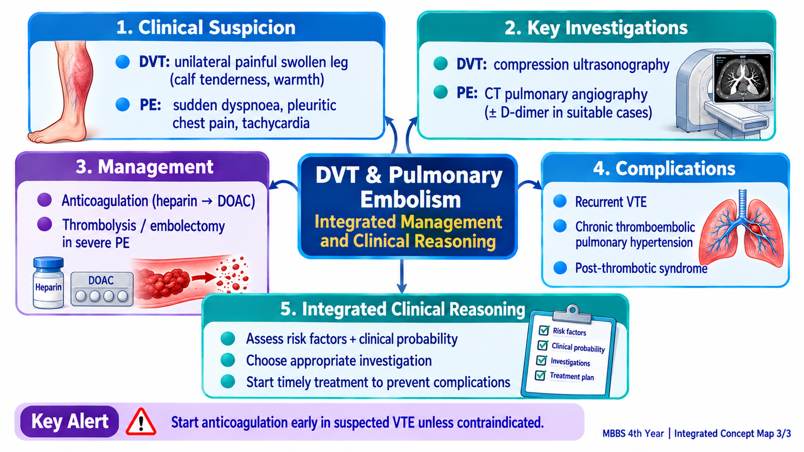
The central treatment for both DVT and most pulmonary emboli is anticoagulation. Anticoagulants do not mechanically remove an established clot; they reduce further thrombin generation and prevent clot extension and new thrombosis while the body’s natural fibrinolytic system gradually resolves the existing thrombus.
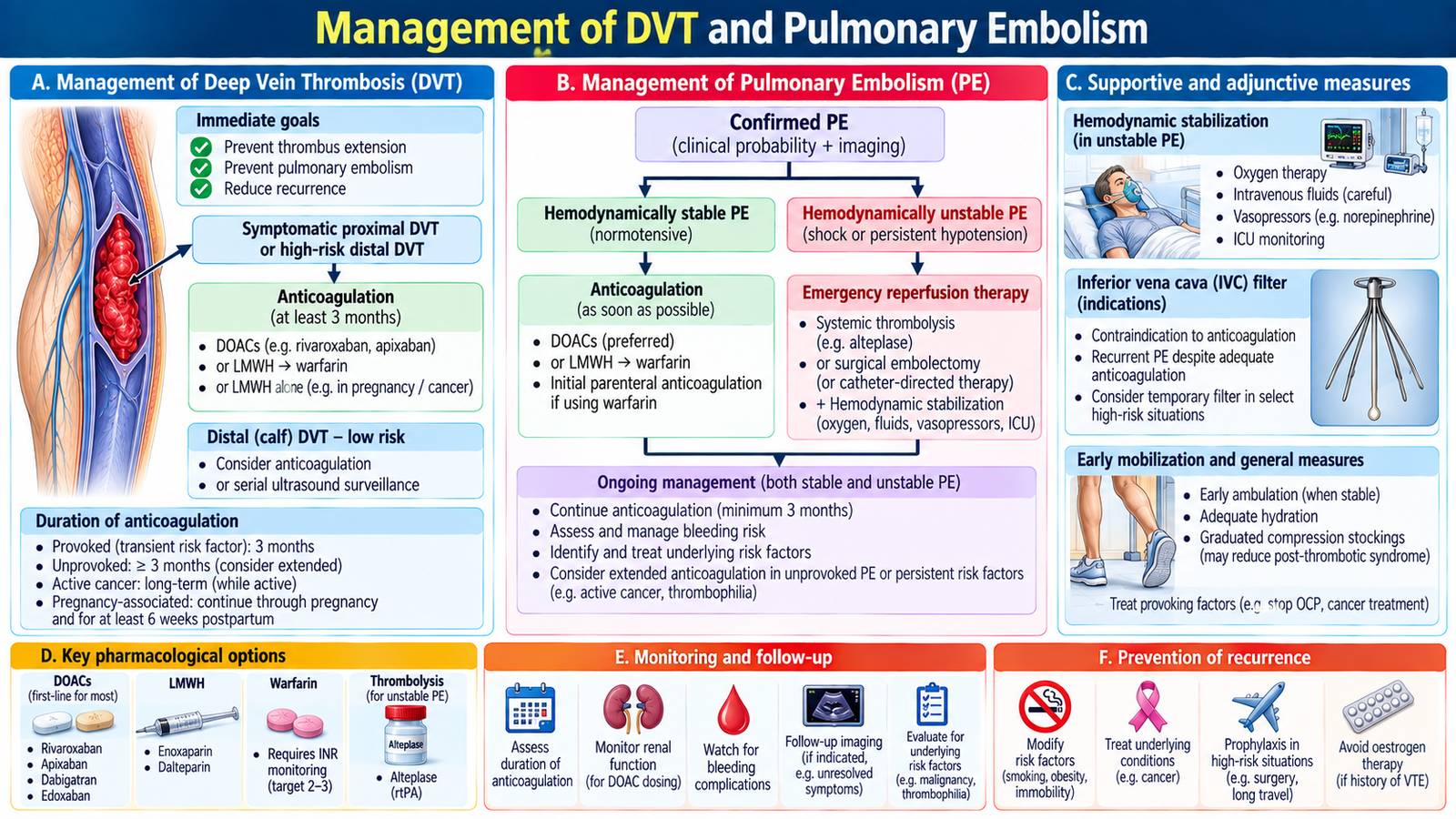
Management of confirmed DVT
In a patient with confirmed DVT and no major contraindication, anticoagulation is the main treatment. Common therapeutic approaches use a heparin-based anticoagulant or a direct oral anticoagulant according to the clinical setting, patient factors and local practice. The duration of anticoagulation depends on whether the episode was provoked, unprovoked, recurrent, associated with cancer or accompanied by continuing risk factors.
Practical anticoagulation: Apixaban or rivaroxaban are preferred oral anticoagulants for most adults with confirmed proximal DVT. Low-molecular-weight heparin, such as enoxaparin, is an alternative when appropriate; pregnancy requires a different anticoagulation approach. Treatment generally continues for at least 3 months, with longer treatment considered for unprovoked or recurrent VTE. If DVT is clinically likely and diagnostic imaging is delayed, start interim therapeutic anticoagulation when appropriate after assessing bleeding risk.
Therapeutic logic:
Anticoagulant → suppresses further coagulation → prevents thrombus extension and additional emboli → reduces recurrence while endogenous fibrinolysis acts on the existing clot.
Management of pulmonary embolism
Management begins by asking one question: Is the patient stable?
- Stable PE: anticoagulation is the principal treatment, with clinical monitoring for deterioration.
- Unstable PE with shock or major hemodynamic compromise: urgent stabilization is required and reperfusion treatment, most commonly systemic thrombolysis where appropriate, should be considered.
- If thrombolysis is unsuitable or unsuccessful: specialist catheter-based or surgical reperfusion may be considered in selected severe cases.
Practical treatment of PE: Stable PE is usually treated with anticoagulation, commonly apixaban or rivaroxaban in eligible patients. High-risk PE with shock requires urgent resuscitation, anticoagulation when appropriate and consideration of systemic thrombolysis using alteplase if bleeding risk permits. Oxygen is administered for hypoxemia, and unstable patients require urgent specialist or critical-care management. Routine thrombolysis is not indicated in hemodynamically stable PE.
When anticoagulation cannot be used
When there is a major contraindication to anticoagulation, an inferior vena cava filter may be considered in selected patients to reduce the risk of further emboli reaching the pulmonary circulation. It is not a routine substitute for anticoagulation.
Monitoring and prevention of complications
Patients require monitoring for recurrent VTE, progression of symptoms and bleeding related to anticoagulant therapy. Follow-up should also reconsider whether the event was provoked by a temporary risk factor or whether a continuing risk factor remains.
In hospitalized and other high-risk patients, prevention is based on recognizing VTE risk, encouraging appropriate mobilization and using mechanical or pharmacological thromboprophylaxis when indicated and safe.
Integrated Mechanism Flow
Immobility, surgery, malignancy or hypercoagulability
Stasis, injury or increased coagulation
Painful unilateral swelling
Thrombus travels through right heart
Pulmonary arterial obstruction
Hypoxemia, RV strain ± shock
AIM High-Yield Review
- ⭐ VTE is one disease spectrum: DVT is commonly the source, and pulmonary embolism is the potentially life-threatening consequence of embolization.
- Virchow’s triad explains thrombus formation: venous stasis + endothelial injury + hypercoagulability.
- A sudden unilateral painful swollen leg in a patient with VTE risk factors should trigger structured assessment for DVT.
- ⭐ D-dimer is sensitive but nonspecific. Its main value is helping exclude VTE in appropriately selected patients with low clinical probability; a positive test does not confirm VTE.
- Compression ultrasonography is the key diagnostic test for lower-limb DVT; failure of a deep vein to compress supports thrombosis.
- Sudden unexplained dyspnea, pleuritic chest pain, tachypnea or tachycardia should raise suspicion of PE, particularly when VTE risk factors or DVT signs are present.
- ⭐ CT pulmonary angiography is the principal imaging test for confirming PE in most patients; pulmonary arterial filling defects support the diagnosis.
- Hypotension, shock or collapse in PE suggests severe hemodynamic compromise from acute right ventricular strain and requires emergency management.
- ⭐ Anticoagulation is the central treatment for DVT and most PE because it prevents further thrombus propagation and recurrent embolization.
- Thrombolysis is not routine for every PE. It is mainly a reperfusion strategy for selected patients with severe hemodynamic compromise.
- An IVC filter is not routine therapy. It is reserved for selected situations, particularly when effective anticoagulation cannot be given.
- Always connect the leg and lungs: a patient with DVT symptoms who develops sudden breathlessness or chest pain must be evaluated for pulmonary embolism.
Venous Thromboembolism: Deep Vein Thrombosis & Pulmonary Embolism
Review the clinical connection between DVT and pulmonary embolism, including presentation, diagnosis and management.
Suggested focus: DVT risk factors and presentation → diagnostic approach → pulmonary embolism recognition → treatment principles.