
This chapter follows the KMU outcomes for infective endocarditis and rheumatic fever. First understand how infection, inflammation, cardiac damage, clinical findings and management connect; use the High-Yield Review at the end for rapid revision.
Topic 8. Infective Endocarditis & Rheumatic Fever Across the Lifespan
Understanding two important causes of acquired cardiac disease: infection of the endocardial surface and immune-mediated cardiac injury following streptococcal infection.
Topic Introduction
Infective endocarditis (IE) is a microbial infection involving the endocardial surface of the heart, most importantly the valves, whereas acute rheumatic fever (ARF) is an immune-mediated inflammatory disease that follows group A streptococcal pharyngitis. Both can damage cardiac valves, but their mechanisms, diagnostic approach and management are different. In Final Year, the key is to connect risk factors and pathogenesis with clinical findings, investigations, diagnostic criteria, treatment, prevention and counselling. In children, careful examination and family education are particularly important.
A. Infective Endocarditis: Risk Factors, Etiology & Pathogenesis
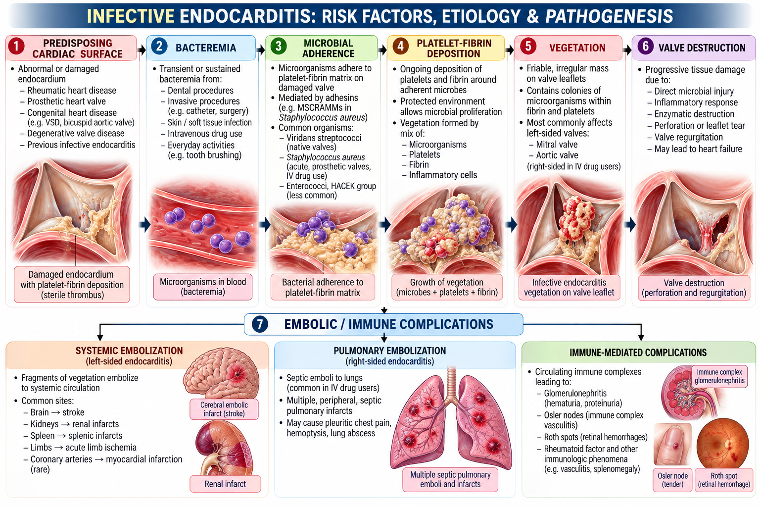
Infective endocarditis develops when microorganisms enter the bloodstream and establish infection on a susceptible endocardial surface. The usual site is a cardiac valve. The resulting infected mass of platelets, fibrin, microorganisms and inflammatory material is called a vegetation.
Who is at risk?
The likelihood of IE rises when the endocardial surface is abnormal or when significant bacteremia occurs. Important clinical risk settings include:
- previous infective endocarditis;
- prosthetic cardiac valves or prosthetic material used in valve repair;
- selected congenital heart disease;
- structural or valvular heart disease;
- intravascular devices and repeated vascular access;
- injection drug use;
- sources of bacteremia, including invasive infection.
Important organisms
Staphylococcus aureus is an important cause of acute IE and may infect previously normal valves. Viridans streptococci classically cause a more indolent infection, particularly in susceptible valves. Enterococci and coagulase-negative staphylococci are also important in appropriate clinical settings, especially prosthetic material and healthcare-associated infection.
How does the vegetation form?
Vegetations can therefore cause disease in two ways: locally, by damaging valves and producing regurgitation or heart failure, and systemically, when embolic or immunological phenomena affect distant organs.

B. Infective Endocarditis: Clinical Presentation & Physical Examination
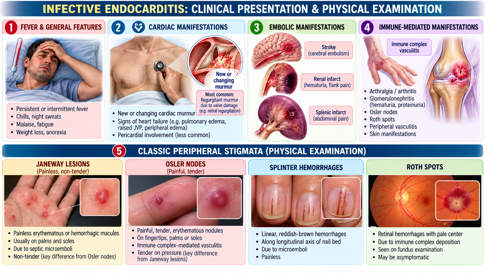
The presentation varies from an acute toxic illness to a more prolonged febrile disease. Clinical findings arise from persistent infection, valvular damage, embolization and immune-mediated injury.
Core clinical pattern
- Fever is the commonest presenting feature.
- Fatigue, malaise, anorexia and weight loss may occur in a more indolent illness.
- A new or changing regurgitant murmur suggests valvular involvement.
- Dyspnea, pulmonary congestion or peripheral edema may indicate developing heart failure.
- Embolization may produce neurological, renal, splenic, limb or pulmonary manifestations depending on the site of infection.
Peripheral findings and what they mean
| Finding | Clinical significance |
|---|---|
| Petechiae | Small hemorrhagic lesions; supportive but not specific. |
| Splinter hemorrhages | Linear nail-bed hemorrhages; may occur in IE but are not diagnostic alone. |
| Janeway lesions | Painless erythematous or hemorrhagic lesions on palms or soles; associated with embolic/infective phenomena. |
| Osler nodes | Painful nodules on fingers or toes; associated with immune phenomena. |
| Roth spots | Retinal hemorrhagic lesions with pale centers; supportive rather than specific. |
Examination in a child with suspected IE
Examination should assess the child’s general condition and fever, look for pallor and peripheral stigmata, evaluate the pulse and circulation, carefully auscultate for a murmur, assess for signs of heart failure and examine for systemic complications such as neurological abnormalities or splenomegaly. The aim is not simply to find a murmur, but to determine whether infection has produced cardiac or extracardiac consequences.
C. Infective Endocarditis: Diagnostic Approach & Diagnostic Criteria
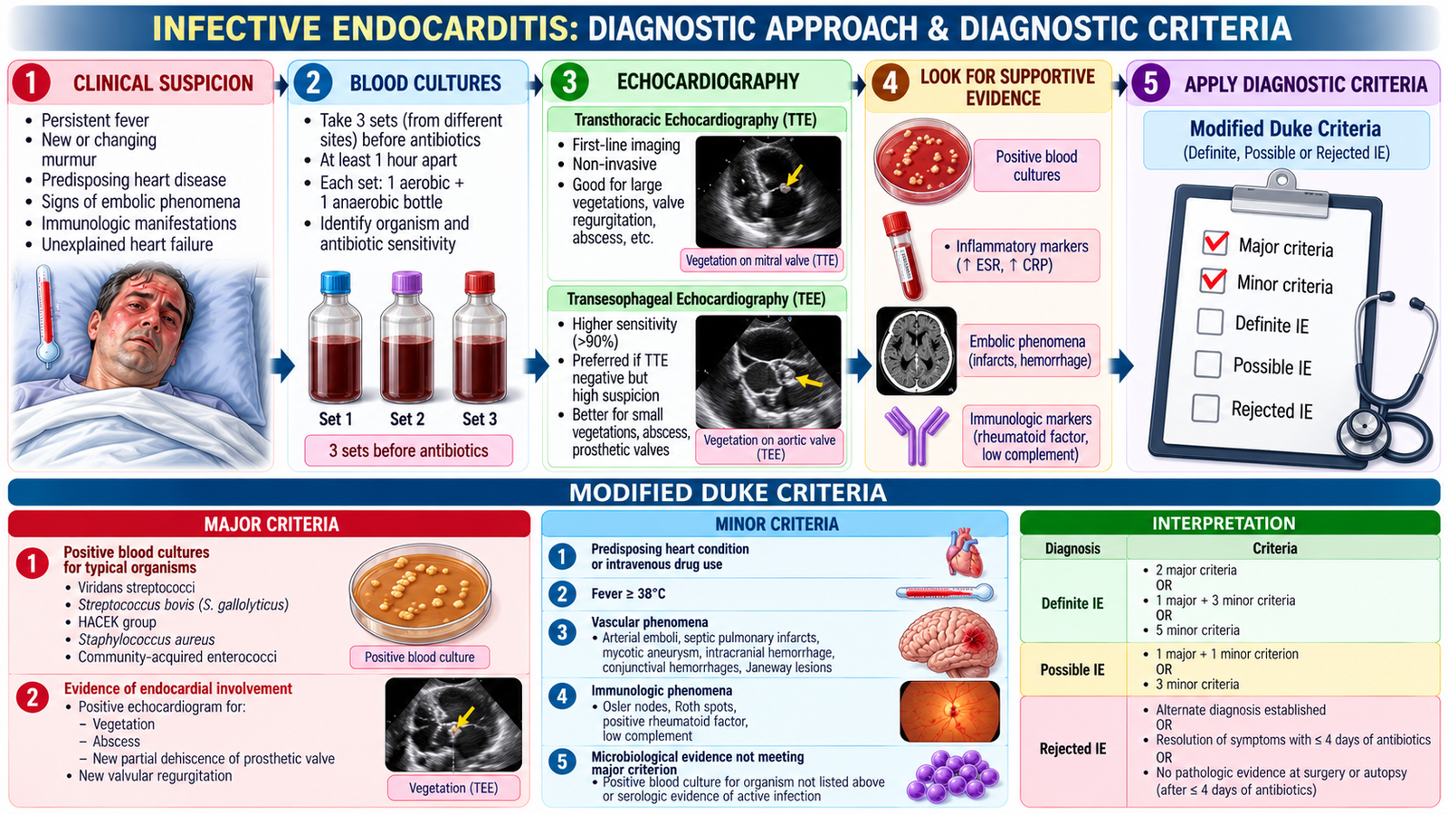
Diagnosis combines the clinical probability of IE with microbiological evidence and evidence of endocardial involvement. Two investigations are especially important: blood cultures identify the causative organism, while echocardiography looks for cardiac involvement such as vegetation or valve complications.
Blood cultures
When the patient is clinically stable, multiple blood-culture samples should be obtained from separate venipuncture sites before antimicrobial therapy. This improves organism detection and permits susceptibility-directed treatment. Prior antibiotics can reduce culture yield and contribute to culture-negative IE.
Echocardiography
Transthoracic echocardiography (TTE) is commonly the initial cardiac imaging test. Transesophageal echocardiography (TEE) provides greater sensitivity for many vegetations and complications and is particularly valuable when clinical suspicion remains high despite a nondiagnostic TTE or when prosthetic material is present.
Diagnostic criteria: understand the logic
Formal diagnostic criteria integrate major evidence of typical bloodstream infection or endocardial involvement with minor clinical evidence such as predisposition, fever and vascular or immunological phenomena. The important undergraduate principle is that IE is not diagnosed from one isolated sign: microbiology, cardiac imaging and the clinical picture must be interpreted together.
Persistent or characteristic positive blood cultures provide major evidence of bloodstream infection.
Evidence of endocardial involvement, such as vegetation or relevant valve pathology, strongly supports the diagnosis.
A child with congenital heart disease develops persistent fever, malaise and a new change in the cardiac murmur. Blood cultures are positive and echocardiography demonstrates a valvular vegetation.
D. Infective Endocarditis: Management, Complications & Prevention
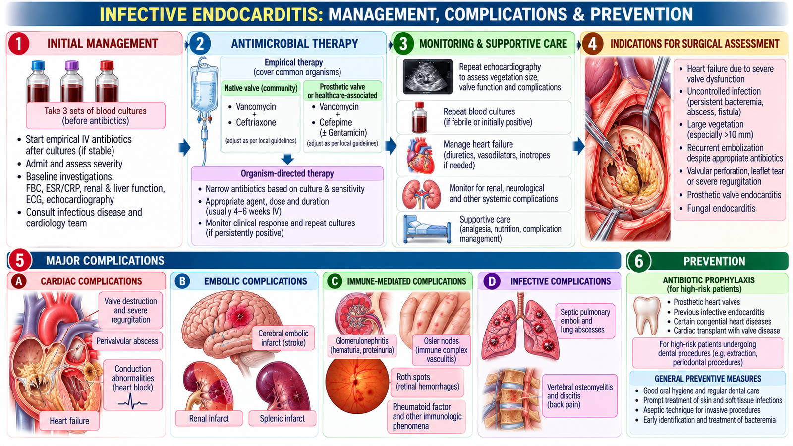
Management has three linked goals: eradicate the infection, identify and manage cardiac damage, and prevent or treat systemic complications. Treatment should be guided by the likely or identified organism and its antimicrobial susceptibility, with specialist input where appropriate.
Antimicrobial treatment
IE usually requires prolonged bactericidal antimicrobial therapy because organisms within vegetations are relatively protected. Empirical therapy is chosen according to the clinical setting after appropriate cultures have been obtained, and is then refined when microbiological results become available.
Specific antibiotic therapy: Treatment usually requires prolonged intravenous antibiotics, commonly for 4–6 weeks, depending on the organism and valve involved. Penicillin G or ceftriaxone may be used for susceptible streptococcal endocarditis; flucloxacillin or cefazolin for methicillin-sensitive Staphylococcus aureus (MSSA); and vancomycin for MRSA when appropriate. Antibiotic selection and duration must be guided by blood cultures, susceptibility results and specialist assessment, including in pediatric patients.
When is surgery considered?
Surgical assessment becomes important when medical treatment alone is unlikely to control the disease—for example, severe valve dysfunction causing heart failure, uncontrolled infection, destructive complications or selected situations with major embolic risk. The Final-Year principle is to recognize why surgery may be required rather than memorize specialist operative protocols.
Major complications
- Valve destruction → acute or progressive regurgitation → heart failure.
- Vegetation fragmentation → systemic embolization from left-sided disease.
- Neurological complications, including embolic cerebral events.
- Renal involvement from embolic or immune mechanisms.
- Perivalvular extension or persistent infection.
- Right-sided disease may produce septic pulmonary embolic complications.
Prevention
Prevention begins with good oral hygiene, appropriate treatment of infections and reduction of avoidable bloodstream infection. Antibiotic prophylaxis is not routine for every patient with a murmur; it is reserved for selected patients at highest risk of serious IE in specific procedural settings according to accepted clinical guidance.
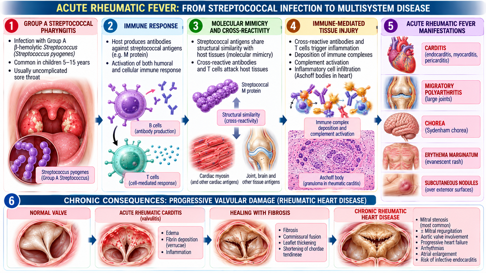
E. Acute Rheumatic Fever: From Streptococcal Infection to Multisystem Disease
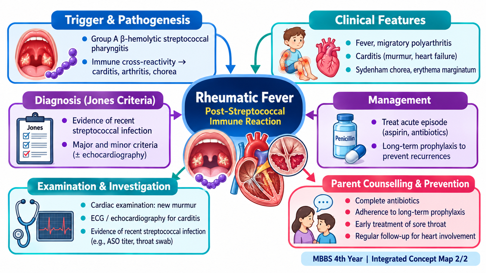
Acute rheumatic fever is a delayed, immune-mediated complication of group A streptococcal pharyngitis. The organism does not directly invade the heart or joints. Instead, an abnormal immune response produces inflammation in susceptible tissues, particularly the heart, joints, skin and central nervous system.
Major clinical manifestations
- Carditis: may involve the endocardium, myocardium and pericardium; valvulitis is clinically important.
- Migratory polyarthritis: typically affects large joints and moves from one joint to another.
- Sydenham chorea: involuntary movements with neurological and behavioural manifestations.
- Erythema marginatum: a characteristic non-pruritic rash.
- Subcutaneous nodules: small, firm nodules associated particularly with significant cardiac disease.
Cardiac importance
Carditis is the manifestation most closely linked to long-term morbidity. Recurrent inflammatory episodes can progressively damage cardiac valves, producing chronic rheumatic heart disease. The mitral valve is most commonly affected, although other valves may also be involved.
F. Rheumatic Fever: Diagnostic Workup & Pediatric Physical Examination
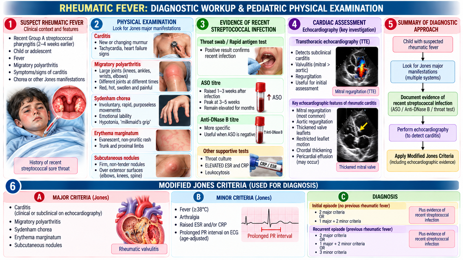
ARF is diagnosed clinically by combining compatible manifestations with evidence of preceding group A streptococcal infection and excluding more likely alternatives. The Jones criteria provide the diagnostic framework.
Major manifestations
Carditis, arthritis, chorea, erythema marginatum and subcutaneous nodules form the major clinical manifestations used in the diagnostic framework. Minor manifestations and supporting investigations contribute to the overall assessment.
Evidence of preceding streptococcal infection
The clinician should seek evidence that a recent group A streptococcal infection preceded the inflammatory illness. Depending on timing and clinical circumstances, this may be supported by throat testing or evidence of an increased/rising streptococcal antibody response such as an antistreptolysin O titre.
Important investigations
- Inflammatory markers support the presence of systemic inflammation.
- ECG may demonstrate conduction abnormalities such as PR prolongation.
- Echocardiography evaluates valvular involvement and may identify carditis even when auscultatory findings are subtle.
- Tests for preceding streptococcal infection support the etiological link.
Physical examination of the child
Assess the child’s general condition and vital signs, then actively look for the multisystem manifestations of ARF. Examine the cardiovascular system for tachycardia, murmurs and signs of heart failure; assess large joints for inflammatory arthritis; observe for choreiform movements; and inspect the skin for erythema marginatum and subcutaneous nodules. Examination findings should be interpreted together rather than in isolation.
G. Pediatric Rheumatic Fever: Management, Prevention & Parent Counselling
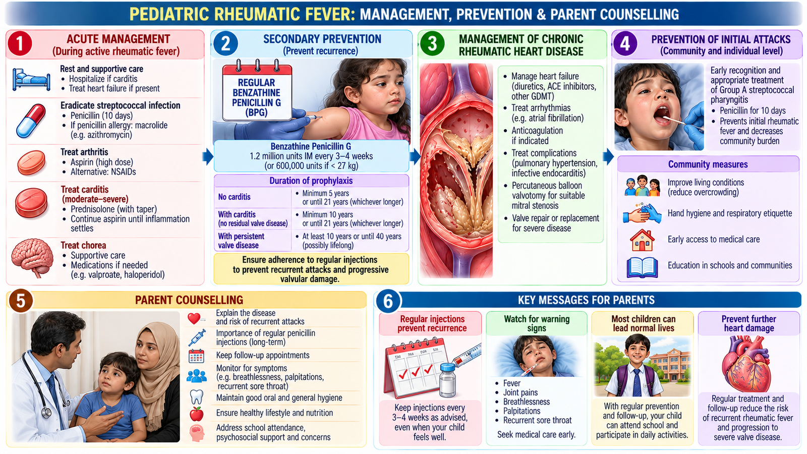
Management addresses the current inflammatory illness while also preventing future streptococcal infections and recurrent rheumatic fever. This long-term preventive component is essential because repeated attacks increase the risk of progressive valvular damage.
Management principles
- Eradicate group A streptococcal infection with appropriate antibiotic therapy.
- Control inflammatory symptoms, particularly arthritis, with appropriate anti-inflammatory treatment.
- Assess and manage carditis and heart failure when present.
- Manage chorea supportively and pharmacologically when clinically significant.
- Plan follow-up according to cardiac involvement and recurrence risk.
Specific treatment: Group A streptococcal infection is eradicated with penicillin, commonly intramuscular benzathine penicillin G. Arthritis is treated with anti-inflammatory medicines such as naproxen or aspirin when appropriate. Rheumatic carditis with heart failure requires specialist management, including diuretics such as furosemide when indicated. Corticosteroids may be considered in selected severe cases of carditis.
Secondary prevention
After ARF, prevention of recurrent streptococcal infection is a major therapeutic goal. Secondary antibiotic prophylaxis reduces recurrent attacks and therefore reduces further immune-mediated valve injury. The duration depends on factors such as previous carditis and residual valvular disease and should follow the appropriate clinical protocol.
Secondary prophylaxis regimen: Intramuscular benzathine penicillin G every 3–4 weeks is the preferred preventive treatment; oral penicillin V is an alternative when appropriate. Prophylaxis must continue for several years, with duration determined by previous carditis, residual valvular disease and recurrence risk. Long-term adherence is essential to prevent recurrent rheumatic fever and progressive valve damage.
What parents need to understand
- ARF occurs after a streptococcal throat infection because of an abnormal immune response.
- Cardiac involvement can lead to long-term valve disease.
- Completing treatment and adhering to secondary prophylaxis are important.
- Future sore throats should receive appropriate clinical assessment rather than being ignored or self-treated.
- Follow-up is important, especially when carditis or residual valve disease is present.
- Parents should seek medical review if the child develops breathlessness, edema, marked exercise intolerance, persistent fever or other concerning symptoms.
Counselling in pediatric infective endocarditis
For a child with IE, explain that the infection involves the heart lining or valve and requires adequate antimicrobial treatment and follow-up. Parents should understand the importance of completing therapy, monitoring for cardiac or systemic complications, maintaining good oral health and informing relevant clinicians of important cardiac history. Prevention advice should be individualized rather than implying that every child with heart disease requires antibiotic prophylaxis before every procedure.
AIM High-Yield Review
- ⭐ IE: bacteremia + susceptible endocardial surface → infected vegetation → valve destruction, embolization and systemic manifestations.
- In suspected IE, obtain appropriate blood cultures before antibiotics when clinically feasible; cultures identify the organism while echocardiography assesses endocardial involvement.
- ⭐ Fever + compatible cardiac findings + positive cultures + echocardiographic evidence is a powerful IE pattern; no single peripheral sign is diagnostic.
- Janeway lesions are painless; Osler nodes are painful.
- Severe valve dysfunction with heart failure, uncontrolled infection or destructive complications should prompt early surgical assessment.
- IE prophylaxis is selective, not routine for every patient with a cardiac murmur.
- ⭐ Acute rheumatic fever is immune mediated and follows group A streptococcal pharyngitis; it is not direct bacterial invasion of the heart.
- The major ARF manifestations are carditis, migratory polyarthritis, chorea, erythema marginatum and subcutaneous nodules.
- Diagnosis of ARF uses the Jones framework plus evidence of preceding streptococcal infection, interpreted in the appropriate clinical setting.
- ⭐ Carditis determines much of the long-term risk because recurrent inflammation can progress to chronic rheumatic valvular disease.
- Echocardiography is important in suspected rheumatic carditis because clinically important valvular involvement may be detected even when auscultatory findings are subtle.
- Secondary prophylaxis after ARF prevents recurrent attacks and thereby reduces repeated immune-mediated valve injury.
Infective Endocarditis & Rheumatic Fever Across the Lifespan
Watch this after completing the learning material to reinforce pathogenesis, Jones criteria, infective endocarditis organisms, blood cultures and diagnostic approach.