
Learn this topic as a clinical journey: recognize the respiratory presentation → identify severity → localize the likely infection or suppurative process → choose appropriate investigations → start management and recognize when urgent referral is needed.
Topic 7. Respiratory Infections, Suppurative Lung Disease & Acute Cough/Shortness of Breath Care
CVS Module • Integrated Medicine, Pediatrics & Family Medicine
A clinically integrated approach to pneumonia, pulmonary tuberculosis, bronchiectasis, lung abscess, childhood acute respiratory infections, croup, and the primary-care assessment of acute cough and breathlessness.
Topic Introduction
Acute cough, fever and shortness of breath are among the commonest respiratory presentations, but they range from uncomplicated infection to serious pneumonia, tuberculosis or destructive suppurative lung disease. The important skill is not simply memorizing diseases; it is recognizing the pattern, assessing severity, examining the respiratory system, selecting investigations that answer a clinical question and starting appropriate management. In children, age-specific presentations and deterioration can be rapid, while in primary care the key task is to separate common illness from red flags requiring urgent referral. This chapter connects these problems into one manageable clinical approach.
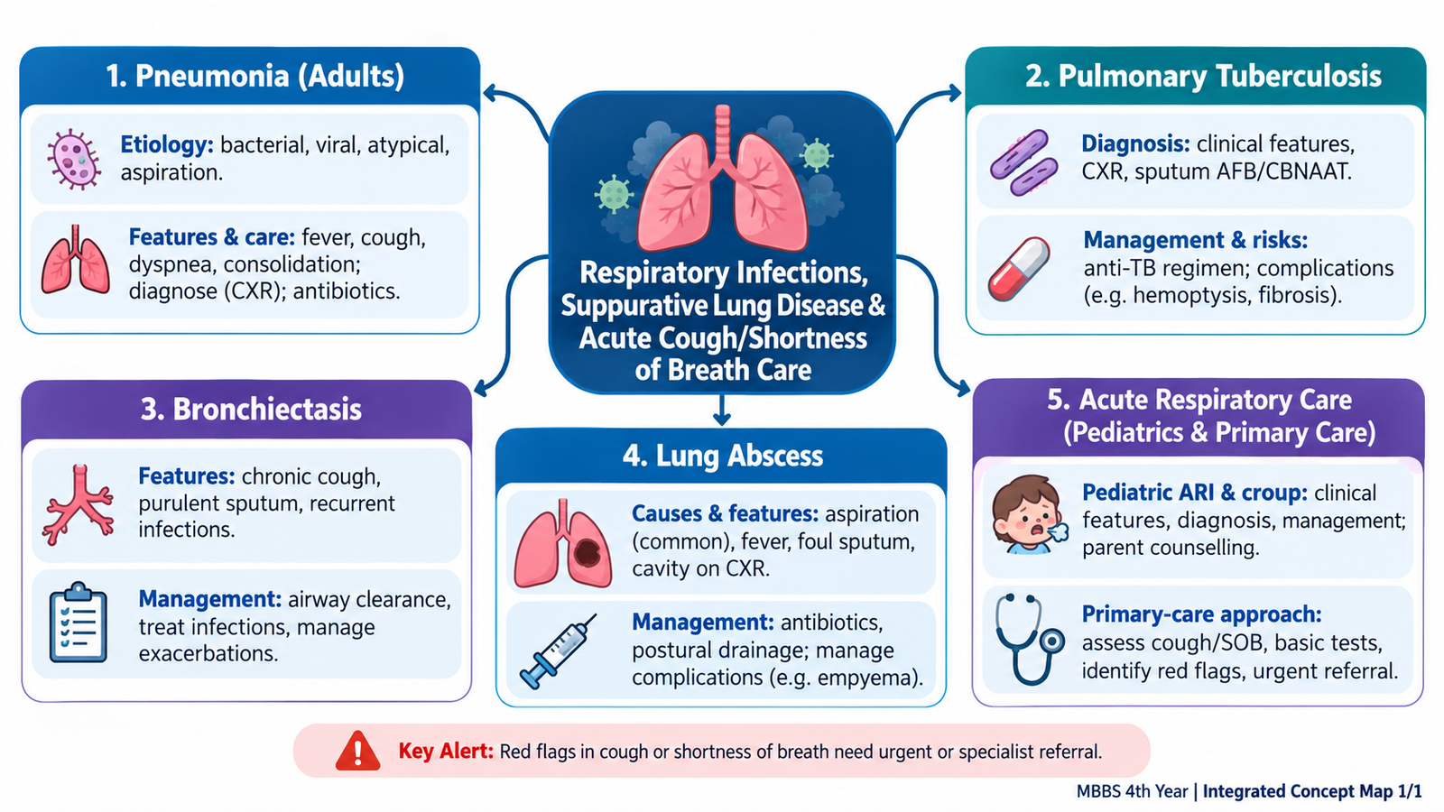
1. Pneumonia in Adults: From Infection to Clinical Management
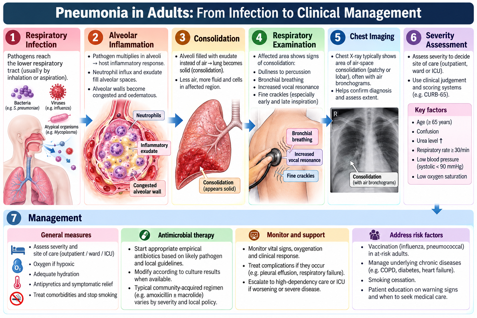
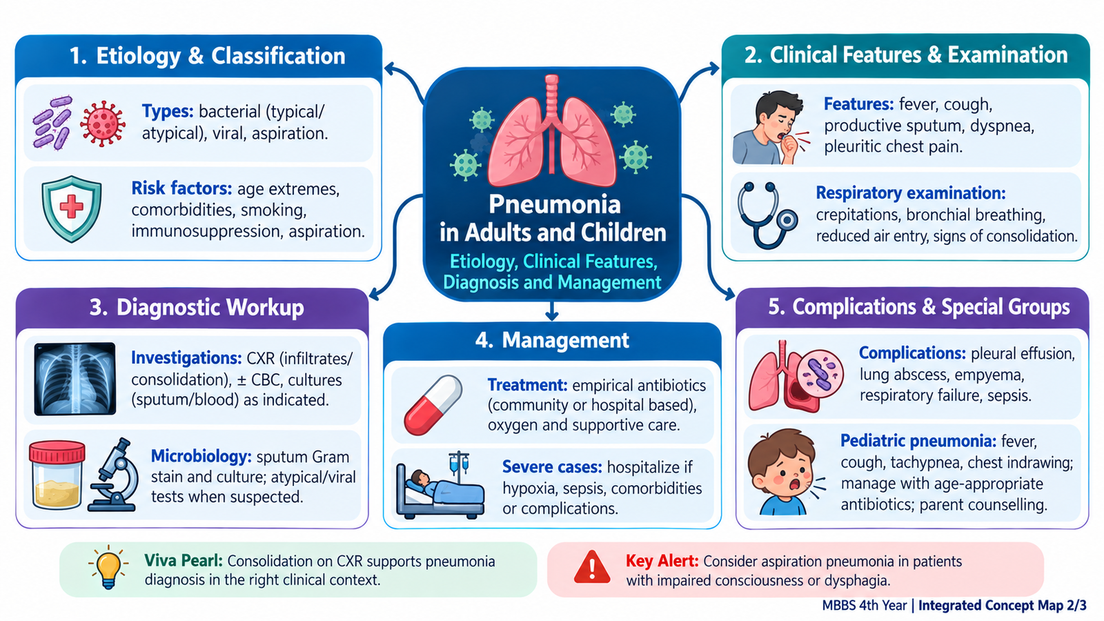
Pneumonia is an acute infection of the lung parenchyma. Infection produces inflammation and filling of alveolar spaces with inflammatory exudate. This reduces normal gas exchange and explains the combination of fever, cough, sputum production, breathlessness and focal chest signs. A useful clinical approach combines the setting of infection, likely cause, severity and patient risk factors.
Etiology and classification
Pneumonia can be approached according to where and how it develops. Community-acquired pneumonia develops outside the hospital setting, while hospital-acquired pneumonia develops in relation to hospitalization. Aspiration can produce pneumonia when contaminated oropharyngeal or gastric material enters the lower respiratory tract. Pneumonia may also be described by its clinical or radiological pattern, such as lobar or bronchopneumonic involvement.
Typical bacterial pathogens commonly produce an acute febrile illness with productive cough and focal consolidation. Other pathogens may produce a less classical pattern with dry cough or more diffuse respiratory and systemic features. The organism cannot be reliably identified from symptoms alone, so microbiological testing is selected according to severity and clinical context.
Risk factors
Risk increases when normal airway protection or host defence is impaired. Important contexts include advanced age, chronic respiratory or systemic disease, impaired consciousness or swallowing, aspiration risk, smoking, reduced immunity and recent healthcare exposure. These factors also influence severity and likely complications.
Clinical presentation and respiratory examination
Common symptoms include fever, cough, sputum production, pleuritic chest pain and dyspnea. The examination must assess both local lung involvement and overall physiological severity.
- General assessment: respiratory rate, oxygenation, pulse, blood pressure, temperature, mental state and signs of respiratory distress.
- Inspection: tachypnea, increased work of breathing or reduced movement of an affected area.
- Palpation: expansion may be reduced over the affected region; vocal fremitus may increase with consolidation.
- Percussion: consolidation may produce dullness.
- Auscultation: crackles, bronchial breathing and increased vocal resonance support alveolar consolidation.
These findings are not isolated examination facts. Alveoli filled with inflammatory material become relatively airless but transmit sound efficiently, producing the characteristic physical signs of consolidation.
Diagnosis and investigations
The diagnosis begins clinically. Chest radiography supports the diagnosis, defines the distribution of disease and may reveal complications such as pleural involvement. Blood tests help assess inflammation, systemic impact and organ dysfunction. Microbiological specimens are particularly useful in severe, complicated, unusual or treatment-unresponsive disease.
Management principles
Management depends on clinical severity, patient factors and likely cause. Core principles are appropriate antimicrobial therapy when bacterial pneumonia is suspected, oxygen for hypoxemia, hydration and supportive care, treatment of relevant comorbidity, and reassessment for deterioration. Severe physiological disturbance, inability to maintain oxygenation or circulation, altered consciousness, major comorbidity or complications should lower the threshold for hospital-level care.
Antibiotic treatment: Mild community-acquired pneumonia is commonly treated with oral amoxicillin; doxycycline is an alternative in suitable adults. Hospitalized patients may require IV ceftriaxone with azithromycin or another appropriate regimen according to severity and local resistance patterns. Assess severity using CRB-65/CURB-65 alongside clinical judgment to guide admission. Review antibiotic response within 48–72 hours.

2. Pulmonary Tuberculosis: Recognition, Diagnosis, Management & Complications
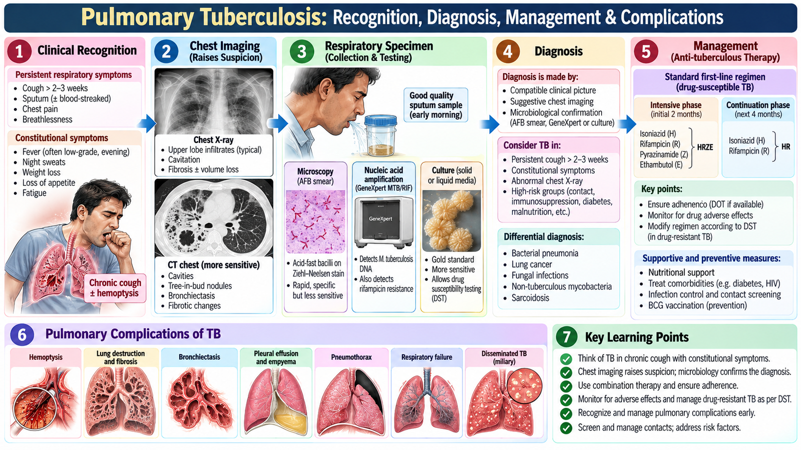
Pulmonary tuberculosis is an important chronic respiratory infection. Unlike the abrupt presentation of many acute pneumonias, pulmonary TB commonly develops more gradually. The key clinical task is to recognize a compatible persistent respiratory and constitutional pattern, establish microbiological evidence where possible, assess the extent of disease and ensure effective treatment and follow-up.
Clinical recognition
Features that should raise suspicion include persistent cough, sputum production, hemoptysis, fever, night sweats, reduced appetite and weight loss. Exposure history, previous TB, immunosuppression and relevant living or social circumstances can increase suspicion. Examination may be relatively nonspecific, so a normal or mildly abnormal chest examination does not exclude disease.
Diagnostic workup
The diagnostic approach connects clinical suspicion → chest imaging → microbiological confirmation. Chest radiography helps identify the pattern and extent of pulmonary involvement, but imaging alone does not establish the organism. Respiratory specimens are therefore central to demonstrating Mycobacterium tuberculosis using appropriate microbiological methods. Drug-susceptibility information becomes important when resistance is suspected or demonstrated.
Management
Treatment requires a combination of effective anti-tuberculous drugs rather than single-drug therapy. The undergraduate principle is to understand why: combination treatment improves eradication of organisms and reduces the emergence of resistance. Treatment should follow the appropriate TB programme or local protocol, with attention to adherence, adverse effects, response and drug resistance.
Patient education is essential. Explain the need to complete treatment, attend follow-up, report important adverse effects and follow infection-control advice. Contacts and public-health measures should be managed according to the appropriate local TB programme.
First-line treatment: Drug-susceptible pulmonary tuberculosis is treated with a standard 6-month regimen (2HRZE/4HR):
-
Intensive phase (2 months): Isoniazid (H), rifampicin (R), pyrazinamide (Z) and ethambutol (E), given daily.
-
Continuation phase (4 months): Isoniazid (H) and rifampicin (R), given daily.
Pyridoxine (vitamin B6) is given with isoniazid to patients at risk of peripheral neuropathy, including pregnant women, patients with diabetes and malnourished patients.
Monitoring and adverse effects:
-
Isoniazid: Hepatotoxicity and peripheral neuropathy.
-
Rifampicin: Hepatotoxicity, drug interactions and orange discoloration of body fluids.
-
Pyrazinamide: Hepatotoxicity and hyperuricemia.
-
Ethambutol: Optic neuritis and impaired color vision.
Follow-up: Ensure medication adherence, monitor clinical response and adverse effects, and assess bacteriological response according to national TB programme guidelines. Provide infection-control advice, contact screening and patient counselling.
Drug-resistant TB: Requires specialist management with susceptibility-guided treatment under the national TB programme. Persistent symptoms or poor response should prompt reassessment for non-adherence, resistance, complications or an alternative diagnosis.
Complications
Pulmonary destruction can lead to hemoptysis, fibrosis, bronchiectatic change and impaired respiratory function. Pleural involvement and dissemination beyond the lungs may also occur. Persistent or worsening symptoms despite treatment should prompt reassessment for adherence problems, complications, an alternative diagnosis or drug resistance.
3. Suppurative Lung Disease: Bronchiectasis & Lung Abscess
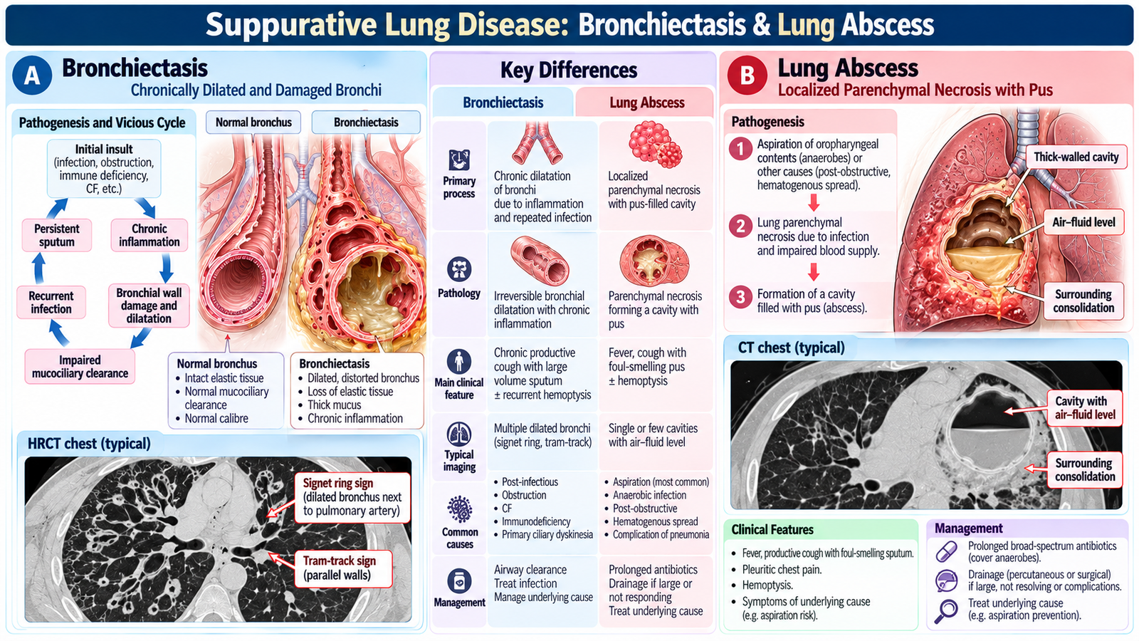
Bronchiectasis and lung abscess both involve persistent or destructive pulmonary infection, but the structural problem differs. Bronchiectasis is characterized by permanently abnormal dilatation of bronchi associated with impaired clearance and recurrent infection. A lung abscess is a localized area of pulmonary suppuration with tissue necrosis and cavity formation.
Bronchiectasis: the vicious cycle
Airway injury impairs mucociliary clearance. Retained secretions then promote infection and inflammation, which cause further airway damage. This creates a self-perpetuating cycle:
The typical clinical pattern is chronic or recurrent productive cough, often with substantial sputum, repeated infective exacerbations and sometimes hemoptysis. Coarse crackles may be present. Imaging, particularly appropriate cross-sectional imaging, demonstrates the abnormal dilated bronchi and establishes the structural diagnosis.
Management of bronchiectasis
Management aims to interrupt the infection–retention–damage cycle. Important principles include airway clearance, treatment of infective exacerbations, identifying and treating an underlying cause where possible, vaccination and prevention where appropriate, and specialist assessment when disease is severe, recurrent or complicated. Hemoptysis, worsening respiratory function or repeated severe infections require careful reassessment.
Specific treatment: Regular airway clearance physiotherapy is central to long-term management. Acute infective exacerbations require antibiotics guided by sputum culture, commonly amoxicillin or co-amoxiclav when appropriate. Recurrent exacerbations, especially with Pseudomonas aeruginosa, require respiratory specialist assessment for targeted treatment.
Lung abscess
A lung abscess develops when severe localized infection produces necrosis and cavitation. Aspiration is an important mechanism, particularly when airway protection is impaired. Other infections or obstruction may also contribute.
Patients may have fever, systemic illness, cough and purulent or foul-smelling sputum. Imaging typically demonstrates a cavity, often with an air–fluid level. The cavity represents necrotic lung containing gas and infected material.
Management and complications of lung abscess
Management requires appropriate antimicrobial treatment and correction of contributing factors such as aspiration risk. Failure to improve should trigger reassessment for inadequate drainage, an obstructing lesion, resistant or unusual infection, or another cavitating pulmonary disorder. Important complications include pleural spread, sepsis, significant hemoptysis and persistent structural lung damage.
Antibiotic treatment: Lung abscess requires prolonged antibiotics covering oral anaerobes and other likely organisms, commonly IV ampicillin-sulbactam initially, followed by oral amoxicillin-clavulanate when clinically stable. Treatment often continues for several weeks according to clinical and radiological response. Failure to improve may require drainage assessment and investigation for bronchial obstruction or empyema.
4. Acute Respiratory Infections & Pneumonia in Children
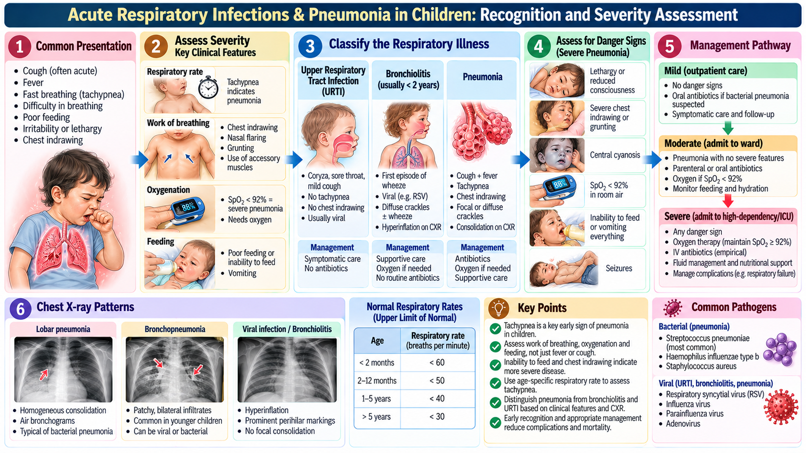
Children with acute respiratory infection may present with cough, fever, nasal symptoms, noisy breathing, poor feeding or breathlessness. The priority is to decide whether the illness is predominantly upper or lower respiratory, determine how unwell the child is, and recognize signs suggesting hypoxemia, respiratory distress or systemic deterioration.
Clinical presentation and assessment
Assessment must be age-appropriate. In infants, serious respiratory illness may present with poor feeding, irritability, lethargy or apnea rather than a clear verbal complaint of breathlessness. In older children, cough, fever, chest discomfort and breathing difficulty may be more obvious.
Important examination features include:
- respiratory rate and overall work of breathing;
- chest recession or use of accessory muscles;
- oxygenation and color;
- ability to feed, drink and interact;
- air entry, crackles or other focal chest findings;
- mental state, hydration and circulation.
The examination is not simply about naming chest sounds. A child who is tiring, hypoxemic, poorly perfused, unable to feed or becoming less responsive may have severe disease even before the exact pathogen is known.
Pediatric pneumonia
Pneumonia should be considered when an acute febrile respiratory illness is associated with lower-respiratory features such as tachypnea, increased work of breathing, hypoxemia or focal chest findings. Investigations are guided by severity. Mild uncomplicated disease can often be assessed clinically, whereas severe, atypical or complicated disease may require imaging and laboratory or microbiological evaluation.
Management
Management combines supportive care with treatment directed at the likely cause. Oxygen is important when hypoxemia is present. Hydration and feeding need attention because respiratory distress can interfere with oral intake. Appropriate antimicrobial therapy is used when bacterial pneumonia is suspected. The need for admission or urgent escalation depends more on physiological severity and the child’s ability to maintain oxygenation, hydration and feeding than on the label of pneumonia alone.
Pediatric pneumonia treatment: Oral amoxicillin is first-line treatment for uncomplicated bacterial community-acquired pneumonia. Severe pneumonia requires admission, oxygen when hypoxemic and parenteral antibiotics such as IV ampicillin or ceftriaxone according to age, severity and local guidelines. Viral upper respiratory infections generally require supportive care without routine antibiotics.
Parent counselling
Parents should understand the expected course of the illness, how prescribed treatment should be used, the importance of fluids and feeding, and which warning signs require urgent reassessment. Counselling should particularly emphasize worsening breathing difficulty, reduced intake, increasing drowsiness, color change, persistent deterioration or other signs that the child is becoming seriously unwell.
5. Croup: Recognizing Upper-Airway Obstruction in a Child
Croup is an acute upper-airway illness in young children in which inflammation and edema around the laryngeal and subglottic region narrow an already small pediatric airway. This explains the characteristic combination of barking cough, hoarse voice and inspiratory stridor.
Why stridor occurs
Upper-airway narrowing creates turbulent airflow, producing the harsh inspiratory sound of stridor. As obstruction becomes more severe, the child develops increased work of breathing. Stridor at rest, marked recession, exhaustion, hypoxemia or reduced responsiveness indicates more serious airway compromise.
Diagnostic approach
Croup is usually recognized clinically from the characteristic presentation and assessment of severity. The child should be kept as calm as possible because distress can worsen upper-airway obstruction. Routine investigations are not the priority in a typical presentation; atypical features or diagnostic uncertainty should prompt consideration of other causes of acute upper-airway obstruction.
Management principles
Treatment is directed at reducing airway inflammation and supporting breathing. Corticosteroid therapy reduces airway edema. Children with more significant obstruction may require additional acute therapy and close observation. Severe airway compromise requires urgent escalation and appropriately skilled airway support.
Specific treatment: Give oral dexamethasone 0.15–0.6 mg/kg according to croup severity and local protocol. Moderate-to-severe croup with significant stridor or respiratory distress may require nebulized adrenaline under monitoring. Observe for recurrence after adrenaline; impending respiratory failure requires urgent specialist airway management. Routine antibiotics are not indicated for uncomplicated viral croup.

🩺 AIM Clinical Bridge
A patient presents with cough and shortness of breath. The first useful question is not simply “Which infection is this?” First determine whether the patient is physiologically stable, then use the time course, sputum pattern, constitutional symptoms, examination and imaging to narrow the diagnosis.
Think clinically: Acute fever + focal consolidation → think pneumonia.
Think clinically: Persistent cough + weight loss/night sweats → consider pulmonary TB.
Think clinically: Chronic copious sputum/recurrent infection → consider bronchiectasis.
6. Primary-Care Approach to Acute Cough & Shortness of Breath
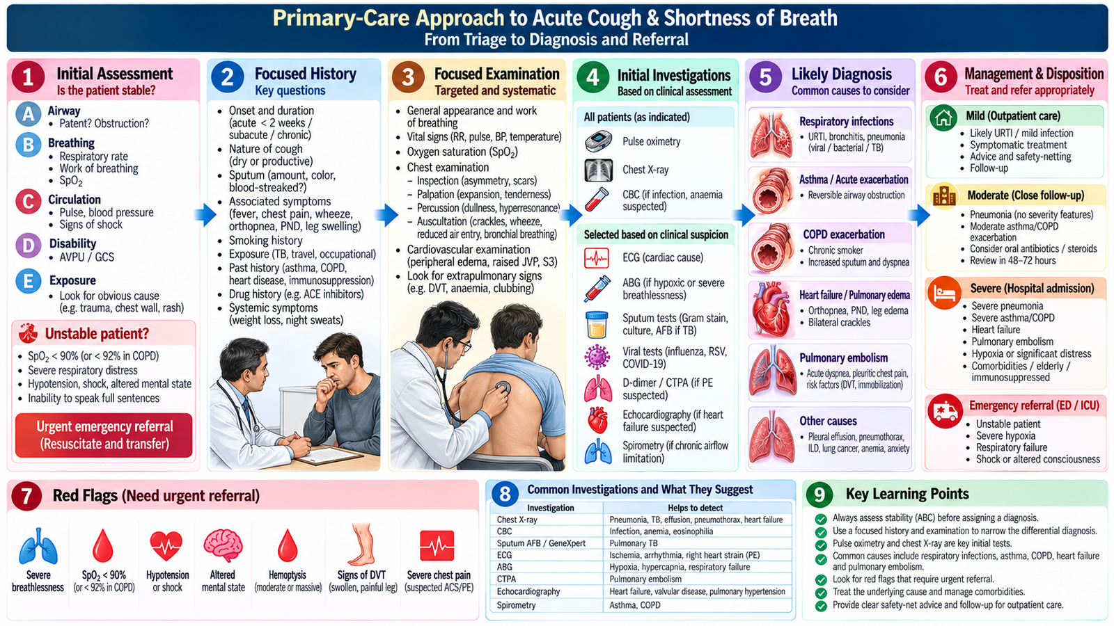
In family practice, cough and shortness of breath are presentations rather than diagnoses. The task is to identify the likely cause while rapidly separating stable patients suitable for primary-care management from those who need urgent investigation, emergency treatment or specialist referral.
Step 1 — Establish urgency
Begin with the patient’s overall appearance, airway, breathing and circulation. Assess respiratory distress, oxygenation, respiratory rate, pulse, blood pressure, mental state and ability to speak or function normally. Severe breathlessness, cyanosis or hypoxemia, hemodynamic instability, altered consciousness, major hemoptysis or rapidly progressive symptoms are red flags.
Step 2 — Use the history to define the pattern
The time course immediately narrows the problem. Acute cough with fever and sputum suggests respiratory infection, while pleuritic pain may accompany pneumonia or pleural disease. Sudden breathlessness requires consideration of important acute cardiopulmonary causes rather than assuming infection. Persistent cough, constitutional symptoms or hemoptysis should trigger evaluation for important chronic pulmonary disease, including TB where clinically appropriate.
Ask about sputum, fever, wheeze, chest pain, hemoptysis, orthopnea, exposure to infection, smoking, aspiration risk, previous respiratory or cardiac disease, medications and immune status. The history should guide examination rather than produce an indiscriminate list of tests.
Step 3 — Focused examination
Look for respiratory distress and systemic illness, then examine the chest for asymmetry, reduced expansion, percussion changes, altered breath sounds, crackles or wheeze. Cardiac examination and assessment for peripheral edema may be important when breathlessness could have a cardiovascular cause.
Step 4 — Select investigations according to the clinical question
Not every patient with cough needs extensive investigation. Pulse oximetry helps assess oxygenation. Chest radiography is useful when pneumonia, significant structural disease or another important intrathoracic process is suspected. Blood tests and microbiological studies are selected according to severity, duration and diagnostic suspicion. Persistent or unexplained symptoms require reassessment rather than repeated symptomatic treatment without a diagnosis.
Step 5 — Treat, safety-net or refer
Stable uncomplicated illness may be managed in primary care with appropriate treatment, supportive advice and planned reassessment. Patients with severe physiological disturbance, significant diagnostic uncertainty, major comorbidity, suspected serious disease, failure to improve or complications should be referred to the appropriate level of care.
7. Connecting the Respiratory Presentation to the Correct Diagnosis
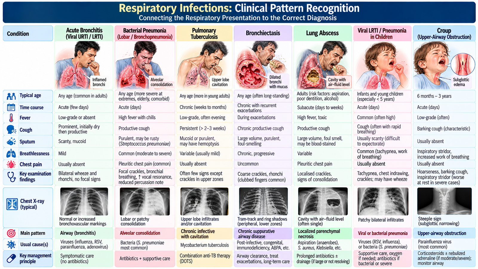
The final clinical skill is pattern recognition without premature closure. Several disorders in this topic cause cough and breathlessness, but their time course, sputum pattern, systemic features, examination and imaging point in different directions.
| Pattern | Pneumonia | Pulmonary TB | Bronchiectasis | Lung Abscess |
|---|---|---|---|---|
| Typical course | Acute | Usually persistent/subacute or chronic | Chronic with exacerbations | Acute/subacute suppurative illness |
| Key clue | Fever + focal respiratory findings | Persistent cough + constitutional symptoms | Recurrent infection + chronic productive cough | Toxic illness + purulent/foul sputum |
| Structural basis | Inflamed, exudate-filled alveoli | Chronic mycobacterial lung disease | Permanently dilated damaged bronchi | Necrotic cavitary lung lesion |
| Important investigation link | Chest imaging ± selected microbiology | Imaging + respiratory microbiology | Imaging demonstrates bronchial dilatation | Cavity ± air–fluid level on imaging |
The reasoning principle
Do not allow one feature—such as cough, fever or an abnormal chest radiograph—to make the entire diagnosis. Combine the tempo of illness + risk context + examination + investigation pattern. At the same time, severity assessment runs in parallel: a patient can require urgent stabilization before the final diagnostic label is established.
Integrated Mechanism Flow
AIM High-Yield Review
- ⭐ Pneumonia: alveolar inflammation and exudate explain crackles, bronchial breathing and impaired gas exchange.
- Adult pneumonia assessment: diagnosis matters, but physiological severity determines urgency and site of care.
- ⭐ Pulmonary TB: persistent respiratory symptoms plus constitutional features should lead to imaging and microbiological evaluation rather than repeated treatment as an acute infection.
- Bronchiectasis: airway damage → impaired clearance → retained secretions → recurrent infection → further airway damage.
- ⭐ Lung abscess: pulmonary necrosis and cavitation can produce purulent/foul sputum and a cavitary lesion with an air–fluid level.
- Pediatric respiratory infection: feeding, behavior, oxygenation and work of breathing are major indicators of severity.
- ⭐ Croup: barking cough + hoarseness + inspiratory stridor reflect upper-airway narrowing; stridor at rest or exhaustion indicates greater severity.
- Respiratory examination: interpret findings through the underlying structural change rather than memorizing isolated signs.
- Primary care: acute cough or breathlessness must first be screened for physiological instability and red flags before routine diagnostic categorization.
- Imaging: consolidation suggests alveolar disease, bronchial dilatation supports bronchiectasis, and a cavitary air–fluid pattern supports abscess in the correct clinical context.
- Microbiology: use it when identifying the organism changes diagnosis or management, particularly in TB and significant or complicated infection.
- Safety-netting: worsening breathlessness, hypoxemia/cyanosis, altered consciousness, poor perfusion, major hemoptysis or rapid deterioration require urgent reassessment or referral.
Respiratory Infections, Suppurative Lung Disease & Acute Cough/Shortness of Breath Care
Watch these focused videos alongside the AIM Learning Material to reinforce the major clinical concepts.
1. Adult Pneumonia
Causes, pathophysiology, clinical features, diagnosis, imaging and management.
2. Pulmonary Tuberculosis
Microbiology, pathophysiology, clinical presentation, diagnosis and treatment principles.
3. Bronchiectasis
Etiopathogenesis, clinical features, diagnostic approach and management.
4. Pediatric Croup
Barking cough, stridor, severity assessment, treatment and warning signs.