🩺 Station 11 — Pathology Sampling and Lab Processing Identification
📋 Complete OSPE Station Content
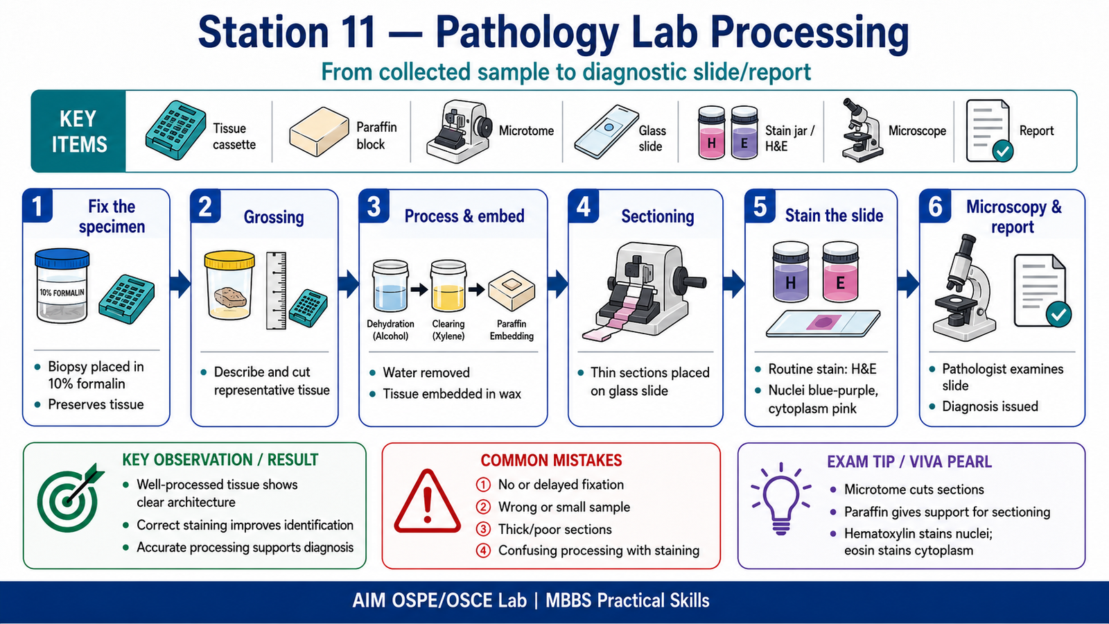
OSPE Station Name
Station 11 — Pathology Sampling and Lab Processing Identification
Learning Target
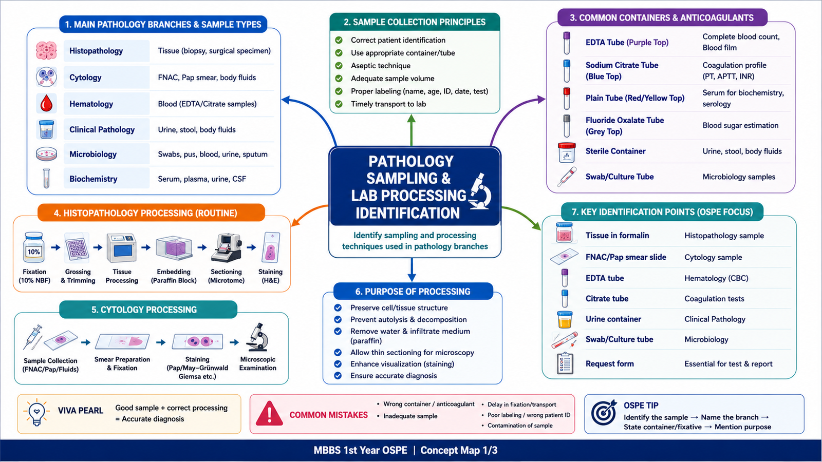
- Identify common pathology samples and processing techniques used in histopathology, cytology, hematology, clinical pathology, and microbiology.
- Explain the basic purpose of fixation, labeling, transport, processing, staining, and reporting in pathology laboratory diagnosis.
Required Material
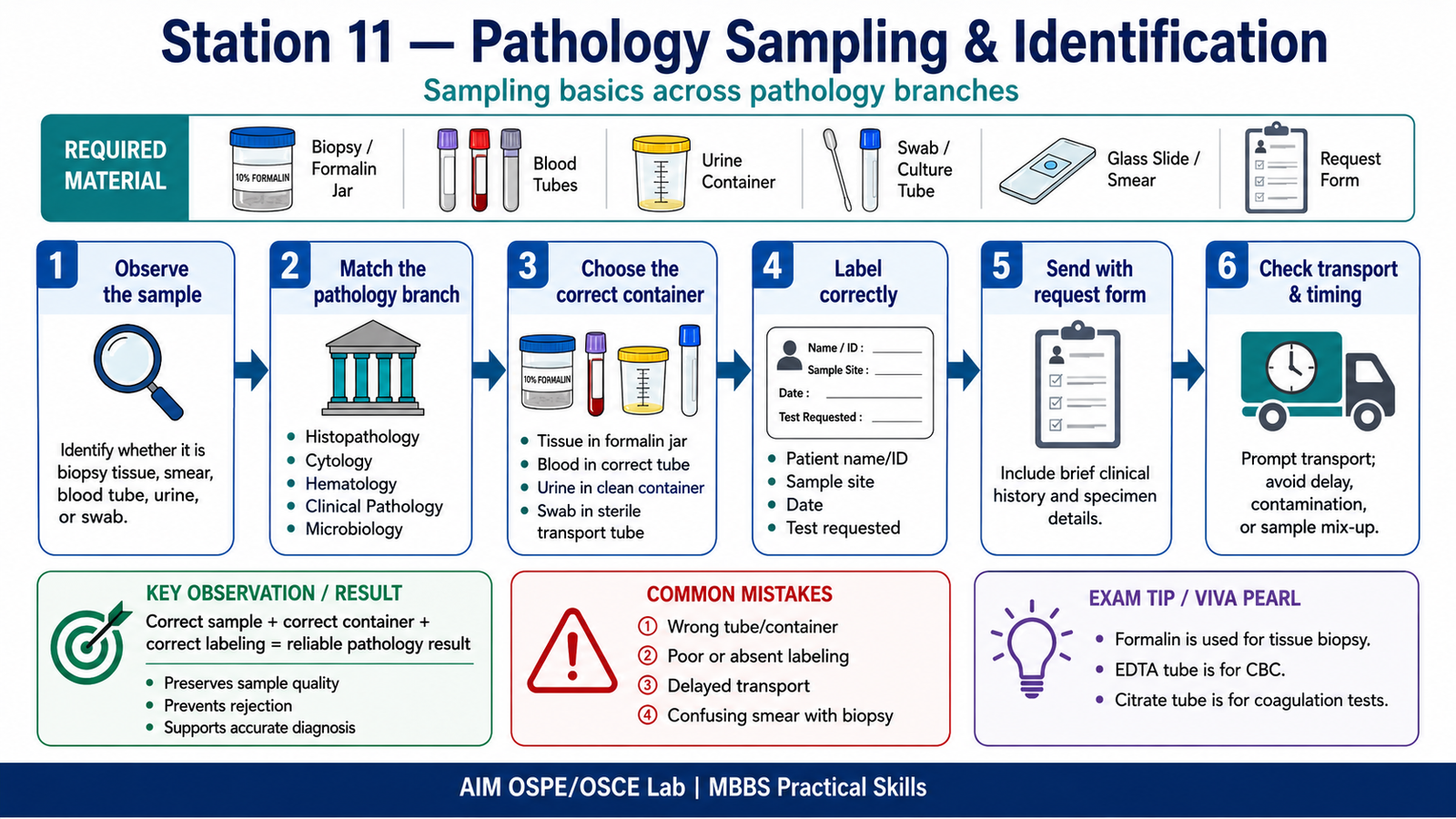
Labeled or unlabeled sample containers
Biopsy specimen container with formalin
Blood collection tubes: EDTA, citrate, plain tube
Urine container
Swab container or culture tube
Glass slides for smear preparation
FNAC/Pap smear slide image or model
Paraffin block or tissue cassette
Microtome image/model
H&E stained slide image
Pathology request form
Pointer
Student Task / Procedure
- Observe the given sample, container, tube, slide, or instrument.
- Identify the pathology branch related to it.
- Name the sample or processing technique.
- Mention the correct fixative, anticoagulant, or transport method where relevant.
- State the basic purpose of the technique.
- Identify one common error that can affect the final laboratory result.
- Mention one clinical use of the sample or technique.
Observation / Identification Points
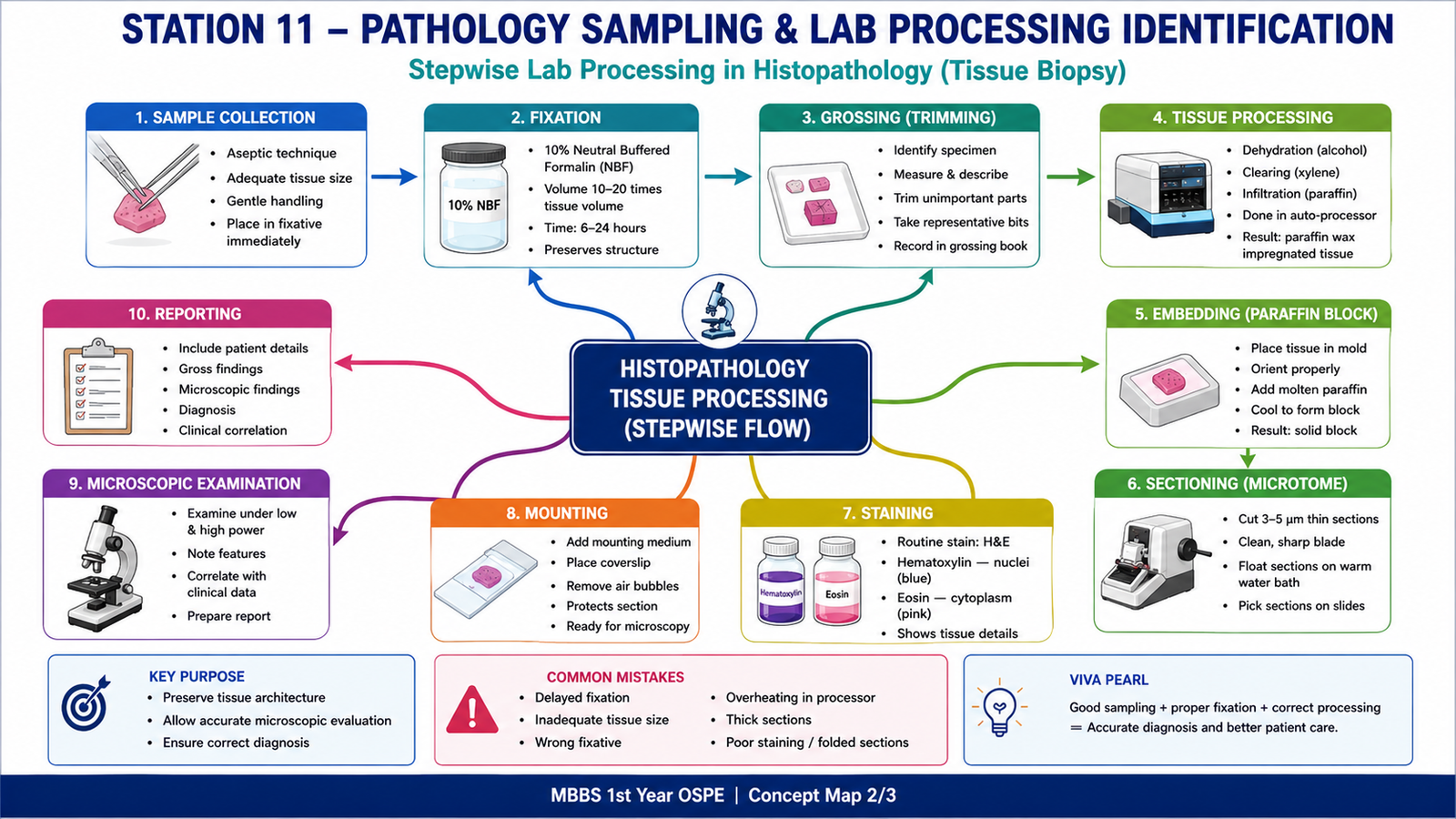
Biopsy specimen → histopathology
Formalin container → tissue fixation
Tissue cassette/paraffin block → tissue processing and embedding
Microtome → thin tissue section cutting
H&E slide → routine histopathological staining
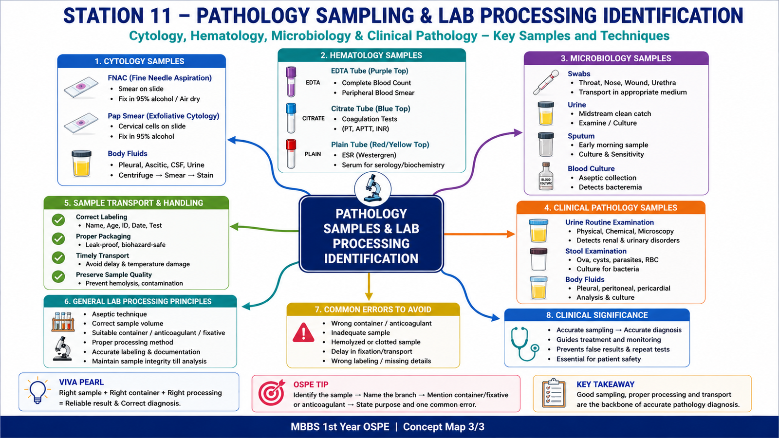
FNAC smear → cytology
Pap smear → exfoliative cytology
EDTA tube → complete blood count and blood smear
Citrate tube → coagulation profile
Plain tube → serum biochemistry or serology
Urine container → routine urine examination
Sterile swab/culture container → microbiology sample collection
Request form → patient identification, clinical history, sample site, and required test
Result / Interpretation
Correct sampling and processing are essential for accurate pathology diagnosis. A properly collected, labeled, fixed, transported, and processed sample preserves cellular and tissue details and prevents false results.
Clinical significance:
Wrong container, delayed fixation, inadequate sample, wrong anticoagulant, or poor labeling can lead to tissue autolysis, clotting, hemolysis, contamination, rejected samples, or incorrect diagnosis.
Common Student Mistakes
- Confusing histopathology biopsy with cytology smear.
- Forgetting that formalin is used for tissue fixation.
- Using EDTA tube for coagulation studies instead of citrate tube.
- Ignoring patient identification and sample labeling.
- Confusing tissue processing steps with staining steps.
AIM Feedback
To improve, first identify the sample type, then link it to the correct pathology branch. Remember the basic chain: sample collection → labeling → fixation or anticoagulant → transport → processing → staining or analysis → report. In OSPE, examiners usually check whether you can recognize the sample/container and explain why the correct handling method is important.
Most Important Viva Questions for This Topic
- What is the purpose of pathology sampling?
Ideal answer: To obtain a suitable specimen from the patient for laboratory examination and diagnosis. - What is the difference between histopathology and cytology?
Ideal answer: Histopathology studies tissue architecture from biopsy or surgical specimens, while cytology studies individual cells from smears or fluids. - Which fixative is commonly used for biopsy specimens?
Ideal answer: 10% neutral buffered formalin. - Why is fixation important in histopathology?
Ideal answer: It preserves tissue structure, prevents autolysis, and maintains cellular details for microscopic examination. - What is the usual sequence of routine histopathology processing?
Ideal answer: Fixation, grossing, tissue processing, embedding, sectioning, staining, and microscopic reporting. - What is the purpose of paraffin embedding?
Ideal answer: It provides support to the tissue so thin sections can be cut using a microtome. - What is a microtome used for?
Ideal answer: It is used to cut thin sections of tissue from a paraffin block. - Which routine stain is commonly used in histopathology?
Ideal answer: Hematoxylin and eosin stain. - What does hematoxylin stain?
Ideal answer: It stains nuclei blue-purple. - What does eosin stain?
Ideal answer: It stains cytoplasm and extracellular proteins pink. - Which tube is used for complete blood count?
Ideal answer: EDTA tube. - Which tube is used for coagulation profile?
Ideal answer: Sodium citrate tube. - Why is correct labeling important in pathology samples?
Ideal answer: It ensures correct patient identification and prevents reporting errors. - What can happen if a biopsy specimen is not placed in formalin on time?
Ideal answer: Tissue autolysis may occur, causing poor preservation and unreliable microscopic diagnosis. - What is FNAC?
Ideal answer: Fine needle aspiration cytology; it uses a thin needle to obtain cells from a lump or lesion for cytological examination.
🖼️ Visual / Image Support
🧩 Concept Map / Interpretation Support