🩺 Station 7 — Tissue Processing and H&E Staining
AIM OSPE/OSCE Lab — Practical Station | KMU Style | MBBS Practical + Viva
📋 Complete OSPE Station Content
OSPE Station Name
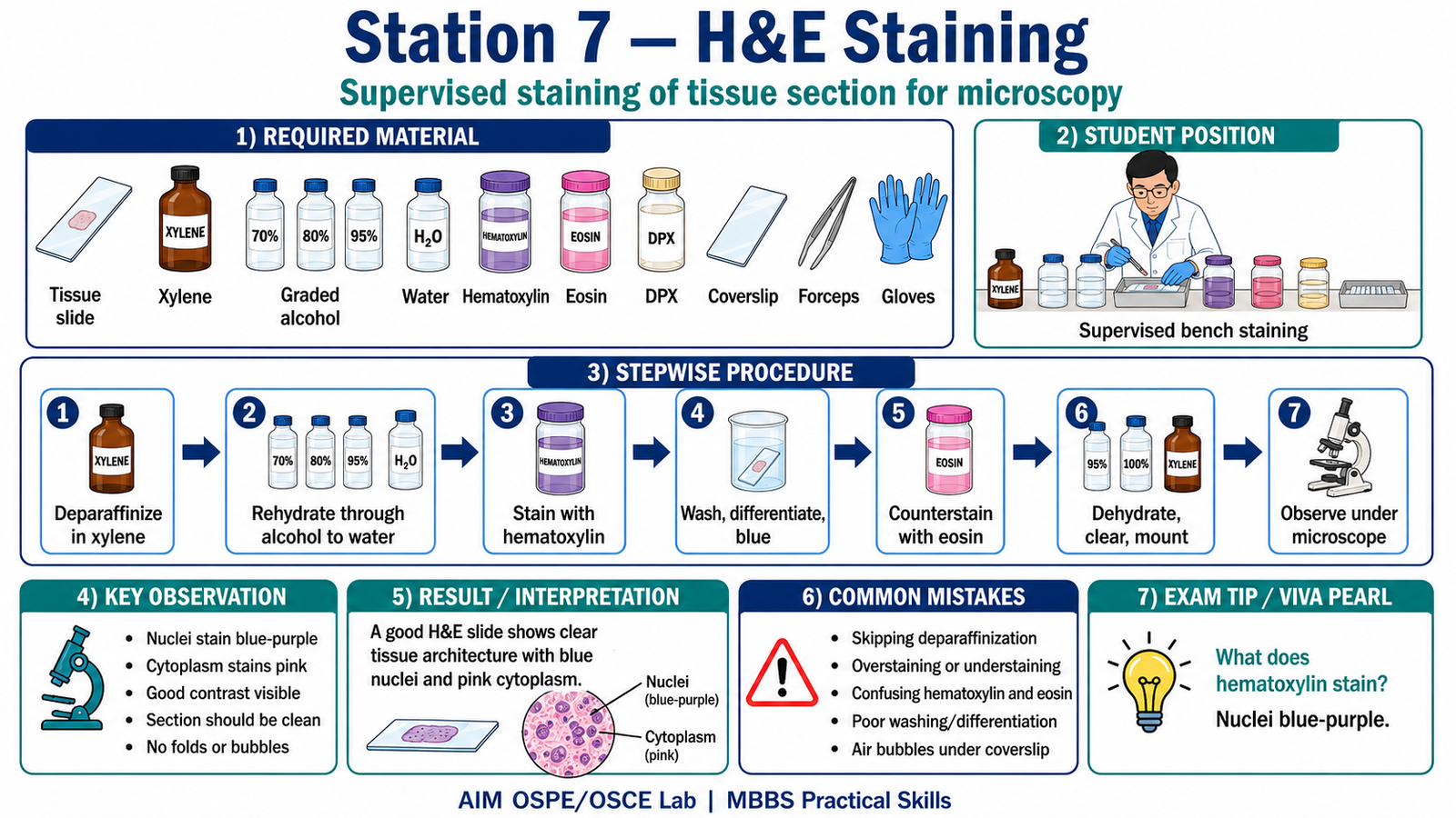
Station 7 — Tissue Processing and H&E Staining
By the end of this station, the student should be able to:
- Describe the basic steps of tissue processing for histopathology.
- Perform or demonstrate the main supervised steps of H&E staining and identify the expected staining result.
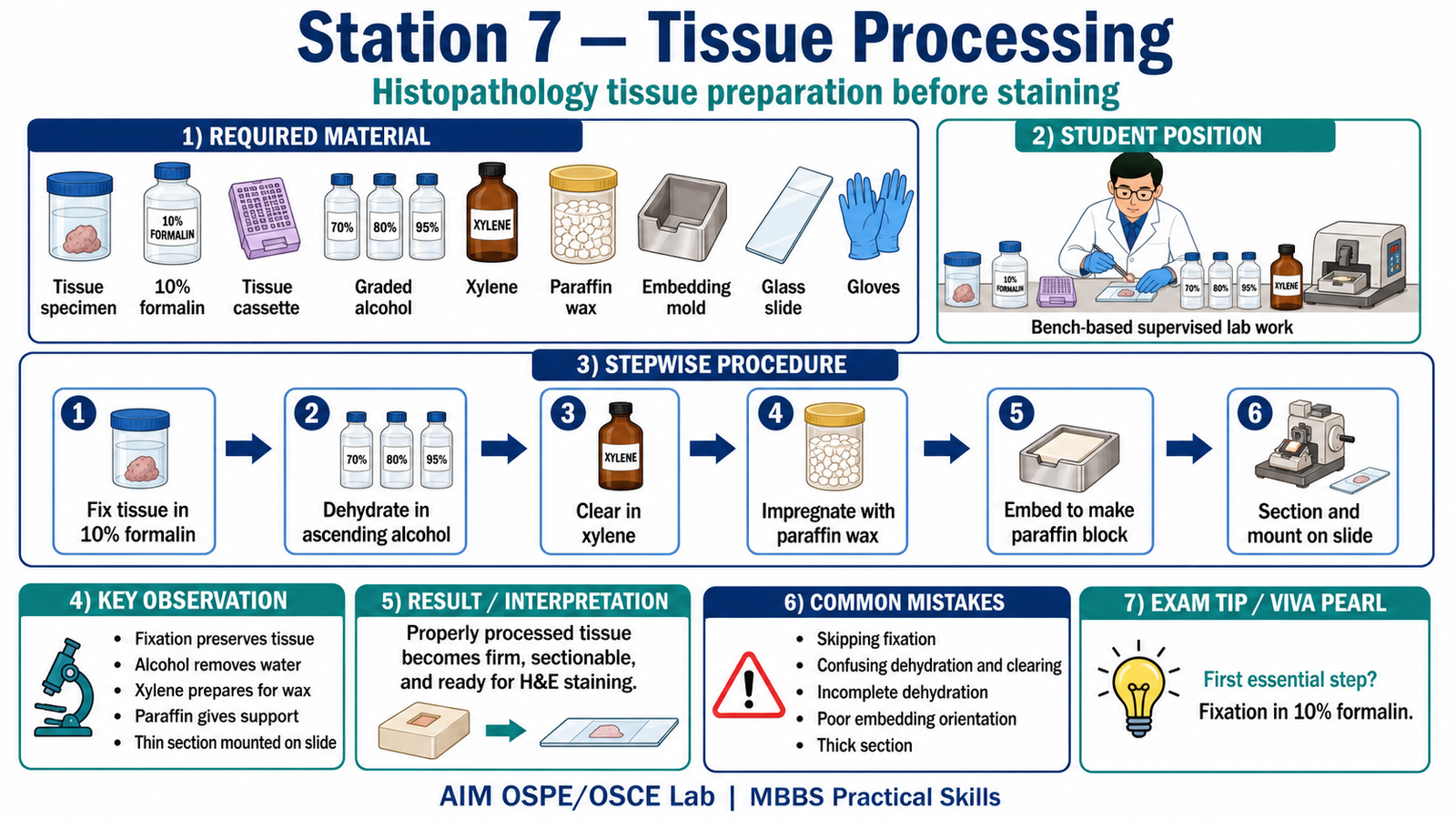
Required Material
- Tissue specimen
- 10% formalin fixative
- Tissue cassette
- Alcohol grades for dehydration
- Xylene or clearing agent
- Paraffin wax
- Embedding mold
- Microtome section slide, already prepared if cutting is not required
- Glass slides
- Hematoxylin stain
- Eosin stain
- Distilled water
- Acid alcohol, if differentiation is included
- Ammonia water or alkaline water, if bluing is included
- DPX or mounting medium
- Coverslip
- Forceps
- Staining jars
- Gloves
- Lab coat
- Waste container
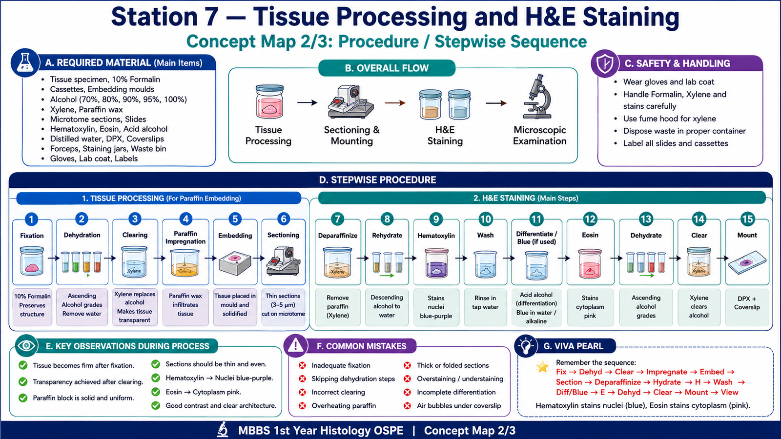
Student Task / Procedure
- Wear lab coat and gloves.
- Identify the tissue specimen and tissue cassette.
- State the first step: fixation in 10% formalin.
- Explain dehydration using ascending grades of alcohol.
- Explain clearing using xylene.
- Explain impregnation with molten paraffin wax.
- Explain embedding of tissue in paraffin block.
- Identify the prepared thin tissue section on a glass slide.
- Deparaffinize the slide using xylene, if required.
- Rehydrate the section through descending grades of alcohol to water.
- Stain the section with hematoxylin.
- Wash in water.
- Differentiate and blue the section, if included in the lab protocol.
- Counterstain with eosin.
- Dehydrate through ascending grades of alcohol.
- Clear in xylene.
- Mount with DPX and coverslip.
- Observe the stained slide under microscope.
Observation / Identification Points
The student should observe, identify, or demonstrate:
- Tissue is fixed before processing.
- Formalin preserves tissue architecture.
- Alcohol removes water from tissue.
- Xylene clears alcohol and makes tissue ready for wax.
- Paraffin supports tissue for thin section cutting.
- Thin sections are mounted on glass slides.
- Hematoxylin stains nuclei blue-purple.
- Eosin stains cytoplasm and extracellular matrix pink.
- Proper H&E slide shows clear nuclear and cytoplasmic contrast.
- Slide should be clean, properly mounted, and free from major folds, air bubbles, and excess stain.
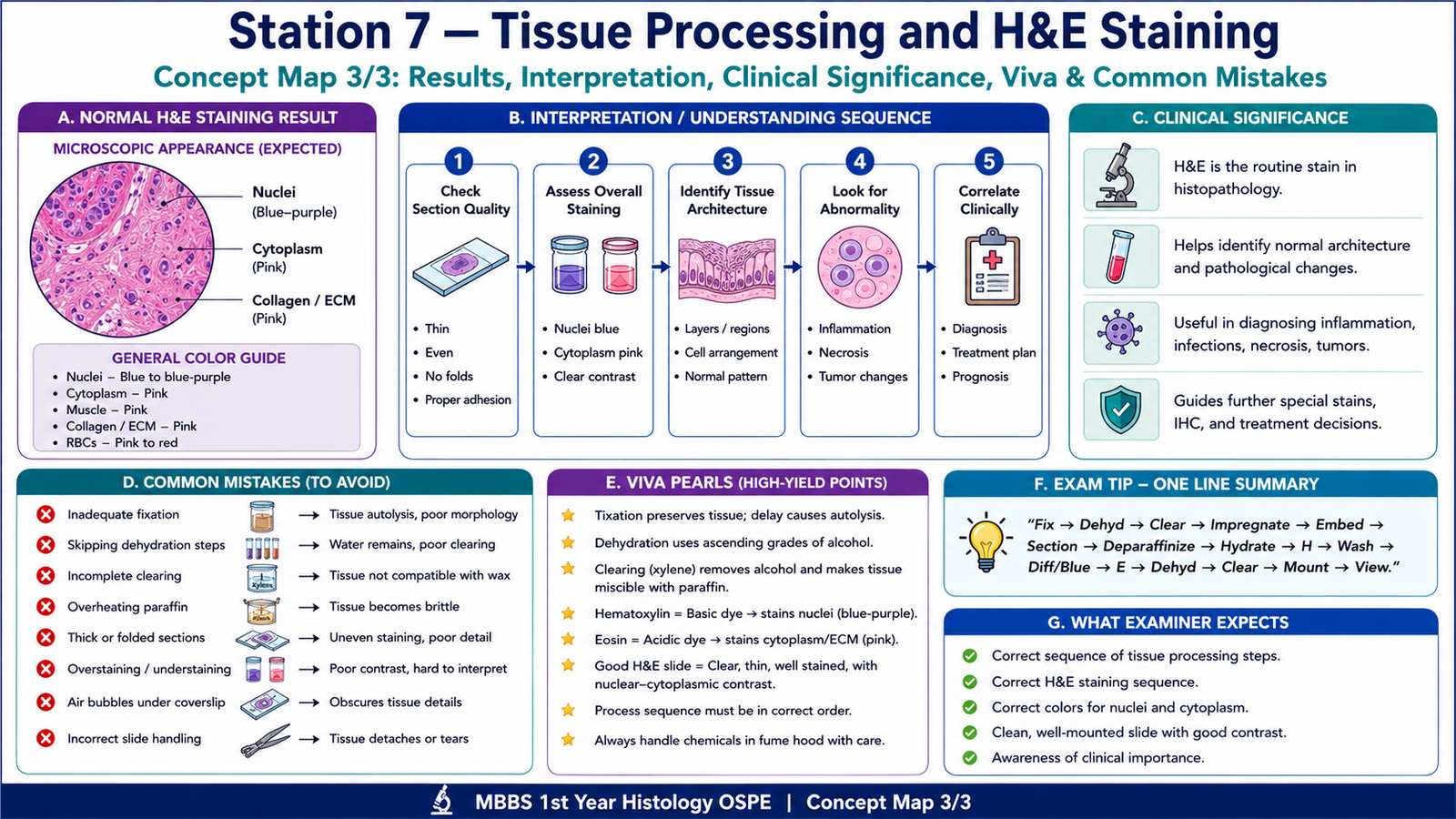
Result / Interpretation
A properly processed and H&E-stained tissue section should show:
- Nuclei: blue-purple
- Cytoplasm: pink
- Muscle/collagen/extracellular matrix: pink shades
- Clear tissue architecture
- Good contrast between nucleus and cytoplasm
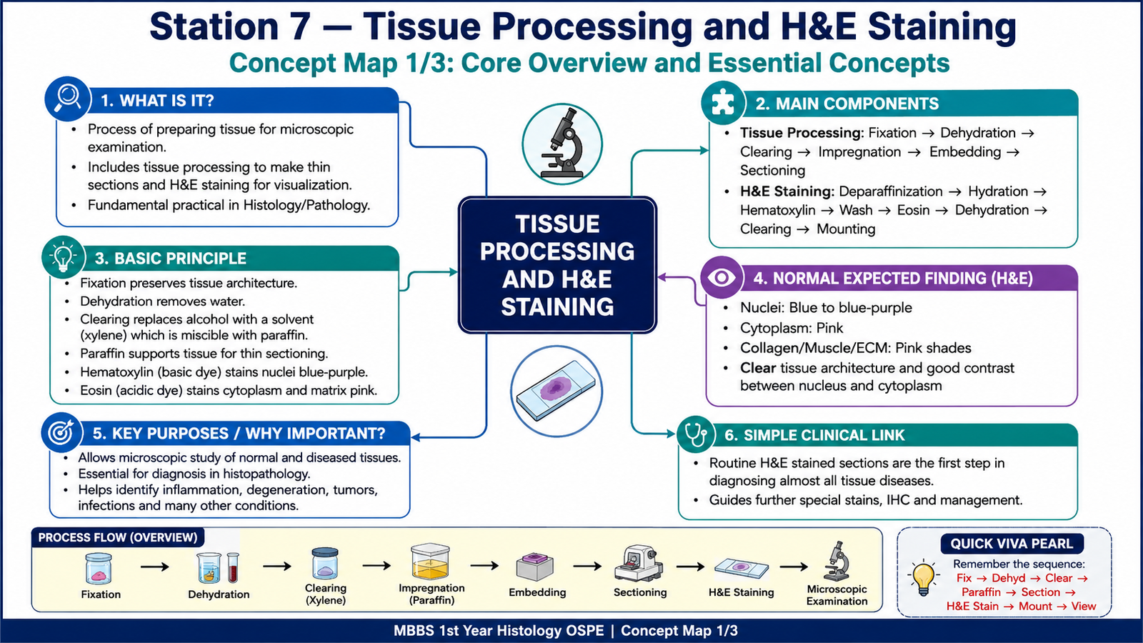
Principle:
Tissue processing converts soft biological tissue into a firm paraffin block so that thin sections can be cut. H&E staining uses hematoxylin as a basic nuclear stain and eosin as an acidic cytoplasmic stain.
Clinical Significance:
H&E staining is the routine basic stain used in histopathology. It helps identify normal tissue architecture, inflammation, necrosis, tumors, and other pathological changes.
Common Student Mistakes
- Confusing dehydration with clearing.
- Forgetting that fixation is the first essential step.
- Saying eosin stains nuclei.
- Saying hematoxylin stains cytoplasm.
- Not knowing the correct color of nuclei and cytoplasm in H&E stain.
- Skipping deparaffinization before staining.
- Overstaining or understaining the section.
- Not mounting the slide properly.
- Producing air bubbles under coverslip.
- Handling xylene/formalin without safety precautions.
AIM Feedback
Tissue processing should be remembered as a sequence: fixation → dehydration → clearing → impregnation → embedding → sectioning → staining → mounting. In H&E staining, the most exam-important point is color interpretation: hematoxylin stains nuclei blue-purple, while eosin stains cytoplasm and extracellular matrix pink. A good slide depends on proper processing, correct staining sequence, and clean mounting.
Most Important Viva Questions for This Topic
- What is tissue processing?
Tissue processing is the preparation of tissue for microscopic examination by fixation, dehydration, clearing, paraffin impregnation, embedding, sectioning, staining, and mounting. - What is the first step in tissue processing?
Fixation. - Which fixative is commonly used in routine histopathology?
10% formalin. - What is the purpose of fixation?
It preserves tissue structure and prevents autolysis and putrefaction. - What is dehydration in tissue processing?
Dehydration is the removal of water from tissue using ascending grades of alcohol. - Why is clearing done?
Clearing removes alcohol and makes tissue compatible with paraffin wax. - Which clearing agent is commonly used?
Xylene. - What is the purpose of paraffin embedding?
Paraffin gives support to the tissue so thin sections can be cut. - Which instrument is used to cut thin tissue sections?
Microtome. - What does hematoxylin stain?
Hematoxylin stains nuclei blue-purple. - What does eosin stain?
Eosin stains cytoplasm and extracellular matrix pink. - What is the clinical importance of H&E staining?
It is the routine stain for studying tissue architecture and diagnosing many pathological conditions such as inflammation, necrosis, and tumors.
🖼️ Visual / Image Support
🧩 Concept Map / Interpretation Support