🩺 Station 8 — Histology of Epithelium and Glands
AIM OSPE/OSCE Lab — Practical Station | KMU Style | MBBS Practical + Viva
📋 Complete OSPE Station Content
OSPE Station Name
Station 8 — Histology of Epithelium and Glands
Learning Target
By the end of this station, the student should be able to:
- Identify simple epithelium, stratified epithelium, and glands under the microscope.
- Describe key microscopic features used to differentiate epithelial types and glandular tissue.
Required Material
- Light microscope
- Prepared histology slides of epithelium
- Slide of simple epithelium
- Slide of stratified epithelium
- Slide of glandular tissue
- Lens tissue
- Pointer / pencil
- Practical notebook
- Histology atlas or LMS reference image
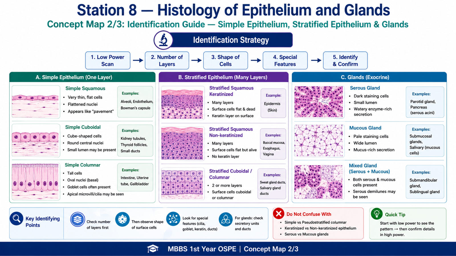
Student Task / Procedure
- Place the given histology slide on the microscope stage.
- Start focusing under low power objective.
- Identify the general arrangement of cells.
- Observe whether the epithelium has one layer or many layers.
- Shift to high power objective for detailed observation.
- Identify the cell shape: squamous, cuboidal, or columnar.
- Look for special features such as cilia, goblet cells, keratin, lumen, ducts, or acini.
- If glandular tissue is present, identify secretory units and ducts.
- Decide whether the gland is mainly serous, mucous, or mixed if visible.
- Write the identification with two key reasons.
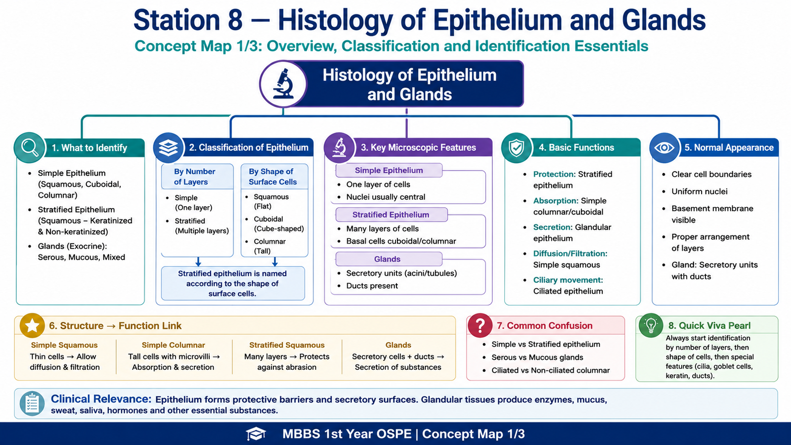
Observation / Identification Points
The student should observe or identify:
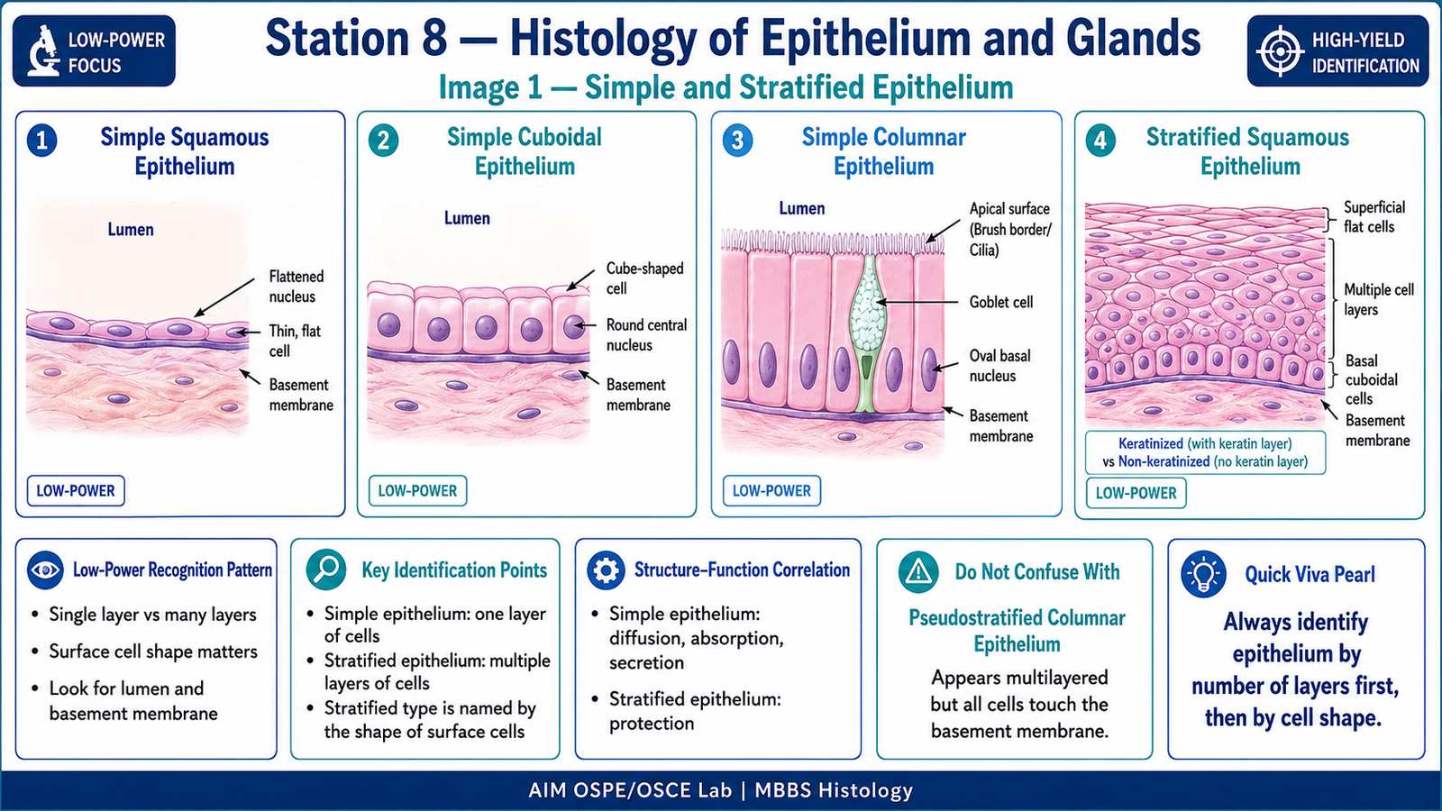
- Simple epithelium: one layer of cells resting on basement membrane.
- Stratified epithelium: multiple cell layers; named according to surface cell shape.
- Simple squamous epithelium: flat cells with flattened nuclei.
- Simple cuboidal epithelium: cube-shaped cells with round central nuclei.
- Simple columnar epithelium: tall cells with oval nuclei, often with goblet cells.
- Stratified squamous epithelium: many layers with flattened surface cells.
- Keratinized stratified squamous epithelium: surface keratin layer present.
- Non-keratinized stratified squamous epithelium: living surface cells, no keratin layer.
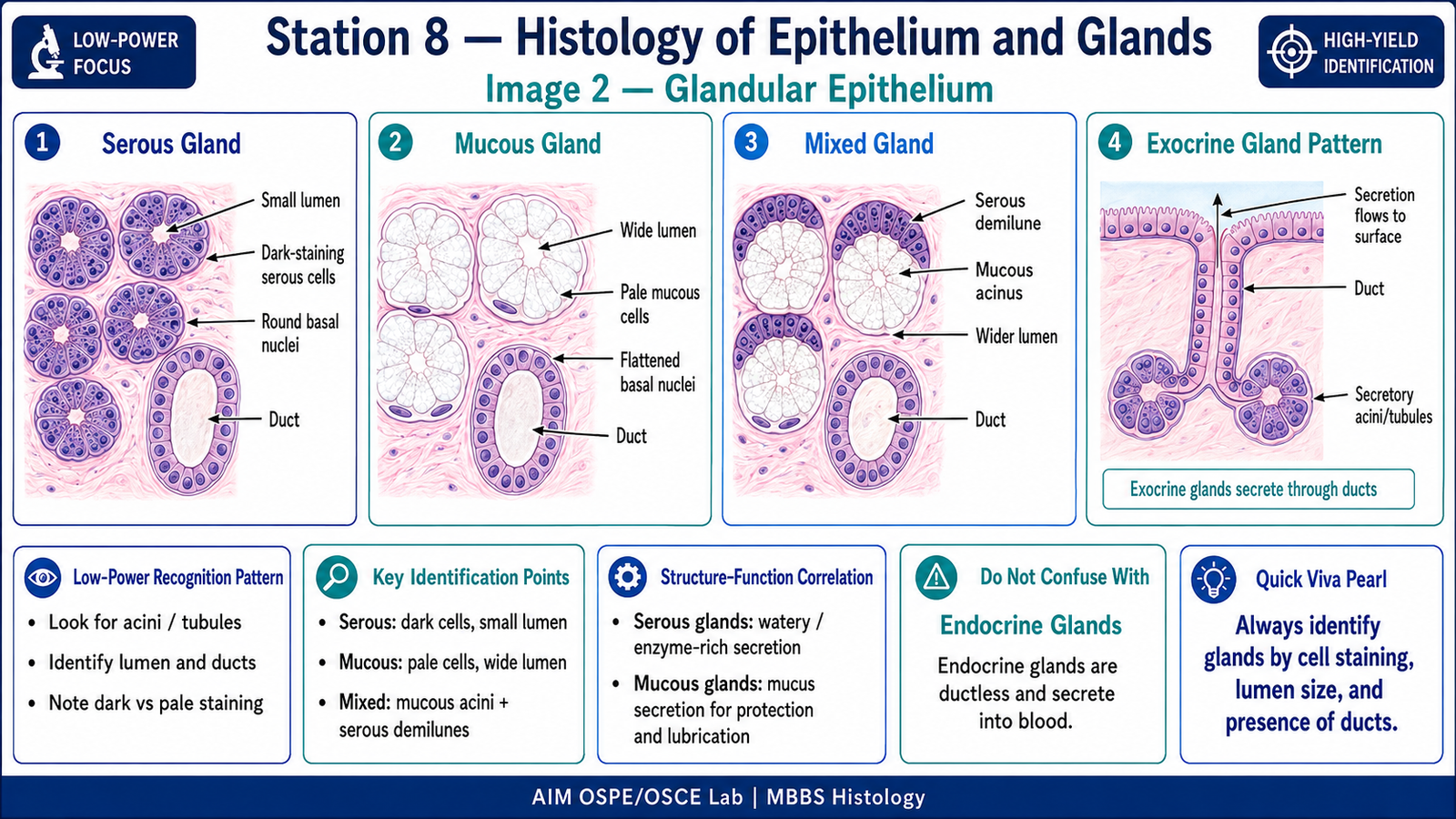
- Glands: secretory epithelial cells arranged as acini, tubules, or ducts.
- Serous glands: dark-staining cells, small lumen.
- Mucous glands: pale-staining cells, wider lumen.
- Mixed glands: both serous and mucous components.
Result / Interpretation
The given tissue is identified by observing the number of cell layers, shape of surface cells, presence of lumen, and glandular arrangement.
Interpretation:
- One cell layer indicates simple epithelium.
- Many layers indicate stratified epithelium.
- Secretory units and ducts indicate glandular epithelium.
- Dark serous acini suggest watery enzyme-rich secretion.
- Pale mucous acini suggest mucus-producing glands.
Clinical significance:
Epithelium forms protective, absorptive, secretory, and lining surfaces. Changes in epithelium, such as metaplasia or dysplasia, are important in pathology and cancer development.
Common Student Mistakes
- Identifying epithelium only by cell shape without checking the number of layers.
- Confusing stratified epithelium with closely packed simple epithelium.
- Calling all glandular tissue “mucous gland” without observing staining and lumen.
- Forgetting that stratified epithelium is named by the shape of the surface cells.
- Starting directly with high power and missing the overall tissue arrangement.
AIM Feedback
For histology identification, always follow a fixed sequence: low power view → number of layers → cell shape → special features → final identification with reasons. In KMU practicals, marks are usually given for correct identification plus two supporting microscopic features. Do not memorize only slide names; learn the visual logic behind each epithelial type.
Most Important Viva Questions for This Topic
1. What is epithelium?
Epithelium is a tissue made of closely packed cells that covers body surfaces, lines cavities, and forms glands.
2. How do you classify epithelium?
Epithelium is classified by the number of layers and shape of cells.
3. What is simple epithelium?
Simple epithelium has a single layer of cells resting on the basement membrane.
4. What is stratified epithelium?
Stratified epithelium has multiple layers of cells and mainly provides protection.
5. How is stratified epithelium named?
It is named according to the shape of the surface cells, not the basal cells.
6. What are the main types of simple epithelium?
Simple squamous, simple cuboidal, simple columnar, and pseudostratified columnar epithelium.
7. Give one example of simple squamous epithelium.
Alveoli, endothelium of blood vessels, and Bowman’s capsule are examples.
8. What is the function of simple cuboidal epithelium?
It is mainly involved in secretion and absorption.
9. What is the function of stratified squamous epithelium?
It provides protection against friction and mechanical stress.
10. What is the difference between keratinized and non-keratinized stratified squamous epithelium?
Keratinized epithelium has a surface keratin layer, while non-keratinized epithelium has living surface cells and remains moist.
11. What is a gland?
A gland is epithelial tissue specialized for secretion.
12. What is the difference between exocrine and endocrine glands?
Exocrine glands secrete through ducts, while endocrine glands release hormones directly into the blood.
13. How can serous and mucous glands be differentiated microscopically?
Serous glands stain darker and have smaller lumens, while mucous glands stain pale and have wider lumens.
14. What are goblet cells?
Goblet cells are unicellular mucous glands that secrete mucus.
15. Why is basement membrane important?
It supports epithelium and separates it from underlying connective tissue.
16. What is pseudostratified epithelium?
It appears multilayered because nuclei are at different levels, but all cells touch the basement membrane.
17. Give one clinical importance of epithelium.
Many cancers, called carcinomas, arise from epithelial tissue.
18. What is metaplasia?
Metaplasia is reversible replacement of one adult epithelial type by another due to chronic irritation.
🖼️ Visual / Image Support
🧩 Concept Map / Interpretation Support