🩺 Station 2 — Microscope Handling and Slide Focusing
AIM OSPE/OSCE Lab — Practical Station | KMU Style | MBBS Practical + Viva
📋 Complete OSPE Station Content
OSPE Station Name
Station 2 — Microscope Handling and Slide Focusing
Learning Target
By the end of this station, the student should be able to:
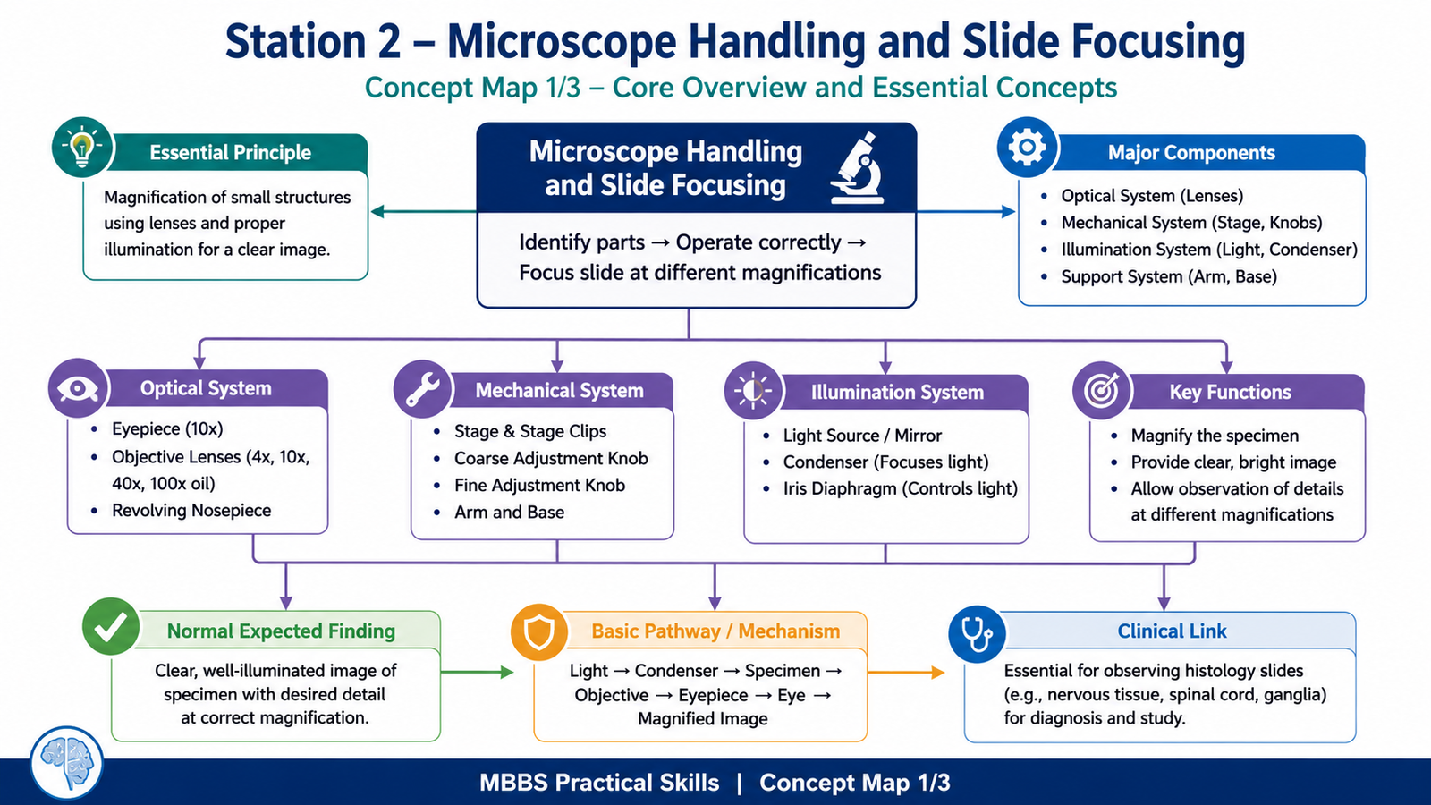
- Identify the main parts of a compound light microscope and explain their basic function.
- Correctly operate the microscope and focus a histology slide under low and high magnification.
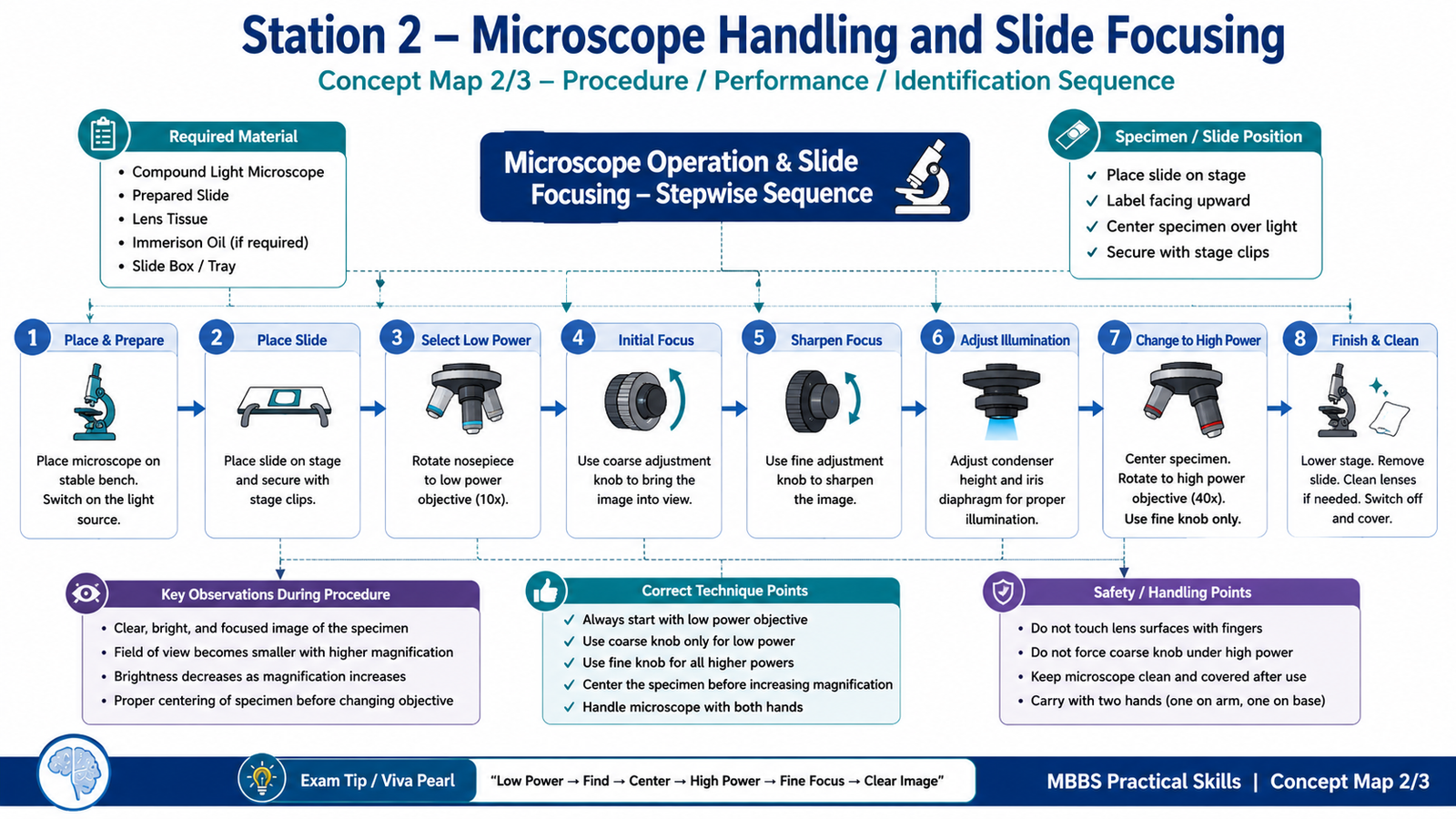
Required Material
- Compound light microscope
- Prepared histology slide
- Slide box / tray
- Lens tissue
- Immersion oil if oil immersion demonstration is required
- Marker pointer / labeled diagram of microscope
- LMS support: short microscope handling video + stepwise focusing image
Student Task / Procedure
- Place the microscope on a stable bench and switch on the light source.
- Identify the main parts of the microscope.
- Place the slide on the stage and secure it with stage clips.
- Start focusing with the low-power objective.
- Use the coarse adjustment knob to bring the image into view.
- Use the fine adjustment knob to sharpen the image.
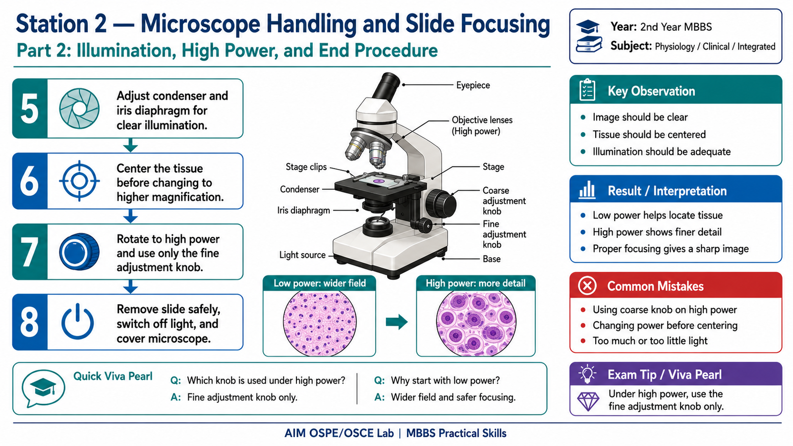
- Adjust the diaphragm/condenser for proper illumination.
- Move to higher magnification by rotating the nosepiece.
- Use only the fine adjustment knob under high power.
- Demonstrate safe removal of the slide and proper microscope handling.
Observation / Identification Points
The student should correctly identify or demonstrate:
- Eyepiece / ocular lens
- Objective lenses: scanning, low power, high power, oil immersion
- Revolving nosepiece
- Stage and stage clips / mechanical stage
- Coarse adjustment knob
- Fine adjustment knob
- Condenser
- Iris diaphragm
- Light source / mirror
- Arm and base
- Correct slide placement
- Proper focusing sequence: low power first, then high power
- Correct use of illumination
- Safe handling with both hands
- Avoiding coarse adjustment under high power
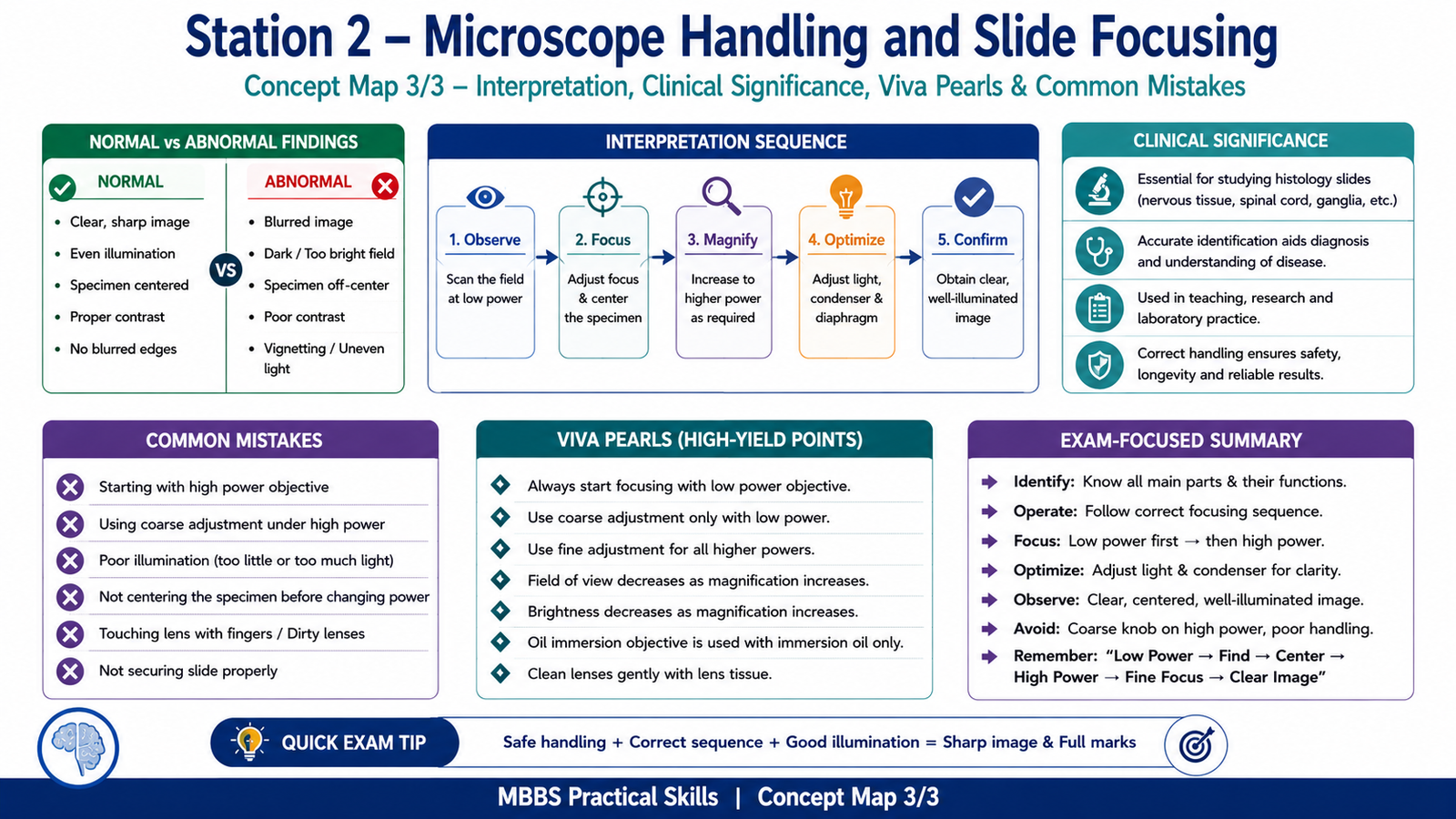
Result / Interpretation
A correctly focused microscopic field should show a clear, centered, well-illuminated image of the tissue section.
Principle:
A compound light microscope magnifies small structures using objective and ocular lenses. Proper focusing begins with low power because it gives a wider field of view and reduces the risk of slide or lens damage. Higher magnification is used only after the tissue is located and centered.
Clinical / Practical Significance:
Correct microscope handling is essential for identifying histological tissues such as nervous tissue, cerebellum, spinal cord, peripheral nerve, ganglia, and other practical slides in the neuroscience module.
Viva Questions
1. Why should focusing begin with the low-power objective?
Because low power provides a wider field of view and makes it easier to locate the tissue safely.
2. Which adjustment knob is used first while focusing under low power?
The coarse adjustment knob.
3. Which adjustment knob should be used under high power?
Only the fine adjustment knob.
4. What is the function of the condenser?
It concentrates light on the specimen.
5. What is the function of the iris diaphragm?
It controls the amount of light passing through the specimen.
Common Student Mistakes
- Starting directly with high power objective.
- Using the coarse adjustment knob under high power.
- Poor illumination due to incorrect diaphragm or condenser adjustment.
- Placing the slide upside down or not securing it properly.
- Touching objective lenses with fingers or dirty lens paper.
AIM Feedback
To perform well in this station, always follow the correct focusing sequence: place slide → start with low power → focus with coarse knob → sharpen with fine knob → center the tissue → shift to high power → use fine knob only.
In OSPE, marks are usually lost when students know the microscope parts but cannot demonstrate safe and correct focusing. Practice the hand movements repeatedly until the sequence becomes automatic.
🖼️ Visual / Image Support

🧩 Concept Map / Interpretation Support