
Course Content
🔬 Station 1 — Identification of Lipoma
Learning Outcomes Identify a lipoma from a provided gross specimen/image or histopathology slide. Identify the key morphological features supporting the diagnosis. Differentiate the lesion from an obviously malignant soft-tissue lesion when asked.
0/1
🔬 Station 2 — Identification of Squamous Cell Carcinoma
Learning Outcomes Identify squamous cell carcinoma from a provided gross/microscopic specimen. Recognize the important morphological features of squamous differentiation. State the diagnosis from the findings provided.
0/1
🔬 Station 3 — Fibroadenoma of Breast: Gross & Microscopic Identification
Learning Outcomes Identify fibroadenoma from a gross specimen or histological image. Recognize the important gross and microscopic points of identification. Give the final pathological identification.
0/1
🧬 Station 4 — Karyogram Preparation & Chromosomal Abnormality Identification
Learning Outcomes Arrange chromosomes appropriately to prepare a karyogram. Determine chromosomal sex from the karyogram. Identify a common numerical chromosomal abnormality from a provided karyogram.
0/1
⚗️ Station 5 — Tissue Organ Bath: Identification, Setup & Tyrode Solution
Learning Outcomes Identify the major components of a tissue organ bath. State the function of the important components. Identify the constituents/purpose of Tyrode's solution. Demonstrate the basic placement/fixation of isolated ileal tissue in the organ bath.
0/1
📈 Station 6 — Acetylcholine Dose–Response & Ceiling Effect
Learning Outcomes Demonstrate the effect of increasing doses of acetylcholine on isolated rabbit ileum. Recognize the ceiling effect. Interpret a provided tracing/dose-response recording. Plot or interpret the corresponding dose-response graph.
0/1
📉 Station 7 — Competitive Antagonism on Isolated Rabbit Ileum
Learning Outcomes Demonstrate/recognize surmountable antagonism using an agonist and antagonist. Interpret the experimental tracing. Explain the change in response after addition of the antagonist. Identify the pharmacological relationship demonstrated.
0/1
👁️ Station 8 — Rabbit Eye Pharmacology: Identify the Unknown Drug
Learning Outcomes Assess/interpret pupil size, conjunctival appearance, corneal reflex and light reflex. Compare the test eye with the control eye. Interpret the pattern produced by an autonomic or local anaesthetic drug. Identify the most likely unknown drug from the observed findings.
0/1
📝 Station 9 — Medico-Legal Autopsy Report
Learning Outcomes Review supplied postmortem findings systematically. Complete the important components of an autopsy report. Formulate an appropriate final opinion from the supplied findings.
0/1
🧪 Station 10 — Toxicology Sample Collection & Preservation
Learning Outcomes Select the appropriate viscera/specimens required for toxicological examination. Select the appropriate preservative/container. Demonstrate or describe correct collection, labelling and handling of the sample. State essential precautions before dispatch.
0/1
⚰️ Station 11 — Identification of Postmortem Changes
Learning Outcomes Identify the postmortem change demonstrated on a model/image/specimen. Describe its characteristic features. Differentiate commonly confused postmortem changes where appropriate. State its basic medico-legal significance, particularly in relation to time since death.
0/1
🚑 Station 12 — Gastric Lavage / Stomach Wash on Manikin
Learning Outcomes Prepare the equipment required for gastric lavage. Demonstrate the correct sequence of gastric lavage on a manikin. Maintain patient safety and correct positioning during the procedure. Recognize situations in which the procedure should not be attempted.
0/1
🧪🔬 AIM OSPE/OSCE Lab — Multisystem-I Module | 3rd Year MBBS
AIM OSCE / OSPE • MBBS

🔬 Station 3 — Fibroadenoma of Breast: Gross & Microscopic Identification

Rapid Revision • Practical Skills • Viva

🎯 Task

CANDIDATE TASK

Examine the provided gross specimen and histological image. Identify the lesion, state the important gross and microscopic features supporting your identification, and give the final pathological diagnosis.

⚡ Model Station Answer

Identification: This is a fibroadenoma of the breast. Gross: It is a well-circumscribed, firm, rounded or lobulated mass with a smooth border and a homogeneous grey-white cut surface; slit-like spaces may be present. Microscopy: A biphasic benign tumour composed of proliferating fibrous stroma and benign breast ducts. The ducts may remain round or become compressed into slit-like spaces. Conclusion: The features are consistent with a benign fibroadenoma.

🔎 Stepwise Procedure / Approach

  1. Inspect the gross specimen systematically and determine whether the lesion is circumscribed or infiltrative.
  2. Assess its shape, surface and border; look for a rounded or lobulated, well-defined mass.
  3. Examine the cut surface for a uniform grey-white appearance and characteristic slit-like spaces.
  4. On microscopy, first assess the overall architecture at low power.
  5. Confirm the presence of both stromal and epithelial components, establishing a biphasic pattern.
  6. Examine the ducts and surrounding stroma; identify round ducts or ducts compressed into elongated slit-like spaces.
  7. Look for benign cytology without significant atypia, destructive invasion or tumour necrosis.
  8. Correlate the gross and microscopic findings and state the final diagnosis as fibroadenoma of the breast.

🖼️ Visual Learning

VISUAL 1

Fibroadenoma — Gross and Microscopic Identification
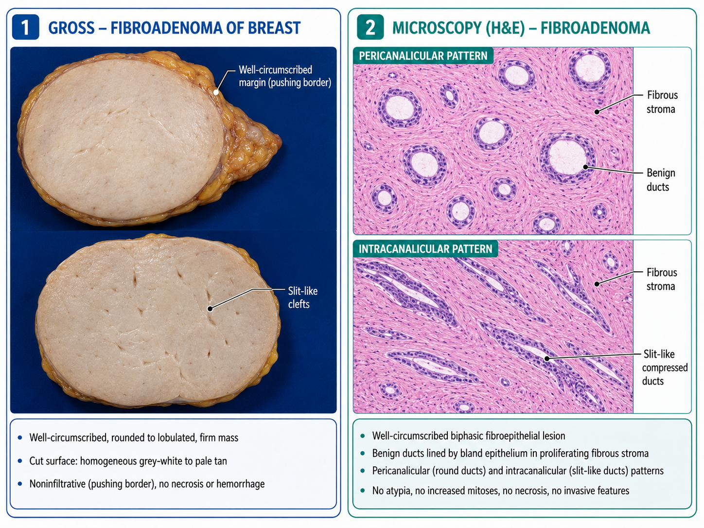
🎥 Practical Demonstration
Fibroadenoma of Breast — Talking Pot & Slide
Source: NUS Pathweb — National University of Singapore

Practical demonstration of fibroadenoma with gross specimen and microscopic features useful for OSPE identification.

▶ Watch Practical Video


📝 Important Viva Questions

Q1. What is a fibroadenoma?

Answer: It is a benign fibroepithelial tumour of the breast composed of both stromal and epithelial components.

“`

Q2. What are the important gross features of fibroadenoma?

Answer: It is typically a well-circumscribed, firm, rounded or lobulated mass with a homogeneous grey-white cut surface and may show slit-like spaces.

Q3. What is meant by the biphasic nature of fibroadenoma?

Answer: The tumour contains two components: benign breast ducts and proliferating fibrous stroma.

Q4. What are the two classical microscopic patterns of fibroadenoma?

Answer: Pericanalicular and intracanalicular patterns.

Q5. What is the intracanalicular pattern?

Answer: Proliferating stroma compresses and distorts the ducts, producing elongated or slit-like spaces.

Q6. What is the pericanalicular pattern?

Answer: Fibrous stroma proliferates around relatively round or open ductal spaces without marked compression.

Q7. Which important lesion may enter the differential diagnosis of a fibroepithelial breast tumour?

Answer: Phyllodes tumour. Compared with a typical fibroadenoma, it generally shows greater stromal cellularity and a characteristic leaf-like architecture.

Scroll to Top
💬 WhatsApp Support