
Identification of Lipoma
Rapid Revision • Practical Skills • Viva
🎯 Task
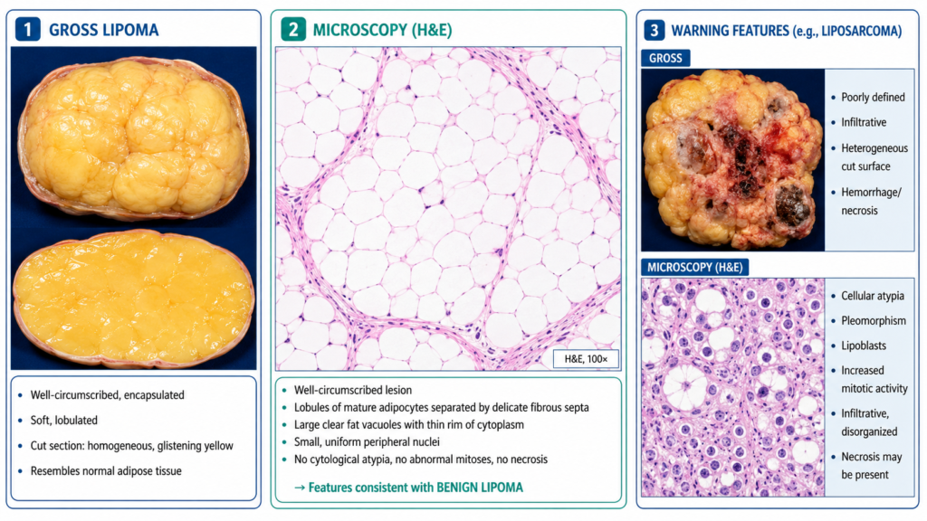
Examine the provided gross specimen/image or histopathology slide. Identify the lesion, state the key morphological features supporting your diagnosis, and indicate features that would raise suspicion of an obviously malignant soft-tissue lesion.
🔎 Stepwise Procedure / Approach
Observe the specimen systematically. Decide whether you are viewing a gross specimen or a histopathology slide.
- Identify the dominant tissue. Look for the characteristic appearance of mature adipose tissue.
- Assess circumscription. A typical lesion is well circumscribed and may be surrounded by a thin fibrous capsule.
- Look for gross identifying features. A typical cut surface is soft, lobulated, homogeneous and yellow, resembling normal fat.
- Look for microscopic identifying features. Identify lobules of mature, uniform adipocytes separated by delicate fibrous septa without significant cytological atypia.
- State the diagnosis. Correlate the fatty appearance, circumscription and mature adipocytes to identify the lesion as a lipoma.
- Classify the lesion. State that it is a benign adipocytic mesenchymal tumour.
- Screen for malignant features. Marked atypia or pleomorphism, increased mitotic activity, infiltrative margins, necrosis, hemorrhage or a heterogeneous solid appearance should raise concern for malignancy.
🖼️ Visual Learning
Lipoma — Gross and Microscopic Identification Features
🎥 Practical Video
🎥 Practical Demonstration — Musculoskeletal System: Lipoma (Gross Pathology)
Demonstrates the salient gross pathological features of lipoma with brief clinicopathological correlation, useful for practical specimen identification. :contentReference[oaicite:0]{index=0}
📝 Important Viva Questions
Q1. What is a lipoma?
Answer: A benign mesenchymal tumour composed of mature adipose tissue.
Q2. What are the characteristic gross features of a lipoma?
Answer: It is usually well circumscribed, soft, lobulated and yellow, with a homogeneous fatty cut surface.
Q3. What is the characteristic microscopic appearance?
Answer: Lobules of mature uniform adipocytes, usually separated by delicate fibrous septa and lacking significant atypia.
Q4. Why does the cut surface of a lipoma appear yellow?
Answer: Because the tumour is predominantly composed of mature adipose tissue.
Q5. Is a lipoma benign or malignant?
Answer: Benign.
Q6. Name the malignant adipocytic tumour that may enter the differential diagnosis.
Answer: Liposarcoma.
Q7. Which features should make you suspicious of an obviously malignant soft-tissue lesion?
Answer: Infiltrative margins, marked cellular atypia or pleomorphism, increased mitoses, necrosis, hemorrhage or a heterogeneous solid appearance are concerning features.