
Course Content
🔬 Station 1 — Identification of Lipoma
Learning Outcomes Identify a lipoma from a provided gross specimen/image or histopathology slide. Identify the key morphological features supporting the diagnosis. Differentiate the lesion from an obviously malignant soft-tissue lesion when asked.
0/1
🔬 Station 2 — Identification of Squamous Cell Carcinoma
Learning Outcomes Identify squamous cell carcinoma from a provided gross/microscopic specimen. Recognize the important morphological features of squamous differentiation. State the diagnosis from the findings provided.
0/1
🔬 Station 3 — Fibroadenoma of Breast: Gross & Microscopic Identification
Learning Outcomes Identify fibroadenoma from a gross specimen or histological image. Recognize the important gross and microscopic points of identification. Give the final pathological identification.
0/1
🧬 Station 4 — Karyogram Preparation & Chromosomal Abnormality Identification
Learning Outcomes Arrange chromosomes appropriately to prepare a karyogram. Determine chromosomal sex from the karyogram. Identify a common numerical chromosomal abnormality from a provided karyogram.
0/1
⚗️ Station 5 — Tissue Organ Bath: Identification, Setup & Tyrode Solution
Learning Outcomes Identify the major components of a tissue organ bath. State the function of the important components. Identify the constituents/purpose of Tyrode's solution. Demonstrate the basic placement/fixation of isolated ileal tissue in the organ bath.
0/1
📈 Station 6 — Acetylcholine Dose–Response & Ceiling Effect
Learning Outcomes Demonstrate the effect of increasing doses of acetylcholine on isolated rabbit ileum. Recognize the ceiling effect. Interpret a provided tracing/dose-response recording. Plot or interpret the corresponding dose-response graph.
0/1
📉 Station 7 — Competitive Antagonism on Isolated Rabbit Ileum
Learning Outcomes Demonstrate/recognize surmountable antagonism using an agonist and antagonist. Interpret the experimental tracing. Explain the change in response after addition of the antagonist. Identify the pharmacological relationship demonstrated.
0/1
👁️ Station 8 — Rabbit Eye Pharmacology: Identify the Unknown Drug
Learning Outcomes Assess/interpret pupil size, conjunctival appearance, corneal reflex and light reflex. Compare the test eye with the control eye. Interpret the pattern produced by an autonomic or local anaesthetic drug. Identify the most likely unknown drug from the observed findings.
0/1
📝 Station 9 — Medico-Legal Autopsy Report
Learning Outcomes Review supplied postmortem findings systematically. Complete the important components of an autopsy report. Formulate an appropriate final opinion from the supplied findings.
0/1
🧪 Station 10 — Toxicology Sample Collection & Preservation
Learning Outcomes Select the appropriate viscera/specimens required for toxicological examination. Select the appropriate preservative/container. Demonstrate or describe correct collection, labelling and handling of the sample. State essential precautions before dispatch.
0/1
⚰️ Station 11 — Identification of Postmortem Changes
Learning Outcomes Identify the postmortem change demonstrated on a model/image/specimen. Describe its characteristic features. Differentiate commonly confused postmortem changes where appropriate. State its basic medico-legal significance, particularly in relation to time since death.
0/1
🚑 Station 12 — Gastric Lavage / Stomach Wash on Manikin
Learning Outcomes Prepare the equipment required for gastric lavage. Demonstrate the correct sequence of gastric lavage on a manikin. Maintain patient safety and correct positioning during the procedure. Recognize situations in which the procedure should not be attempted.
0/1
🧪🔬 AIM OSPE/OSCE Lab — Multisystem-I Module | 3rd Year MBBS
AIM OSCE / OSPE • MBBS

🔬 Station 2 — Identification of Squamous Cell Carcinoma

Rapid Revision • Practical Skills • Viva

🎯 Task

CANDIDATE TASK

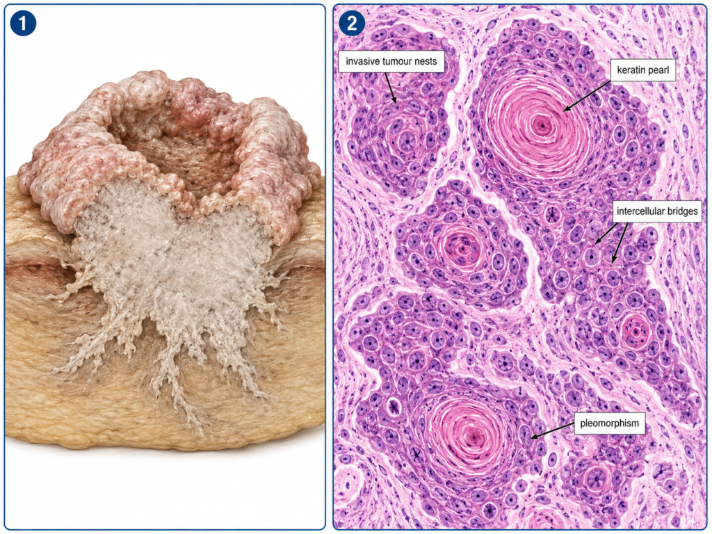
Examine the provided gross specimen and/or H&E-stained microscopic slide. Identify the lesion, state the important features supporting your diagnosis, and indicate the morphological evidence of squamous differentiation.

🔎 Stepwise Procedure / Approach

Orient yourself to the specimen. Determine whether you are being shown a gross specimen, microscopic slide, or both.

  1. Inspect the gross lesion systematically. Look for an irregular ulcerative, infiltrative or proliferative mass and note its surface, margins, consistency and invasion into surrounding tissue where visible.
  2. Scan the microscopic slide at low power. Look for irregular nests, cords or islands of atypical squamous epithelial cells extending into the underlying stroma.
  3. Assess the tumour cells at higher power. Look for cellular and nuclear pleomorphism, hyperchromasia, prominent nucleoli and mitotic activity.
  4. Search for squamous differentiation. Identify keratinization, keratin pearls and/or intercellular bridges where present.
  5. Confirm invasive growth. Demonstration of malignant epithelial nests infiltrating the underlying stroma supports invasive carcinoma rather than an intraepithelial lesion.
  6. Integrate the findings. Correlate malignant epithelial morphology with evidence of squamous differentiation.
  7. State the diagnosis clearly. Give one concise diagnosis and mention one or two characteristic identifying features when asked.

🖼️ Visual Learning

VISUAL 1

Squamous Cell Carcinoma — Gross and Microscopic Identification
🎥 Practical Video

🎥 Practical Demonstration — Squamous Cell Carcinoma Microscopy

Source: NUS Pathweb, National University of Singapore

A talking-slide demonstration of the salient microscopic features of squamous cell carcinoma, useful for rehearsing systematic slide identification before an OSPE station.


▶ Watch Practical Video


📝 Important Viva Questions


Q1. What are the characteristic microscopic features of squamous cell carcinoma?

Answer: Invasive nests or islands of atypical squamous cells showing pleomorphism with variable keratinization, keratin pearls and intercellular bridges.

Q2. What is a keratin pearl?

Answer: It is a concentric laminated collection of keratin produced within a nest of malignant squamous cells.

Q3. What do intercellular bridges indicate?

Answer: They represent desmosomal connections between squamous cells and are an important feature of squamous differentiation.

Q4. What feature confirms that the carcinoma is invasive?

Answer: Infiltration of malignant epithelial nests or cords into the underlying stroma.

Q5. Which microscopic features suggest malignancy in this tumour?

Answer: Cellular and nuclear pleomorphism, hyperchromasia, abnormal or increased mitoses, loss of normal maturation and stromal invasion.

Q6. How does differentiation affect the appearance of squamous cell carcinoma?

Answer: Well-differentiated tumours show more obvious squamous maturation and keratinization, whereas poorly differentiated tumours show less recognizable squamous morphology.

Q7. What is a common OSPE mistake while identifying squamous cell carcinoma?

Answer: Naming the lesion from keratin alone without demonstrating malignant cellular atypia and invasive growth.


Scroll to Top
💬 WhatsApp Support