🩺 Station 1 — Cervical Spinal Cord Histology
AIM OSPE/OSCE Lab — Practical Station | KMU Style | MBBS Practical + Viva
📋 Complete OSPE Station Content
OSPE Station Name
Station 1 — Cervical Spinal Cord Histology
Learning Target
By the end of this station, the student should be able to:
- Identify a transverse section of the cervical spinal cord under the microscope.
- Recognize key histological features of the spinal cord and relate them to basic function.
Required Material
- Prepared histology slide of cervical spinal cord transverse section
- Light microscope
- Pointer or marker on microscope field
- Answer sheet / LMS response box
- Labeled reference image for examiner use
Student Task / Procedure
- Focus the slide under low power.
- Identify the tissue as spinal cord in transverse section.
- Observe the central grey matter and surrounding white matter.
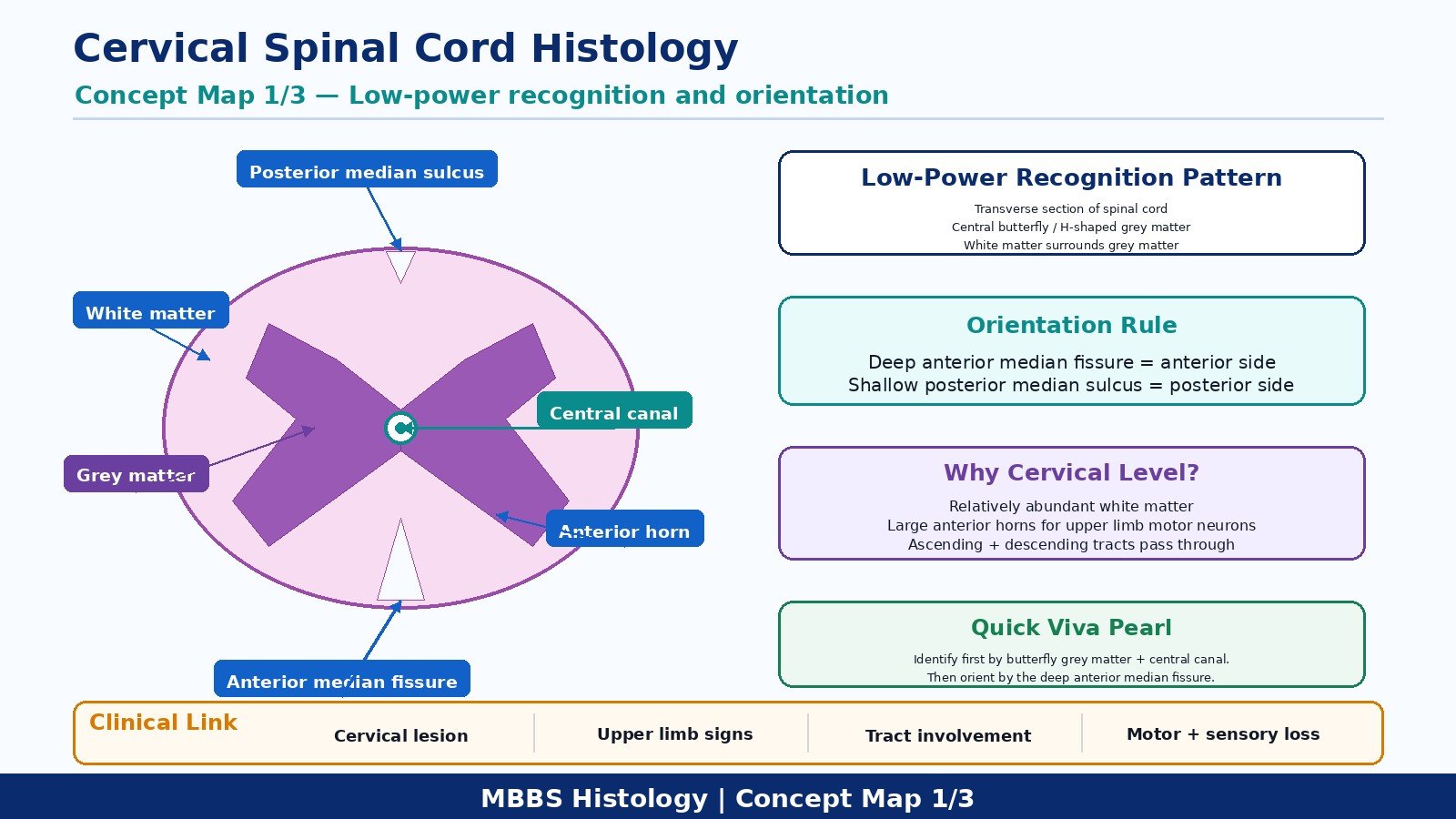
- Identify the anterior median fissure and posterior median sulcus.
- Identify the anterior horn, posterior horn, and central canal.
- State one reason why this section is from the cervical region.
Observation / Identification Points
The student should identify:
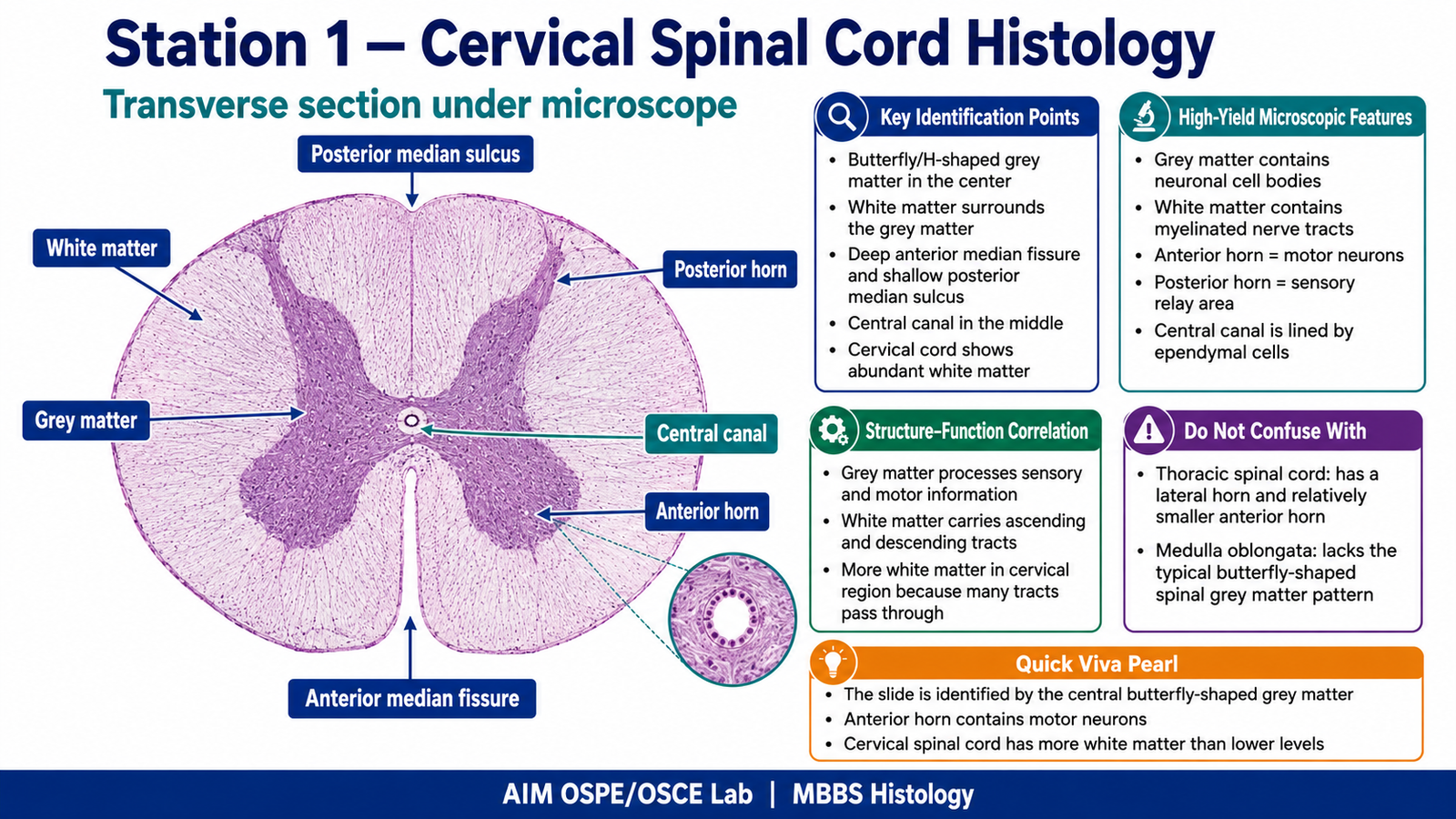
- Butterfly-shaped grey matter in the center
- White matter surrounding the grey matter
- Anterior median fissure: deep groove on anterior side
- Posterior median sulcus: shallow groove on posterior side
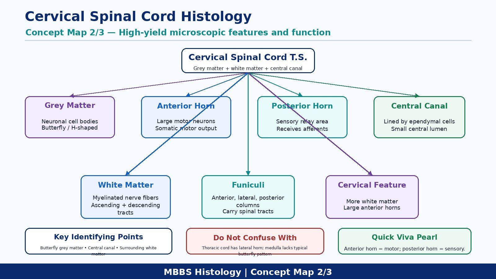
- Anterior horn: broad, rounded motor area
- Posterior horn: narrow sensory area
- Central canal lined by ependymal cells
- Large amount of white matter, suggesting cervical level
- Presence of both ascending and descending tracts in white matter
Result / Interpretation
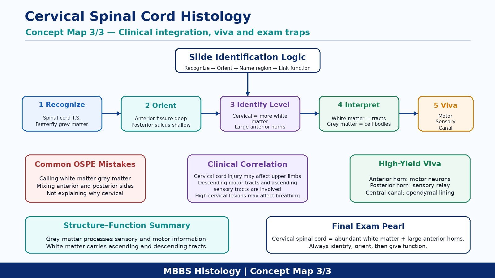
The slide shows a transverse section of the cervical spinal cord.
The cervical spinal cord has relatively more white matter because it contains many ascending sensory tracts from lower body regions and descending motor tracts going to lower spinal levels. The grey matter contains neuronal cell bodies, while the white matter contains myelinated nerve fibers.
Clinical significance:
Damage to the cervical spinal cord can affect motor and sensory function in the upper limbs, trunk, and lower limbs depending on the level and extent of lesion.
Viva Questions
1. How do you identify spinal cord histology under the microscope?
By the central butterfly-shaped grey matter, surrounding white matter, anterior median fissure, posterior median sulcus, and central canal.
2. Why does the cervical spinal cord have more white matter?
Because many ascending and descending tracts pass through the cervical region.
3. What is present in the anterior horn of grey matter?
Motor neurons supplying skeletal muscles.
4. What is the function of the posterior horn?
It receives sensory information from peripheral nerves.
5. What is the central canal lined by?
Ependymal cells.
Common Student Mistakes
- Confusing grey matter with white matter.
- Failing to differentiate anterior median fissure from posterior median sulcus.
- Identifying the section as general spinal cord but not explaining why it is cervical.
AIM Feedback
To improve, first orient the slide by finding the deep anterior median fissure. Then identify the central butterfly-shaped grey matter and surrounding white matter. For cervical spinal cord, remember: more white matter = more tracts passing through. Always connect the histological appearance with function: anterior horn is motor, posterior horn is sensory, and white matter carries ascending and descending pathways.
🖼️ Visual / Image Support
🧩 Concept Map / Interpretation Support