🩺 Station 4 — Lower Limb Deep Tendon Reflexes
AIM OSPE/OSCE Lab — Practical Station | KMU Style | MBBS Practical + Viva
📋 Complete OSPE Station Content
OSPE Station Name
Station 4 — Lower Limb Deep Tendon Reflexes
Learning Target
By the end of this station, the student should be able to:
- Correctly elicit knee jerk and ankle jerk reflexes in the lower limb.
- Interpret lower limb deep tendon reflexes in relation to spinal segments, peripheral nerves, and UMN/LMN lesions.
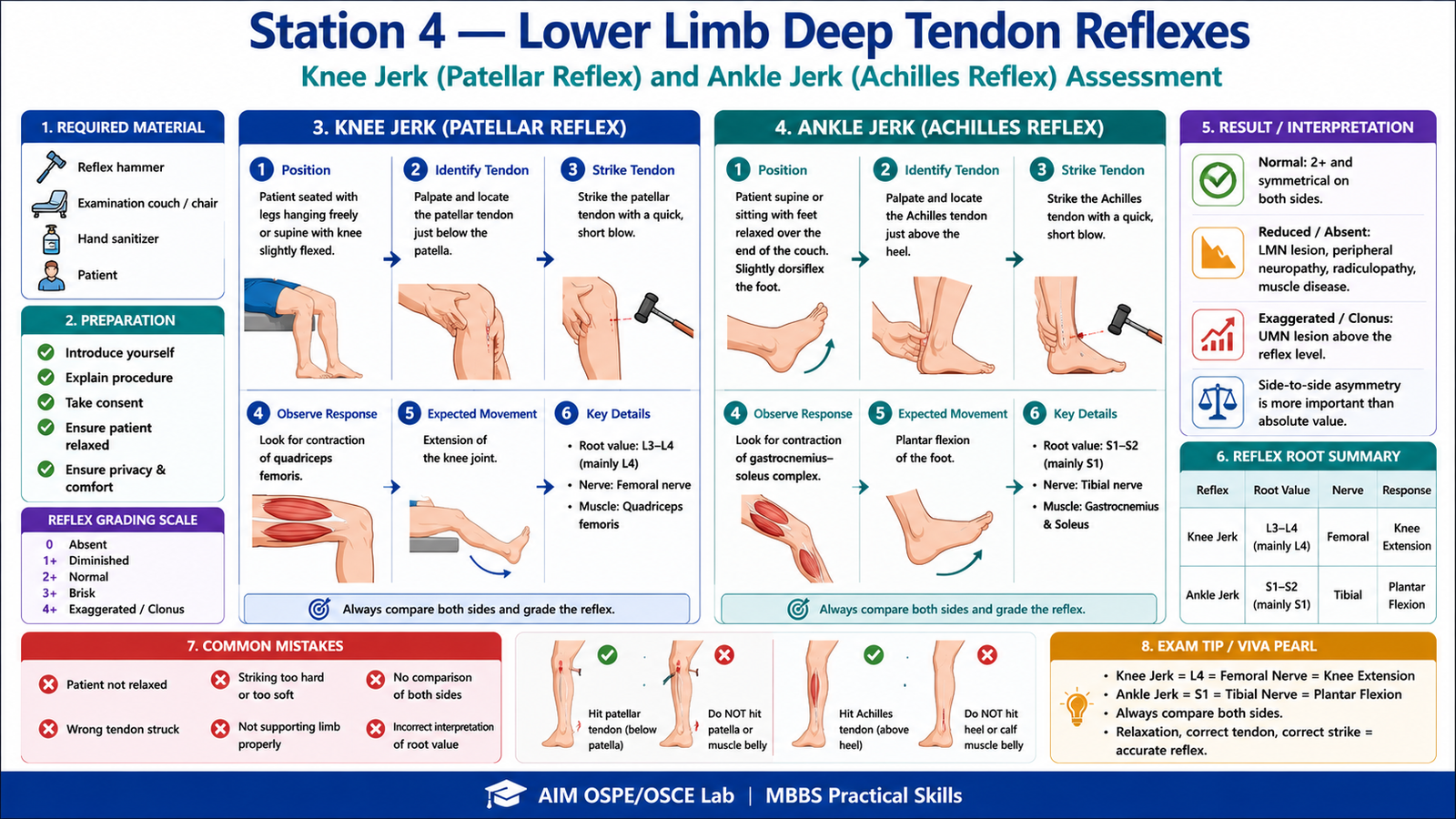
Required Material
- Examination couch / chair
- Reflex hammer
- Patient / simulated patient
- Hand sanitizer
- Answer sheet / LMS response box
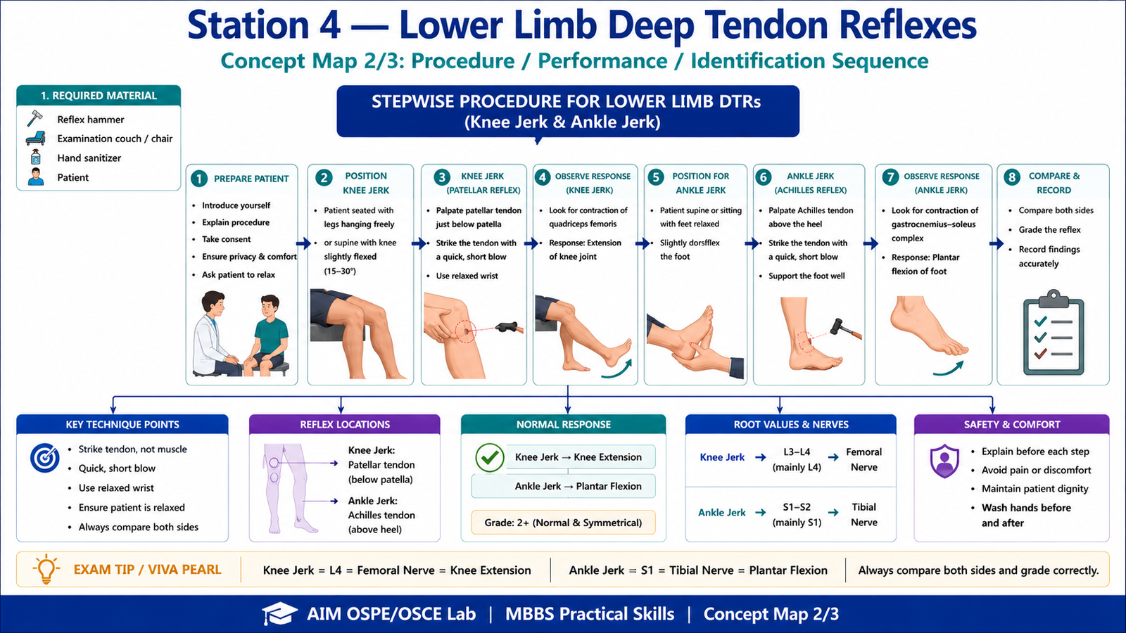
Student Task / Procedure
- Wash hands and introduce yourself to the patient.
- Explain the procedure briefly and take consent.
- Position the patient comfortably and expose the lower limbs appropriately.
- Ask the patient to relax fully.
- Test the knee jerk reflex on both sides.
- Test the ankle jerk reflex on both sides.
- Compare right and left sides.
- State the reflex grade and interpretation.
- Thank the patient and perform hand hygiene.
Observation / Identification Points
The student should observe, perform, or demonstrate the following:
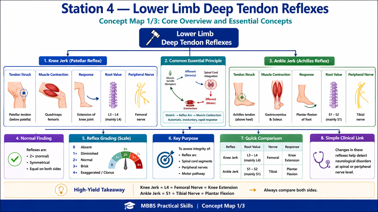
Knee Jerk Reflex
- Patient seated with legs hanging freely, or lying supine with knee slightly flexed.
- Identify the patellar tendon below the patella.
- Strike the patellar tendon with the reflex hammer.
- Observe contraction of quadriceps femoris.
- Expected response: extension of knee joint.
- Reflex root value: L3, L4 mainly L4.
- Peripheral nerve: Femoral nerve.
Ankle Jerk Reflex
- Patient relaxed, foot slightly dorsiflexed.
- Support the foot properly.
- Strike the Achilles tendon above the heel.
- Observe contraction of gastrocnemius–soleus complex.
- Expected response: plantar flexion of foot.
- Reflex root value: S1, S2 mainly S1.
- Peripheral nerve: Tibial nerve.
Reflex Grading
- 0 = absent
- 1+ = diminished
- 2+ = normal
- 3+ = brisk
- 4+ = exaggerated / clonus
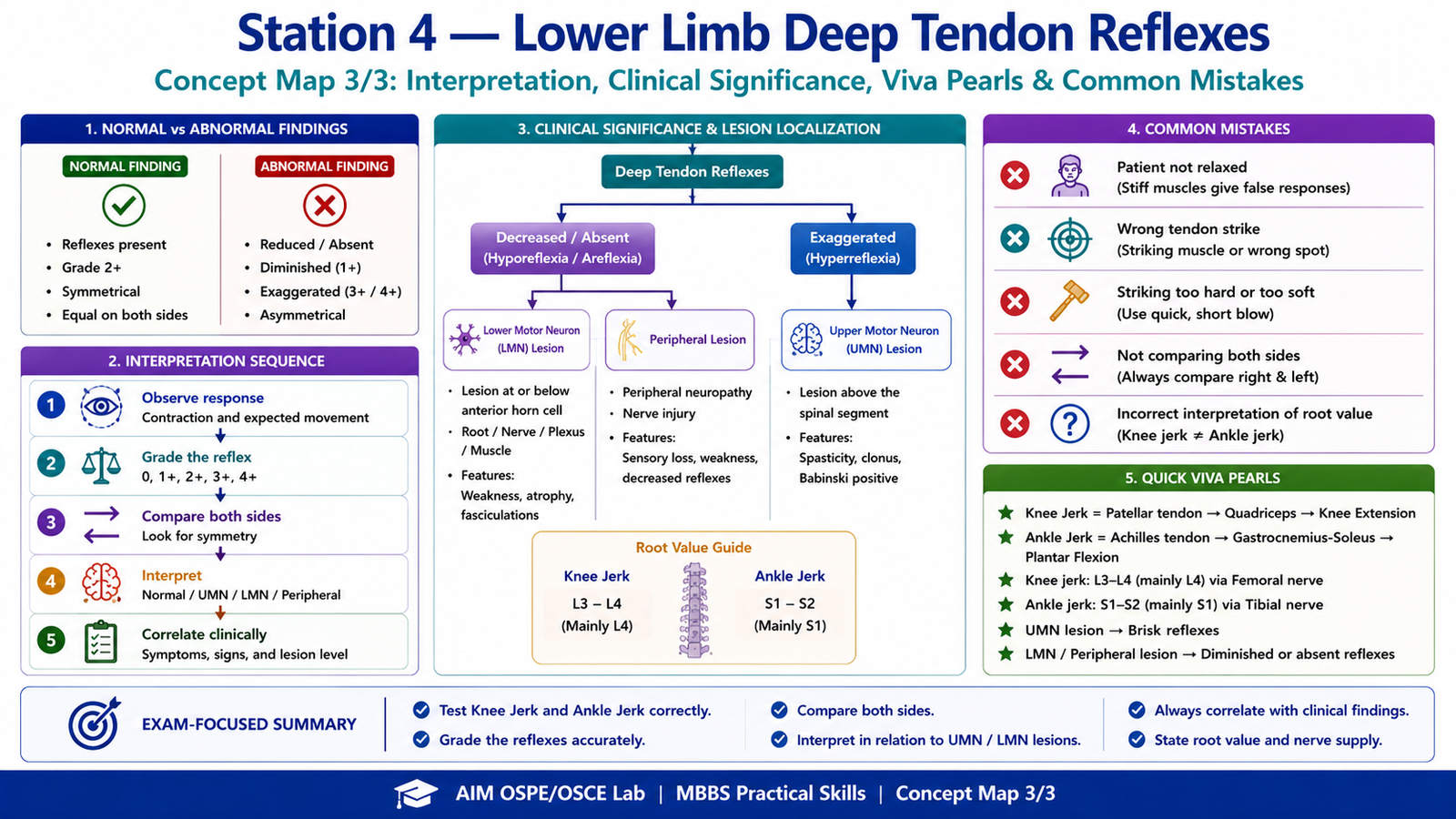
Result / Interpretation
Normal lower limb deep tendon reflexes are graded as 2+ and symmetrical on both sides.
- Knee jerk tests the integrity of the L3–L4 spinal segments, mainly L4, through the femoral nerve.
- Ankle jerk tests the integrity of the S1–S2 spinal segments, mainly S1, through the tibial nerve.
A reduced or absent reflex may suggest a lower motor neuron lesion, peripheral neuropathy, radiculopathy, or muscle disease.
An exaggerated reflex may suggest an upper motor neuron lesion above the reflex level.
Viva Questions
1. What is the receptor involved in deep tendon reflexes?
Answer: Muscle spindle.
2. What is the root value of knee jerk reflex?
Answer: L3–L4, mainly L4.
3. Which nerve is tested in knee jerk reflex?
Answer: Femoral nerve.
4. What is the root value of ankle jerk reflex?
Answer: S1–S2, mainly S1.
5. What does an exaggerated deep tendon reflex suggest?
Answer: Upper motor neuron lesion above the reflex level.
Common Student Mistakes
- Striking the tendon too hard or at the wrong site.
- Not ensuring the patient is relaxed before testing.
- Forgetting to compare both sides.
- Confusing knee jerk root value with ankle jerk root value.
AIM Feedback
To improve your performance, focus on position, relaxation, correct tendon, and correct observation. For knee jerk, remember: patellar tendon → femoral nerve → L3/L4 → knee extension. For ankle jerk, remember: Achilles tendon → tibial nerve → S1/S2 → plantar flexion. Always compare both sides because asymmetry is more clinically important than a single reflex response.
🖼️ Visual / Image Support
🧩 Concept Map / Interpretation Support