🩺 Station 11 — Neural Tissue / Sacral Segment Slides
AIM OSPE/OSCE Lab — Practical Station | KMU Style | MBBS Practical + Viva
📋 Complete OSPE Station Content
OSPE Station Name
Station 11 — Neural Tissue / Sacral Segment Slides
Learning Target
By the end of this station, the student should be able to:
- Identify the sacral spinal cord segment and basic neural tissue structures under the microscope.
- Recognize key microscopic features such as gray matter, white matter, central canal, anterior horn cells, posterior horn, neurons, and nerve fibers.
Required Material
- Prepared histology slide of sacral spinal cord segment
- Prepared nervous tissue slides, if provided:
- Spinal cord section
- Peripheral nerve section
- Multipolar neuron slide
- Dorsal root ganglion slide
- Light microscope
- Pointer or virtual slide marker
- Labeled/unlabeled photomicrograph
- Answer sheet or LMS response box
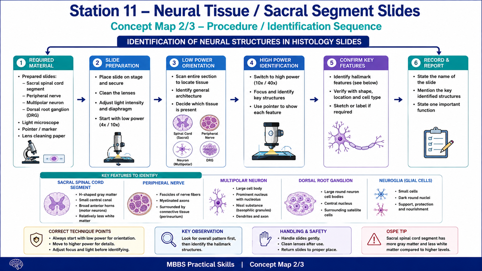
Student Task / Procedure
- Focus the given slide under low power.
- Identify whether the slide shows spinal cord segment or another nervous tissue.
- If spinal cord is shown, identify:
- Gray matter
- White matter
- Central canal
- Anterior horn
- Posterior horn
- Identify the sacral segment pattern using its main features.
- If another neural tissue slide is shown, identify the main structure and one key microscopic feature.
- Write the name of the slide and one identifying point.
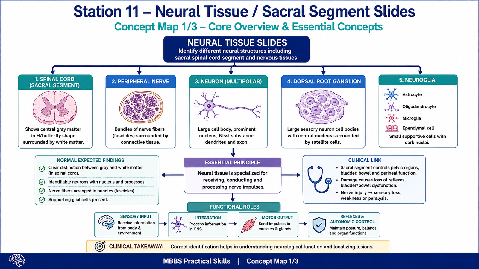
Observation / Identification Points
Students should observe and identify:
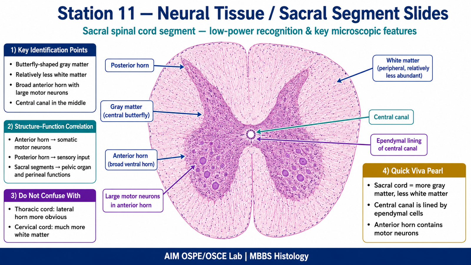
Sacral Spinal Cord Segment
- Gray matter
- Central H-shaped or butterfly-shaped region
- Relatively large compared with white matter
- White matter
- Peripheral lighter-staining region
- Less abundant in sacral segments compared with higher spinal cord levels
- Anterior horn
- Broad ventral part of gray matter
- Contains large motor neurons
- Posterior horn
- Narrow dorsal part of gray matter
- Related to sensory input
- Central canal
- Small canal in the center of gray matter
- Lined by ependymal cells
General Neural Tissue Features
- Neuron
- Large cell body
- Prominent nucleus
- Nissl substance may be visible
- Neuroglia
- Smaller supporting cells
- Darker small nuclei
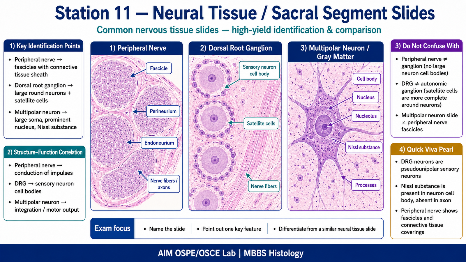
- Peripheral nerve
- Bundles of nerve fibers
- Fascicles surrounded by connective tissue
- Dorsal root ganglion
- Large sensory neuron cell bodies
- Satellite cells around neurons
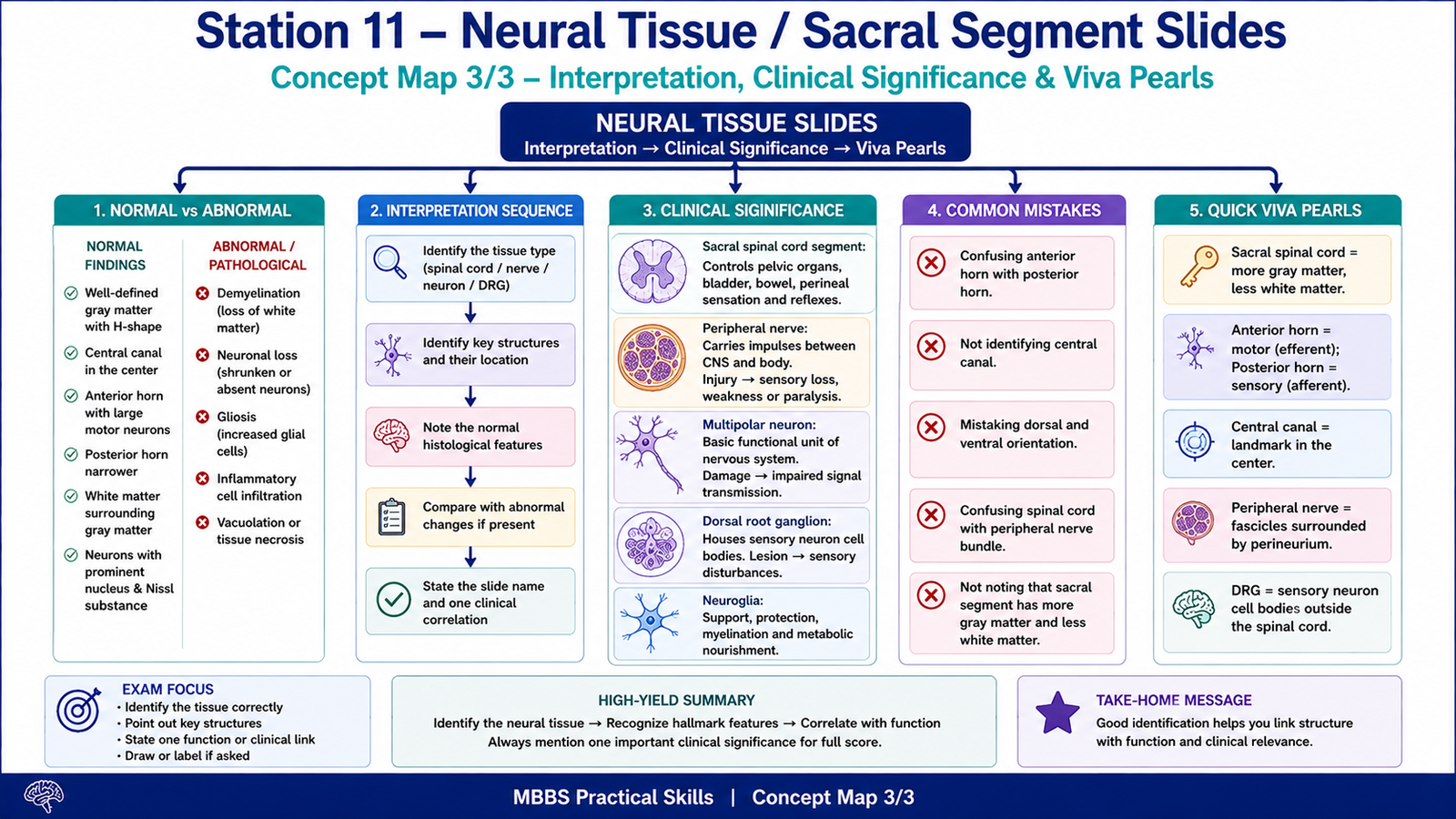
Result / Interpretation
The sacral spinal cord segment is recognized by prominent gray matter with relatively less white matter. The anterior horn contains motor neurons, the posterior horn receives sensory input, and the central canal lies in the center of the gray matter.
Clinical significance:
Sacral spinal cord segments are important for lower limb, pelvic organ, bladder, bowel, and perineal functions. Injury to sacral segments may affect reflexes, bladder control, bowel control, and sensory function in the sacral dermatomes.
Viva Questions
1. What is the central H-shaped area of the spinal cord called?
Answer: Gray matter.
2. What is present in the anterior horn of the spinal cord?
Answer: Large motor neurons.
3. Why does the sacral spinal cord have less white matter than higher segments?
Answer: Because fewer ascending and descending nerve fibers are present at lower spinal cord levels.
4. What lines the central canal of the spinal cord?
Answer: Ependymal cells.
5. What is the function of the posterior horn?
Answer: It receives sensory input.
Common Student Mistakes
- Confusing gray matter and white matter because of poor orientation.
- Calling the anterior horn posterior horn or reversing dorsal and ventral sides.
- Identifying all spinal cord levels as the same without noticing that sacral segments have less white matter.
AIM Feedback
To improve, first locate the central canal and the butterfly-shaped gray matter. Then identify the anterior horn as the broader ventral part containing large motor neurons and the posterior horn as the narrower dorsal part. For sacral segment recognition, remember: more gray matter, less white matter compared with cervical and thoracic levels.
🖼️ Visual / Image Support
🧩 Concept Map / Interpretation Support