🩺 Station 7 — Cerebral Cortex Histology
AIM OSPE/OSCE Lab — Practical Station | KMU Style | MBBS Practical + Viva
📋 Complete OSPE Station Content
OSPE Station Name
Station 7 — Cerebral Cortex Histology
Subject / Integration: Histology with Anatomy, Physiology, and Clinical Correlation
Module: Neuroscience
Year: 2nd Year MBBS
Learning Target
- Identify the cerebral cortex under the microscope and recognize its major histological organization.
- Identify the six layers of the cerebral cortex and relate them briefly to cortical function.
Required Material
- Prepared histology slide of cerebral cortex
- Light microscope
- Pointer or labeled digital slide
- Answer sheet / LMS response box
- Pencil for labeling, if required
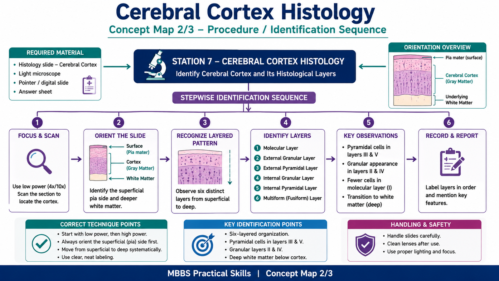
Student Task / Procedure
- Focus the given slide under low power.
- Identify the cerebral cortex.
- Observe the arrangement of cells from superficial to deep layers.
- Identify the main cortical layers.
- Mention one functional or clinical significance of cerebral cortex histology.
Observation / Identification Points
The student should identify:
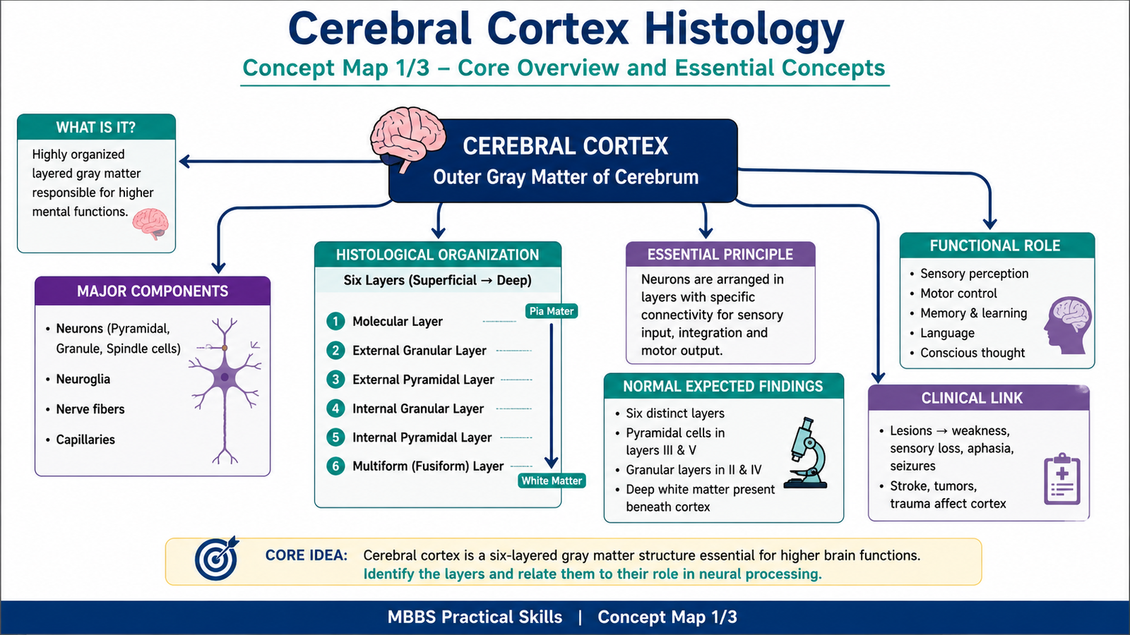
- Cerebral cortex as outer gray matter of cerebrum
- Pia mater side as the superficial surface
- Underlying white matter deep to the cortex
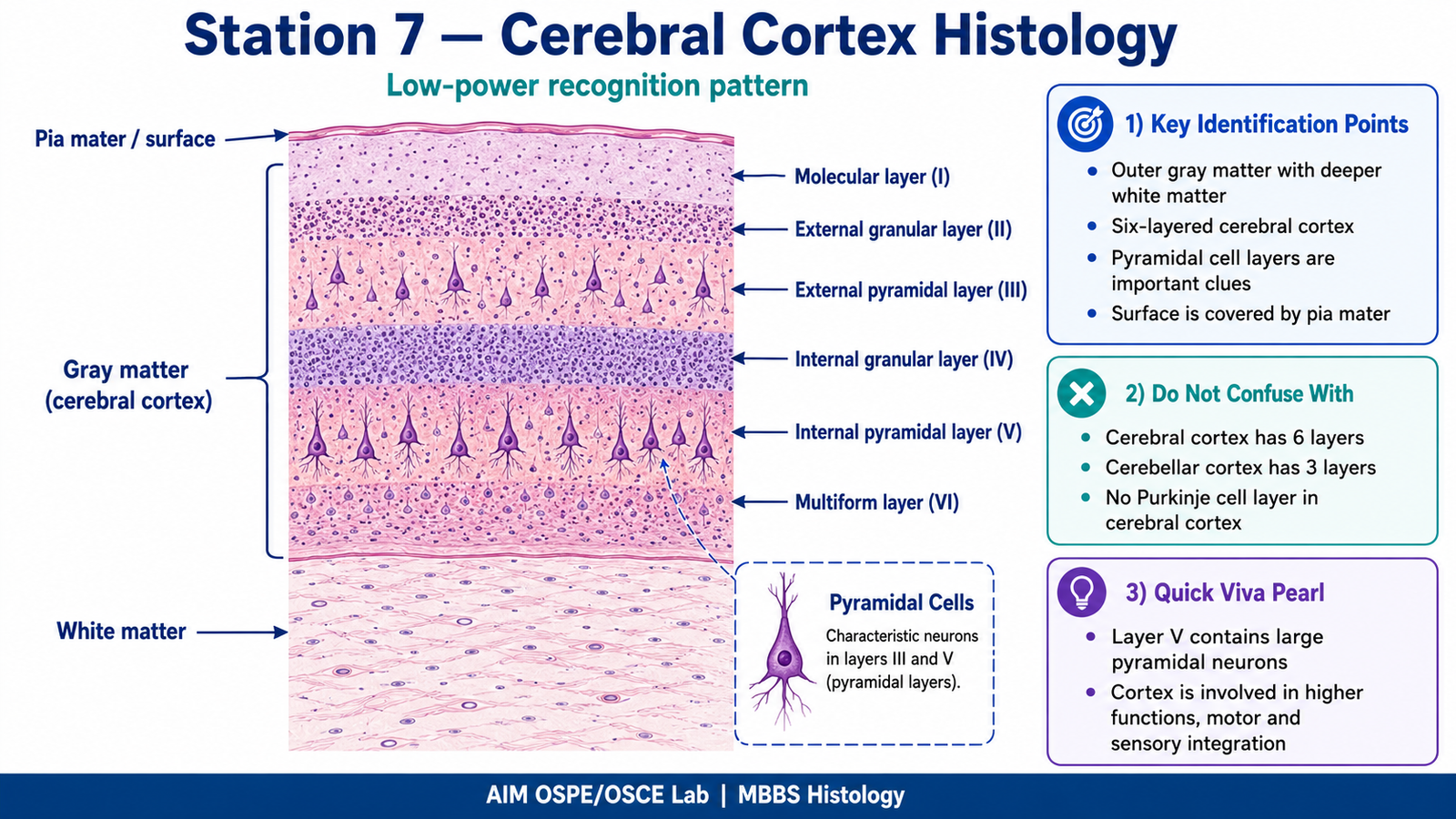
- Six cortical layers, from superficial to deep:
- Molecular layer
- External granular layer
- External pyramidal layer
- Internal granular layer
- Internal pyramidal layer
- Multiform / fusiform layer
- Presence of pyramidal cells, especially in pyramidal layers
- Difference between gray matter cortex and deeper white matter
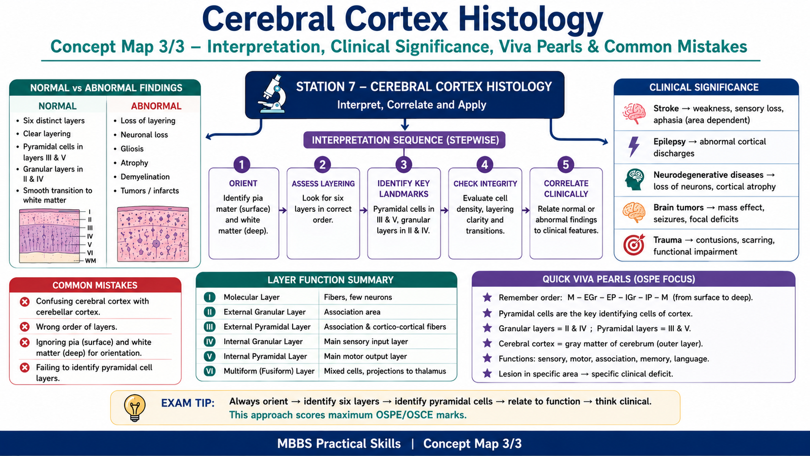
Result / Interpretation
The slide shows cerebral cortex, which is the outer gray matter of the cerebrum. It is arranged in six histological layers containing neurons, neuroglia, and nerve fibers. The cortical layers are important for sensory reception, motor output, association functions, memory, language, and higher mental activity.
Clinically, damage to different cortical areas may produce deficits such as weakness, sensory loss, aphasia, memory impairment, personality changes, or seizures.
Viva Questions
1. What type of tissue forms the cerebral cortex?
Answer: Nervous tissue forming the gray matter of the cerebrum.
2. How many layers are present in the cerebral cortex?
Answer: Six layers.
3. Name the layers of cerebral cortex from superficial to deep.
Answer: Molecular, external granular, external pyramidal, internal granular, internal pyramidal, and multiform layers.
4. Which cells are characteristic of the pyramidal layers?
Answer: Pyramidal neurons.
5. What is the clinical significance of cerebral cortex damage?
Answer: It may cause motor, sensory, language, memory, behavioral, or seizure-related problems depending on the affected cortical area.
Common Student Mistakes
- Confusing cerebral cortex with cerebellar cortex.
- Forgetting the correct order of the six cortical layers.
- Not identifying the superficial pia side and deeper white matter.
- Calling all layers “granular” without recognizing pyramidal cell layers.
AIM Feedback
To improve, first orient the slide by identifying the surface pia mater and the deeper white matter. Then move from superficial to deep and recall the six layers in sequence. Focus especially on recognizing the pyramidal cell layers, because they are important identification points in cerebral cortex histology and commonly asked in viva.
🖼️ Visual / Image Support
🧩 Concept Map / Interpretation Support