🩺Station 3 — Thoracic Spinal Cord Histology
AIM OSPE/OSCE Lab — Practical Station | KMU Style | MBBS Practical + Viva
📋 Complete OSPE Station Content
OSPE Station Name
Station 3 — Thoracic Spinal Cord Histology
Learning Target
By the end of this station, the student should be able to:
- Identify a transverse section of the thoracic spinal cord under the microscope.
- Recognize the key histological features of thoracic spinal cord and relate them to basic function.
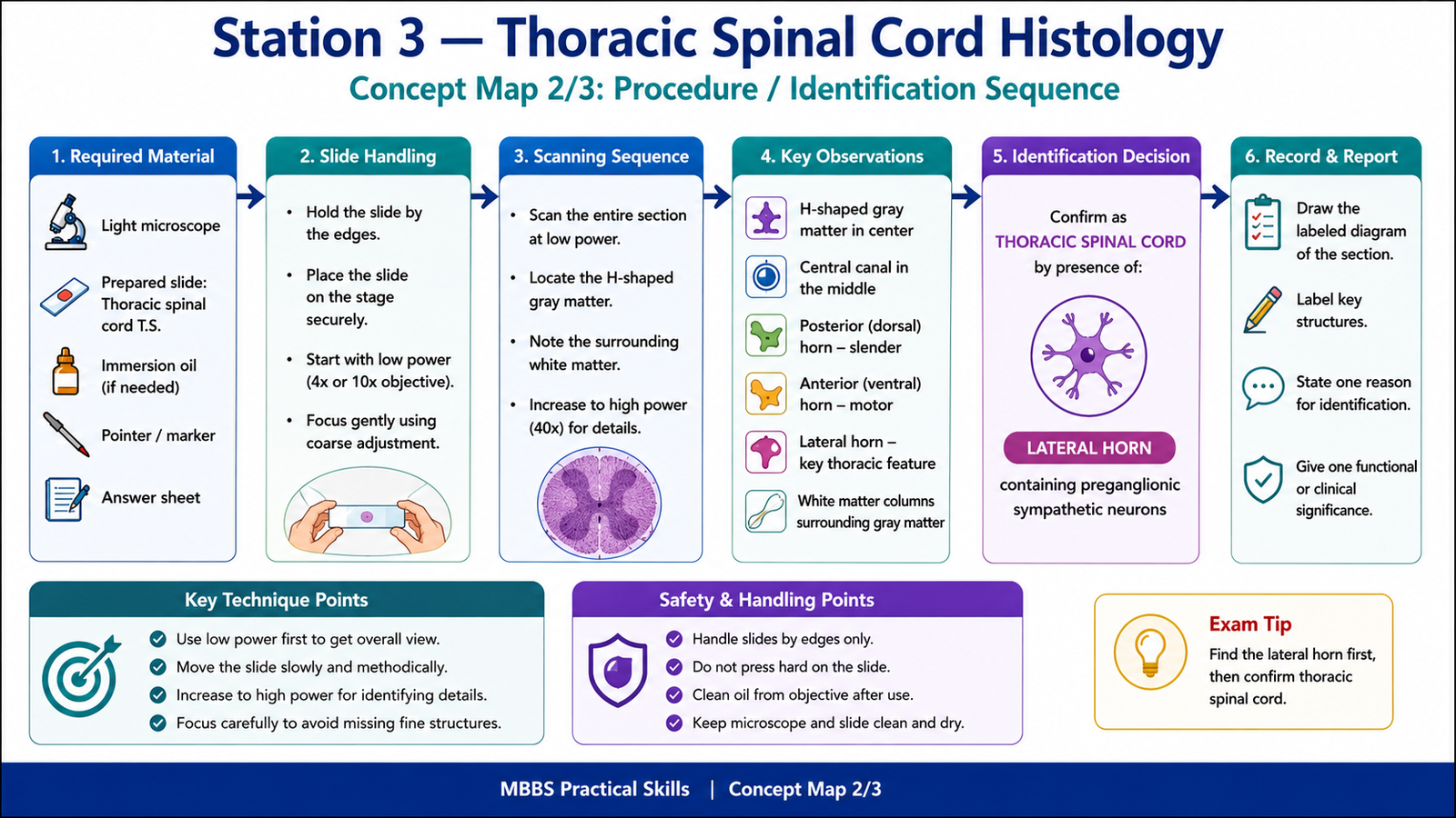
Required Material
- Prepared histology slide of thoracic spinal cord transverse section
- Light microscope / digital microscope image
- Pointer or labeled cursor if using a digital slide
- Answer sheet / LMS response box
- Labeled reference image for examiner use
Student Task / Procedure
- Observe the given microscopic slide carefully.
- Identify the tissue as a transverse section of thoracic spinal cord.
- Point out the main identifying features.
- Mention one reason why this section is thoracic spinal cord.
- State one functional or clinical significance of this region.
Observation / Identification Points
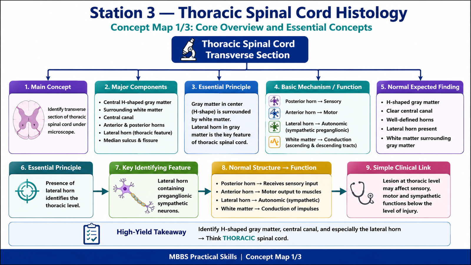
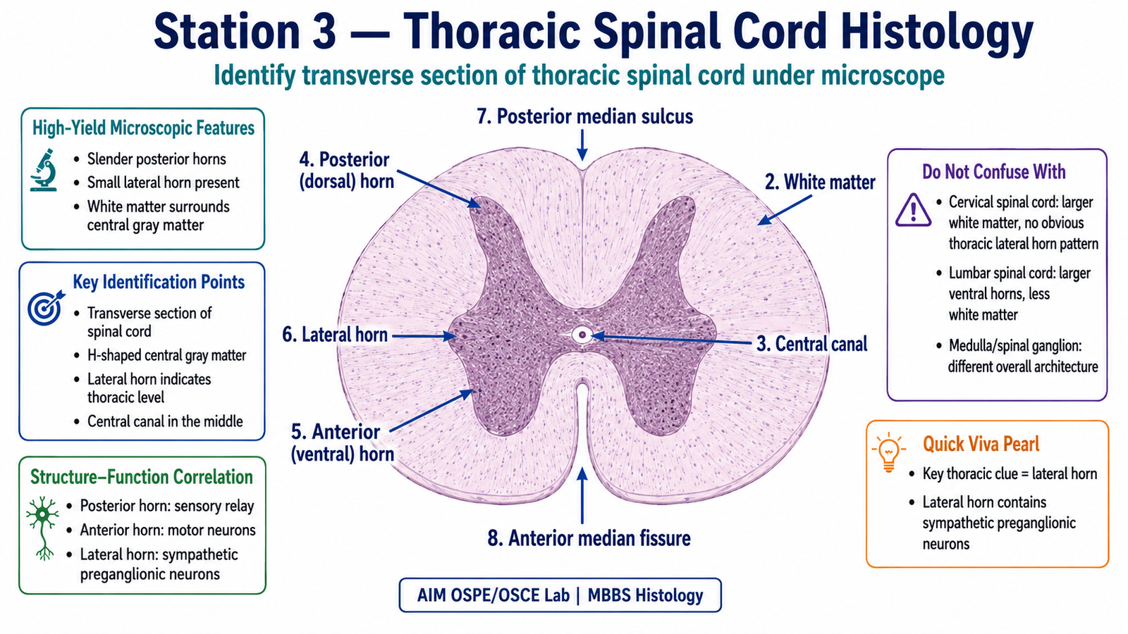
The student should identify or mention the following:
- H-shaped gray matter in the center
- White matter surrounding the gray matter
- Central canal in the middle of gray commissure
- Posterior/dorsal horns — slender and sensory-related
- Anterior/ventral horns — motor-related, less bulky than cervical/lumbar enlargement
- Lateral horn — important identifying feature of thoracic spinal cord
- Posterior median sulcus
- Anterior median fissure
- Posterior, lateral, and anterior white funiculi
- Thoracic clue: presence of lateral horn containing sympathetic preganglionic neurons
Result / Interpretation
The slide shows a transverse section of thoracic spinal cord.
The thoracic spinal cord is identified by the presence of a lateral horn in the gray matter. This lateral horn contains preganglionic sympathetic neurons, which are part of the autonomic nervous system.
Clinically, damage to thoracic spinal cord segments may affect sensory pathways, motor pathways, and sympathetic autonomic functions below the level of lesion.
Viva Questions
1. What is the most important histological feature identifying thoracic spinal cord?
Answer: Presence of the lateral horn in the gray matter.
2. What is present in the lateral horn of thoracic spinal cord?
Answer: Preganglionic sympathetic neurons.
3. What is the function of the posterior horn?
Answer: It receives and processes sensory information entering the spinal cord.
4. What is the function of the anterior horn?
Answer: It contains lower motor neurons supplying skeletal muscles.
5. What is the central canal lined by?
Answer: It is lined by ependymal cells and contains cerebrospinal fluid.
Common Student Mistakes
- Confusing thoracic spinal cord with cervical or lumbar spinal cord.
- Missing the lateral horn, which is the main thoracic identifying feature.
- Calling white matter gray matter because of staining variation under the microscope.
AIM Feedback
To improve, first identify the general pattern of the spinal cord: central H-shaped gray matter with surrounding white matter. Then look specifically for the lateral horn. If a lateral horn is clearly visible, think of the thoracic spinal cord, because this region contains sympathetic preganglionic neurons. Always connect histology with function: posterior horn is sensory, anterior horn is motor, and lateral horn is autonomic.
🖼️ Visual / Image Support
🧩 Concept Map / Interpretation Support