🩺 Station 5 — Lumbar Spinal Cord Histology
AIM OSPE/OSCE Lab — Practical Station | KMU Style | MBBS Practical + Viva
📋 Complete OSPE Station Content
OSPE Station Name
Station 5 — Lumbar Spinal Cord Histology
Learning Target
By the end of this station, the student should be able to:
- Identify the transverse section of the lumbar spinal cord under the microscope.
- Recognize the main histological features of lumbar spinal cord and relate them to basic motor and sensory function.
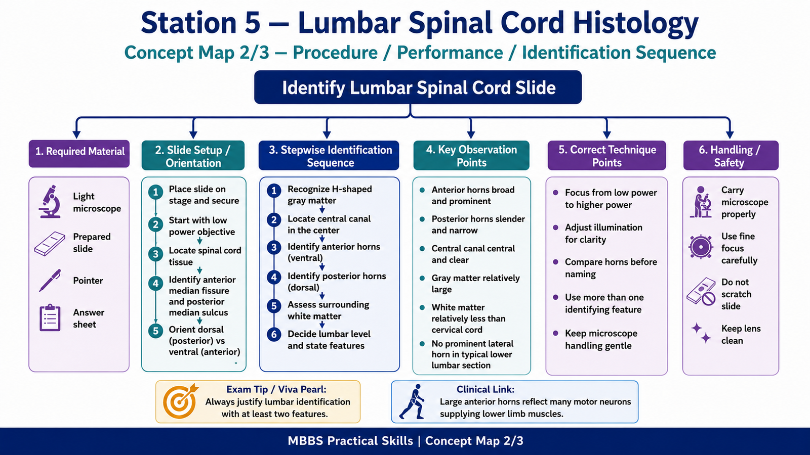
Required Material
Prepared histology slide of transverse section of lumbar spinal cord
Light microscope
Pointer or cursor
Labeled reference diagram/image of spinal cord section
Answer sheet / LMS response form
Student Task / Procedure
- Focus the slide under low power.
- Identify that the tissue is a transverse section of spinal cord.
- Observe the central H-shaped gray matter.
- Identify the anterior horn, posterior horn, central canal, and surrounding white matter.
- State why this section is likely from the lumbar spinal cord.
- Write one functional or clinical significance of the lumbar spinal cord.
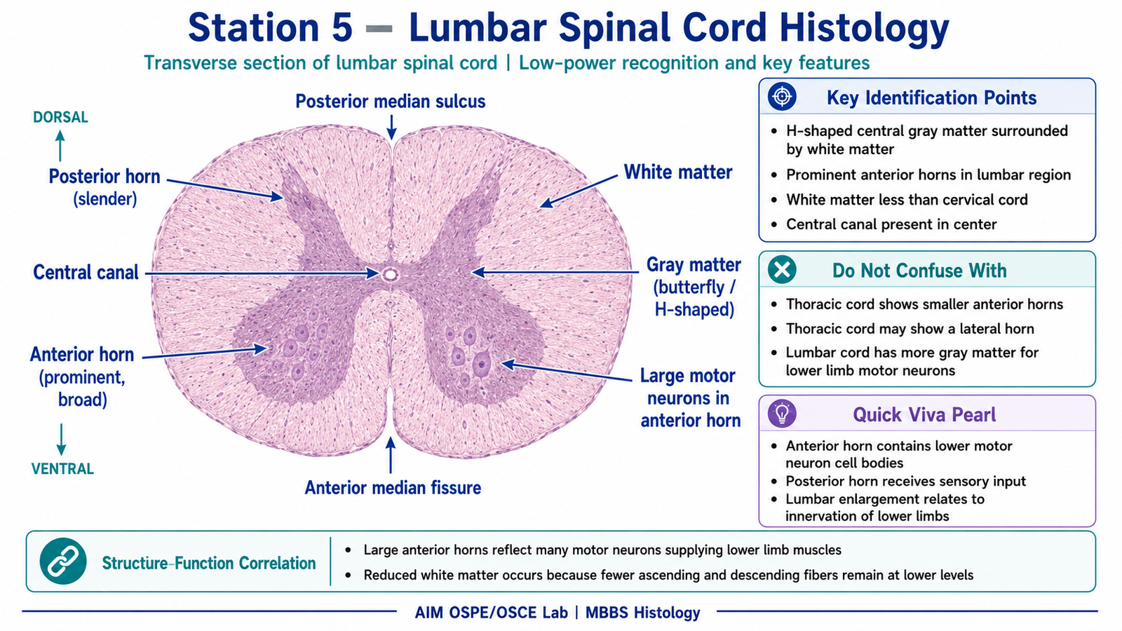
Observation / Identification Points
The student should identify:
Central H-shaped gray matter
Anterior median fissure
Posterior median sulcus
Anterior horn of gray matter
Posterior horn of gray matter
Central canal
Surrounding white matter
Prominent anterior horns due to large motor neurons
Relatively less white matter compared with cervical spinal cord
Absence of a clearly prominent lateral horn in a typical lower lumbar section
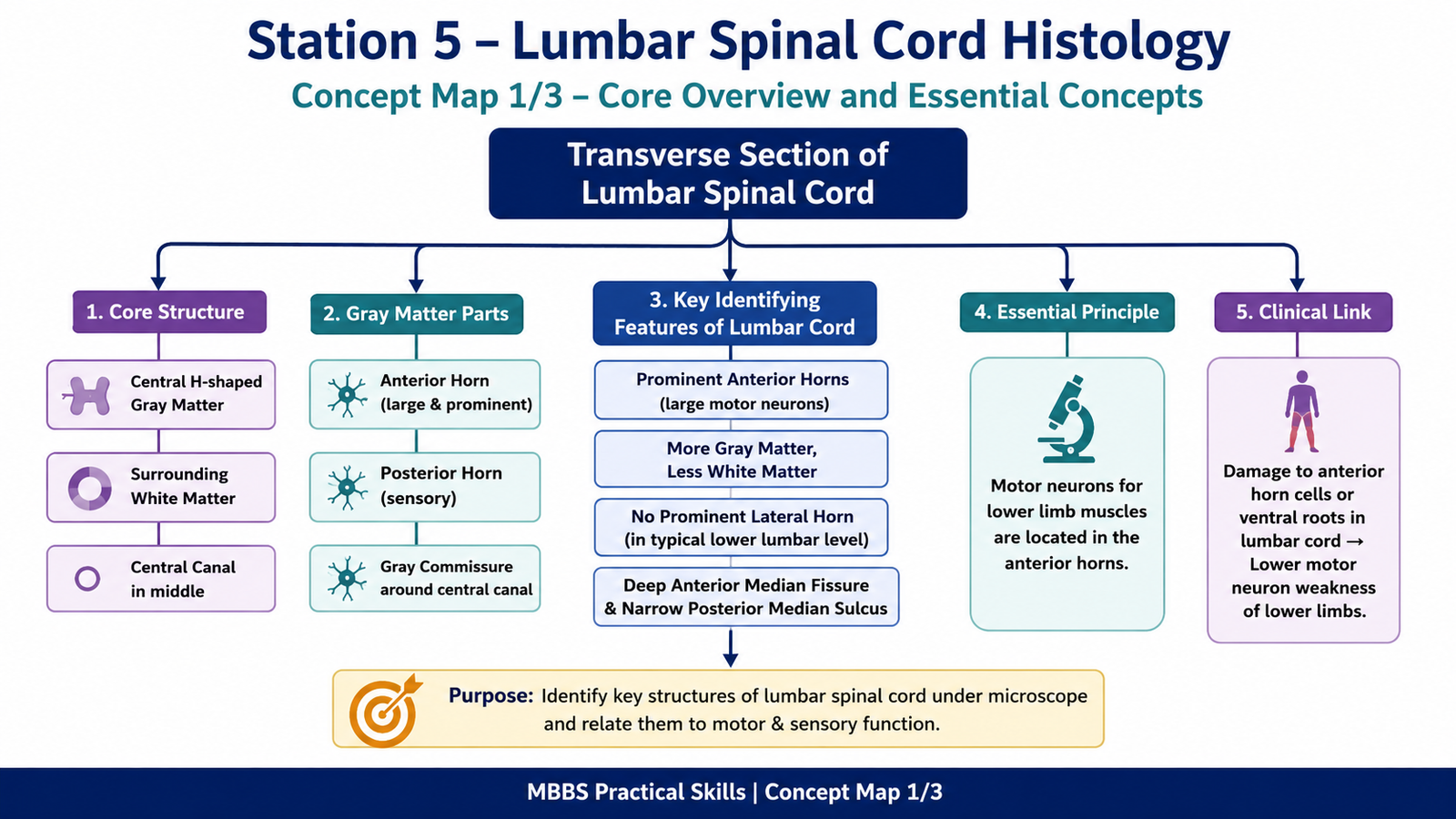
Result / Interpretation
The slide shows a transverse section of the lumbar spinal cord.
The lumbar spinal cord is recognized by its large anterior horns containing motor neuron cell bodies for lower limb muscles. The gray matter is relatively prominent, while the amount of white matter is less than in cervical segments because fewer ascending and descending fibers are present at this lower level.
Clinical significance: Damage to the lumbar anterior horn cells or ventral roots can cause lower motor neuron weakness of lower limb muscles.
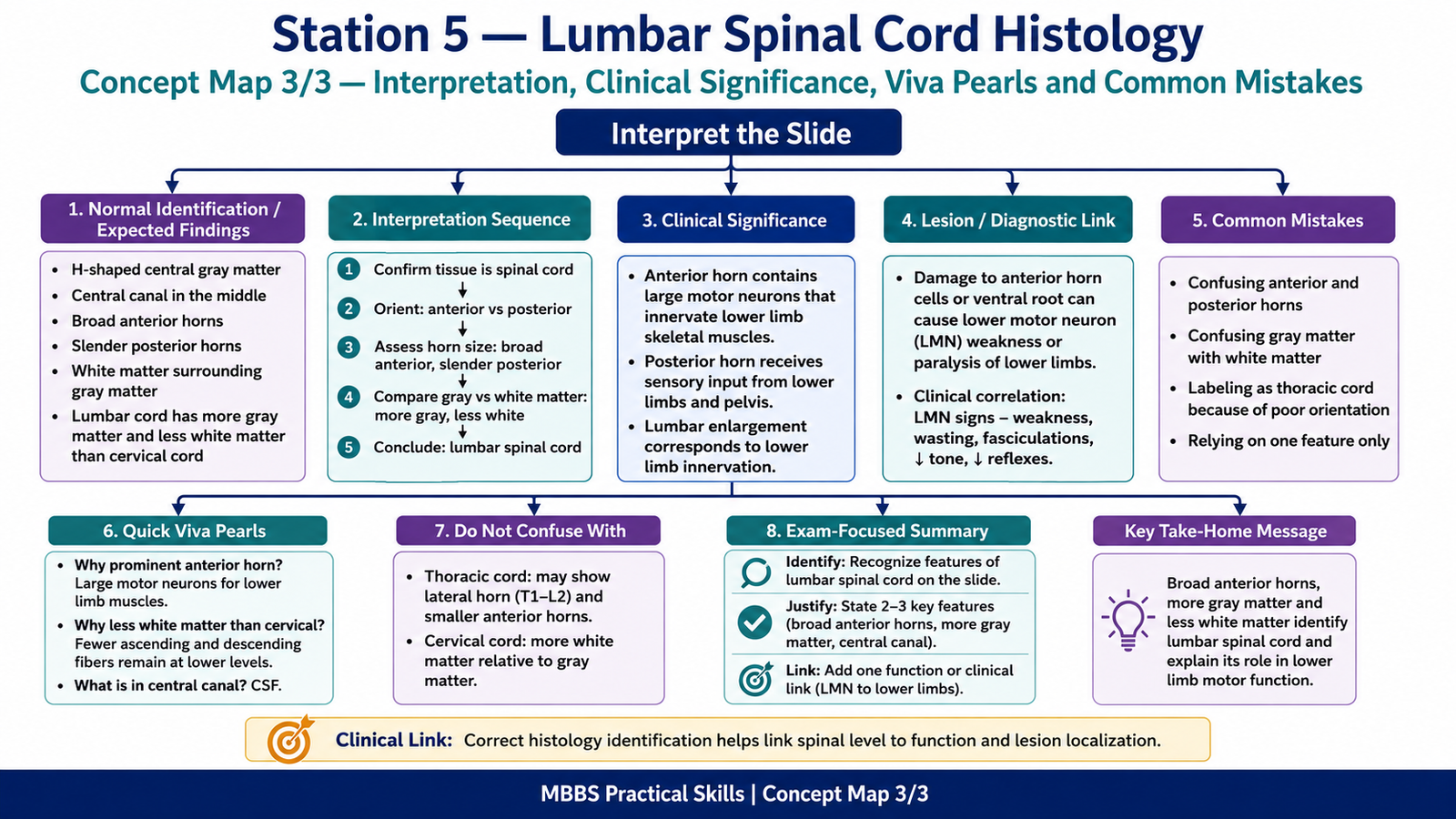
Viva Questions
- How do you identify spinal cord in transverse section?
Ideal answer: By the central H-shaped gray matter surrounded by white matter and the presence of a central canal. - Which horn is more prominent in the lumbar spinal cord?
Ideal answer: The anterior horn is prominent because it contains large motor neurons supplying lower limb muscles. - What is present in the anterior horn?
Ideal answer: Cell bodies of lower motor neurons. - What is the function of the posterior horn?
Ideal answer: It receives and processes sensory information entering through dorsal roots. - How is lumbar spinal cord different from cervical spinal cord histologically?
Ideal answer: Lumbar cord has relatively more gray matter and less white matter, while cervical cord has more white matter.
Common Student Mistakes
Confusing gray matter with white matter.
Identifying the posterior horn as the anterior horn.
Calling every spinal cord section cervical without comparing white matter and horn size.
Missing the central canal as an important identification point.
AIM Feedback
To improve, first identify the spinal cord by looking for the central H-shaped gray matter. Then orient the section using the deep anterior median fissure and the narrower posterior median sulcus. In lumbar cord, focus on the large anterior horns because they contain motor neurons for lower limb muscles. Always support your identification with one reason rather than only naming the slide.
🖼️ Visual / Image Support
🧩 Concept Map / Interpretation Support