🩺 Station 9 — Cerebellar Cortex Histology
AIM OSPE/OSCE Lab — Practical Station | KMU Style | MBBS Practical + Viva
📋 Complete OSPE Station Content
OSPE Station Name
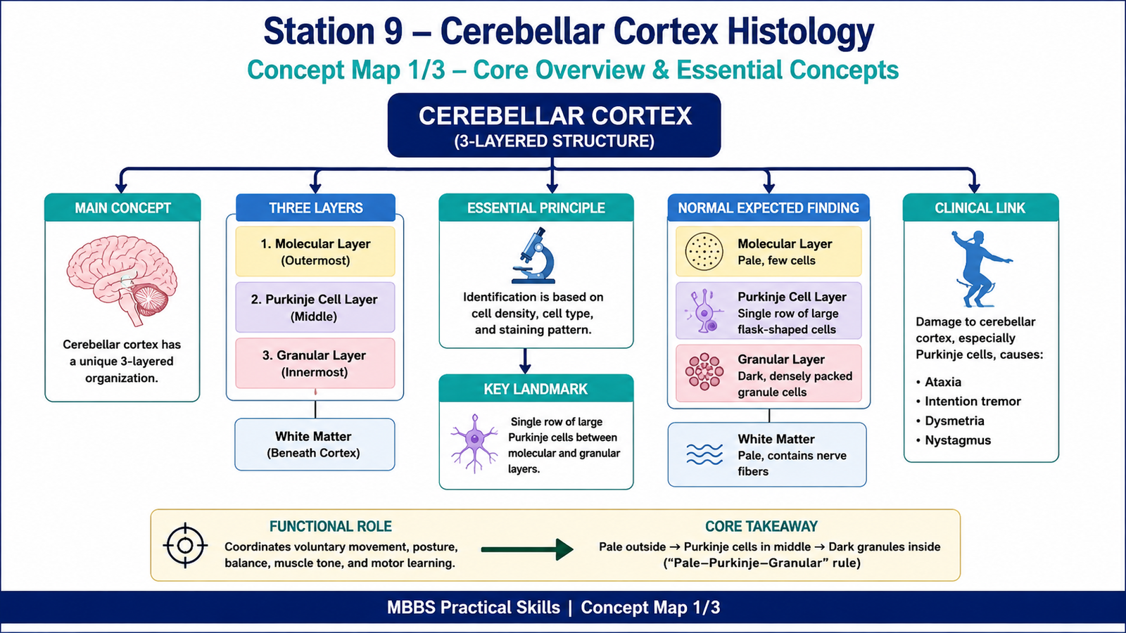
Station 9 — Cerebellar Cortex Histology
Learning Target
By the end of this station, the student should be able to:
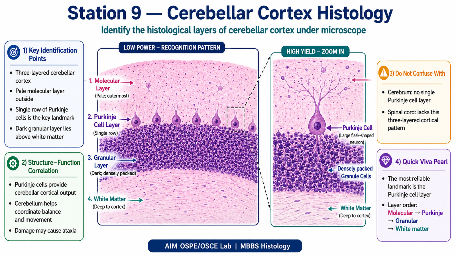
- Identify the three histological layers of the cerebellar cortex under the microscope.
- Differentiate the cerebellar cortical layers using their cell arrangement, staining pattern, and key microscopic features.
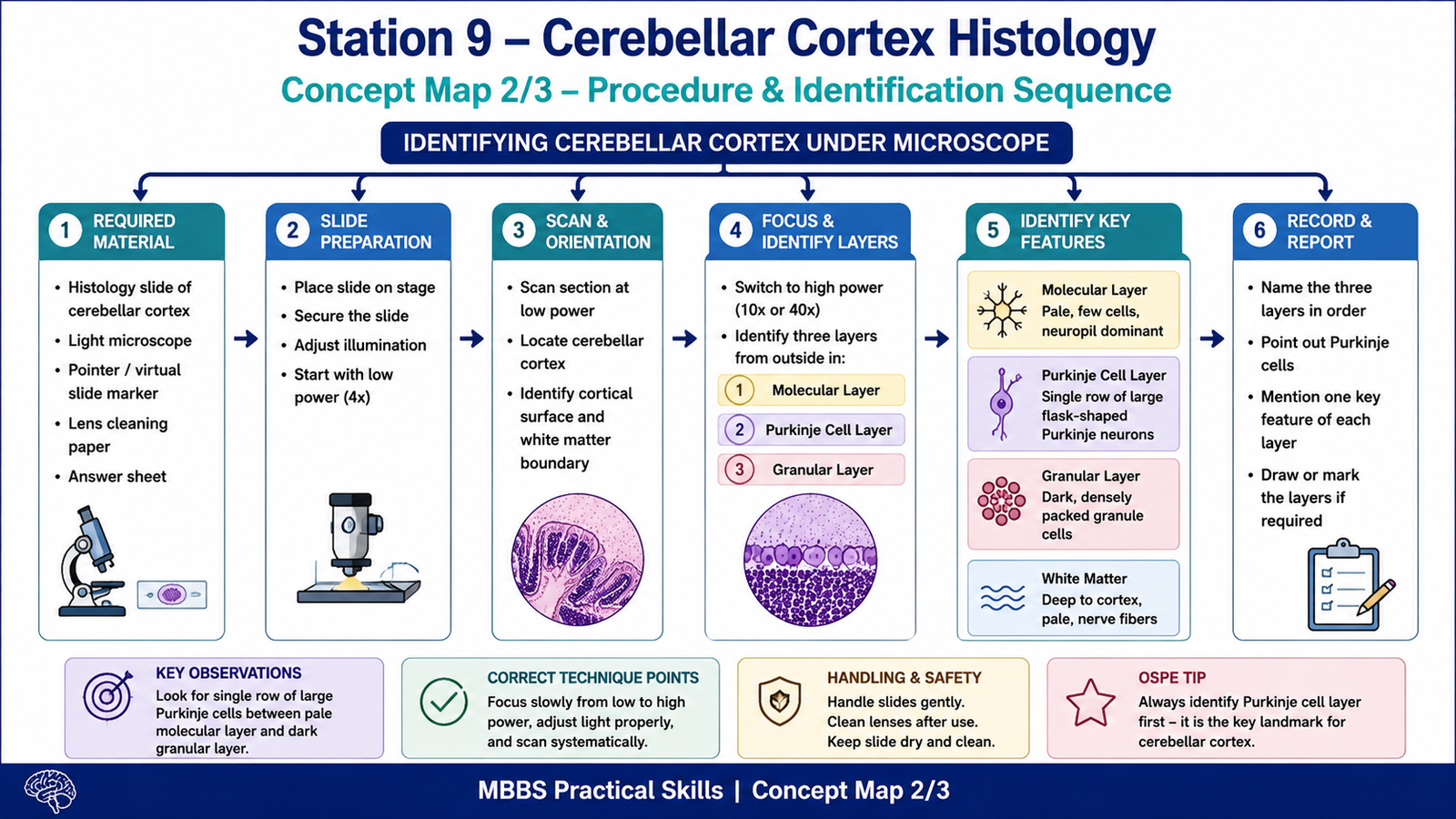
Required Material
- Prepared histology slide of cerebellar cortex
- Light microscope
- Pointer or virtual slide marker
- Labeled/unlabeled photomicrograph of cerebellar cortex
- Answer sheet or LMS response box
Student Task / Procedure
- Focus the cerebellar cortex slide under low power.
- Identify the outer, middle, and inner layers of the cerebellar cortex.
- Name the molecular layer, Purkinje cell layer, and granular layer.
- Point out the Purkinje cells.
- Write one identifying feature of each layer.
Observation / Identification Points
Students should observe and identify:
- Molecular layer
- Outermost pale-staining layer
- Contains fewer cells
- Appears lighter under microscope
- Purkinje cell layer
- Middle single row of large flask-shaped Purkinje neurons
- Located between molecular and granular layers
- Most important identifying landmark
- Granular layer
- Innermost dark-staining layer
- Contains many closely packed granule cells
- Appears deeply basophilic due to high cell density
- White matter
- Present deep to the cortex
- Contains nerve fibers
- Appears lighter than the granular layer
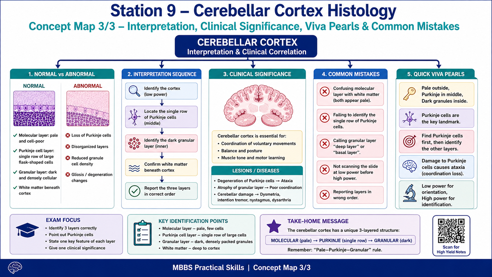
Result / Interpretation
The cerebellar cortex is identified by its three-layered arrangement:
Molecular layer → Purkinje cell layer → Granular layer
The Purkinje cell layer is the key landmark because it contains a single row of large neurons between the pale molecular layer and the dark granular layer.
Clinical significance:
Damage to cerebellar cortical neurons, especially Purkinje cells, may impair coordination, balance, and precision of movement, leading to signs such as ataxia, intention tremor, dysmetria, and nystagmus.
Viva Questions
1. What are the three layers of the cerebellar cortex?
Answer: Molecular layer, Purkinje cell layer, and granular layer.
2. Which layer contains Purkinje cells?
Answer: Purkinje cell layer.
3. Why does the granular layer appear darkly stained?
Answer: Because it contains many densely packed granule cells.
4. Which layer is the outermost layer of the cerebellar cortex?
Answer: Molecular layer.
5. What is the main function of the cerebellum?
Answer: Coordination of voluntary movements, posture, balance, and muscle tone.
Common Student Mistakes
- Confusing the molecular layer with white matter because both appear pale.
- Missing the single row of Purkinje cells, which is the most important identifying feature.
- Calling the granular layer “basal layer” or “deep layer” instead of using the correct histological term.
AIM Feedback
To improve, first locate the single row of Purkinje cells. Once Purkinje cells are identified, the layer above them is the molecular layer, and the layer below them is the granular layer. Remember: pale outside, Purkinje in middle, dark granules inside. This pattern is the key to quickly recognizing cerebellar cortex in OSPE.
🖼️ Visual / Image Support
🧩 Concept Map / Interpretation Support