🩺 Station 1 — Gross Anatomy of Urinary System Models
AIM OSPE/OSCE Lab — Practical Station | KMU Style | MBBS Practical + Viva
📋 Complete OSPE Station Content
OSPE Station Name
Station 1 — Gross Anatomy of Urinary System Models
Module: Renal
Year: 2nd Year MBBS
Subject / Integration: Anatomy with Clinical Integration
Learning Target
By the end of this station, the student should be able to:
-
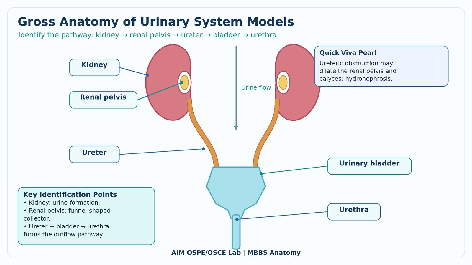
Identify the major gross anatomical parts of the urinary system on models/specimens.
-
Correlate the position and function of kidney, renal pelvis, ureter, urinary bladder, and urethra with common clinical conditions.
Required Material
-
Urinary system model or specimen
-
Kidney model/specimen
-
Model showing renal pelvis and ureter
-
Urinary bladder model
-
Urethra model or pelvic model
-
Pointer/probe
-
Station card/question sheet
-
Checklist marking sheet
Student Task / Procedure
-
Observe the given urinary system model carefully.
-
Identify the kidney.
-
Identify the renal pelvis.
-
Trace and identify the ureter.
-
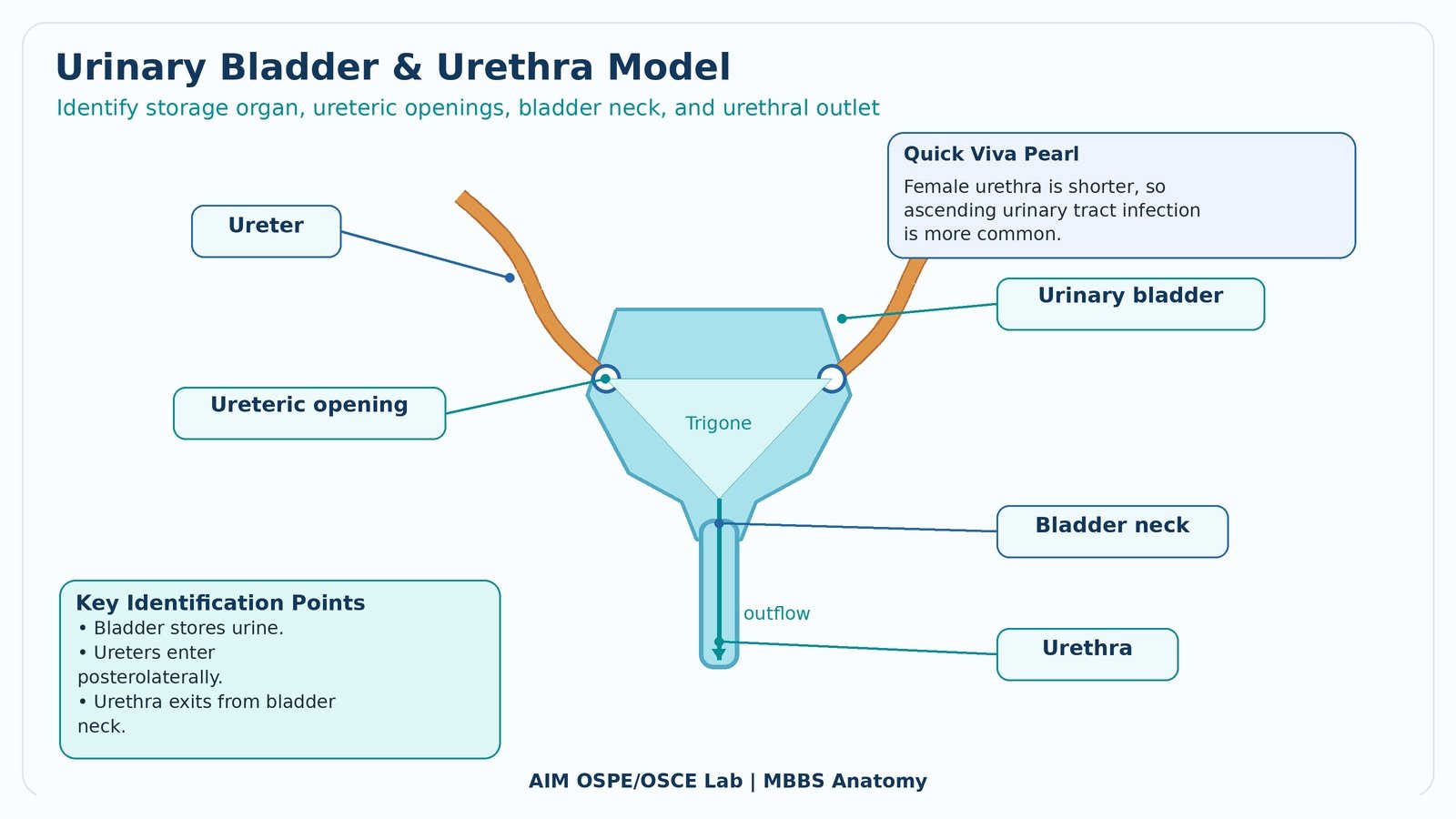
Identify the urinary bladder.
-
Identify the urethra.
-
State one function or clinical significance of any identified structure.
Observation / Identification Points
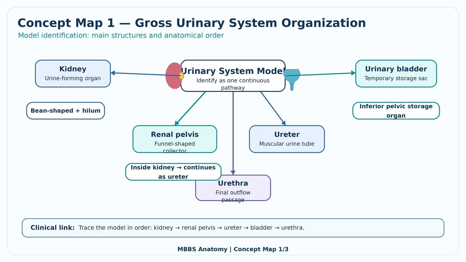
The student should correctly identify:
-
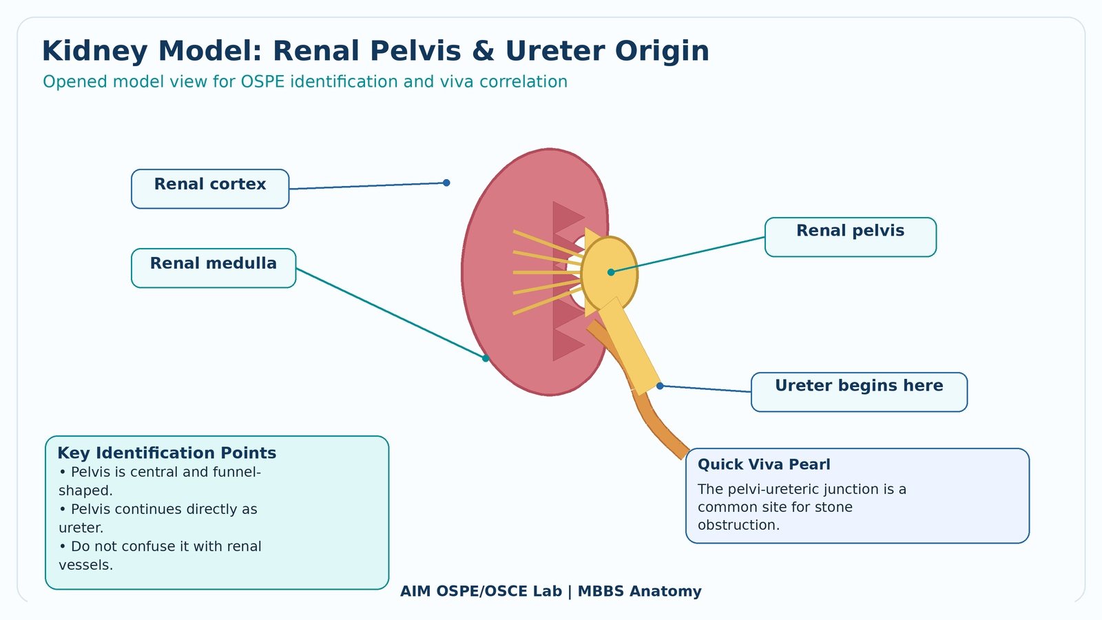
Kidney: bean-shaped organ responsible for urine formation.
-
Renal pelvis: funnel-shaped collecting part of the kidney.
-
Ureter: muscular tube carrying urine from renal pelvis to urinary bladder.
-
Urinary bladder: muscular storage organ for urine.
-
Urethra: terminal passage for urine elimination.
-
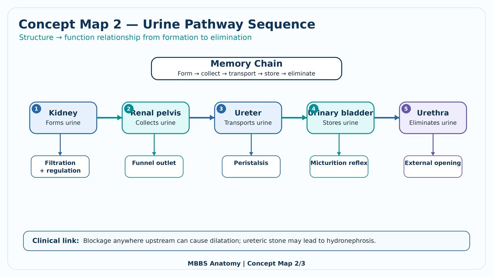
Correct anatomical sequence:
Kidney → Renal pelvis → Ureter → Urinary bladder → Urethra
Result / Interpretation
The urinary system is organized to form, collect, transport, store, and eliminate urine.
Clinical significance:
-
Obstruction in the ureter may cause hydronephrosis.
-
Infection may ascend from the urethra to bladder and kidneys.
-
The renal pelvis and ureter are common sites for urinary stones.
Viva Questions
Q1. What is the function of the kidney?
Answer: It forms urine, regulates fluid and electrolyte balance, and removes metabolic waste.
Q2. What is the renal pelvis?
Answer: It is the funnel-shaped part of the kidney that collects urine from calyces and continues as the ureter.
Q3. What is the function of the ureter?
Answer: It transports urine from the renal pelvis to the urinary bladder.
Q4. What is the main function of the urinary bladder?
Answer: It stores urine temporarily before micturition.
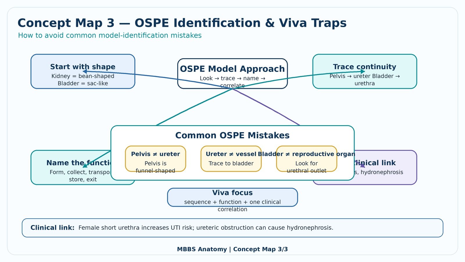
Q5. Why is the female urethra more prone to urinary tract infection?
Answer: Because it is shorter and closer to the external environment, allowing easier bacterial entry.
Common Student Mistakes
-
Confusing renal pelvis with ureter.
-
Identifying the ureter as a blood vessel.
-
Forgetting the correct urine flow sequence.
-
Confusing urinary bladder with pelvic reproductive organs on pelvic models.
Short Caption
Use this labeled guide to revise the gross anatomy of the urinary system before attempting the OSPE station. Focus on identifying each structure and tracing the flow of urine from kidney to urethra.
🖼️ Visual / Image Support
🧩 Concept Map / Interpretation Support