🩺 Station 10 — Histology of Urinary System
AIM OSPE/OSCE Lab — Practical Station | KMU Style | MBBS Practical + Viva
📋 Complete OSPE Station Content
OSPE Station Name
Station 10 — Histology of Urinary System
Module: Renal
Year: 2nd Year MBBS
Subject / Integration: Histology + Anatomy + Physiology + Clinical Integration
Learning Target
- Identify microscopic slides of kidney, ureter, urinary bladder, and urethra using key histological features.
- Correlate the epithelial lining and wall structure of urinary organs with their function and common clinical relevance.
Required Material
- Prepared H&E slides of:
- Kidney
- Ureter
- Urinary bladder
- Urethra
- Light microscope
- Pointer / slide marker
- Histology atlas or labeled reference image
- Answer sheet
- Pencil / pen
Student Task / Procedure
- Focus the given slide under low power.
- Identify the organ shown.
- Observe the epithelial lining, lumen, glands, tubules, or muscle layer.
- Write two key identifying features of the slide.
- Mention one functional or clinical significance.
- Answer the viva questions briefly.
Observation / Identification Points
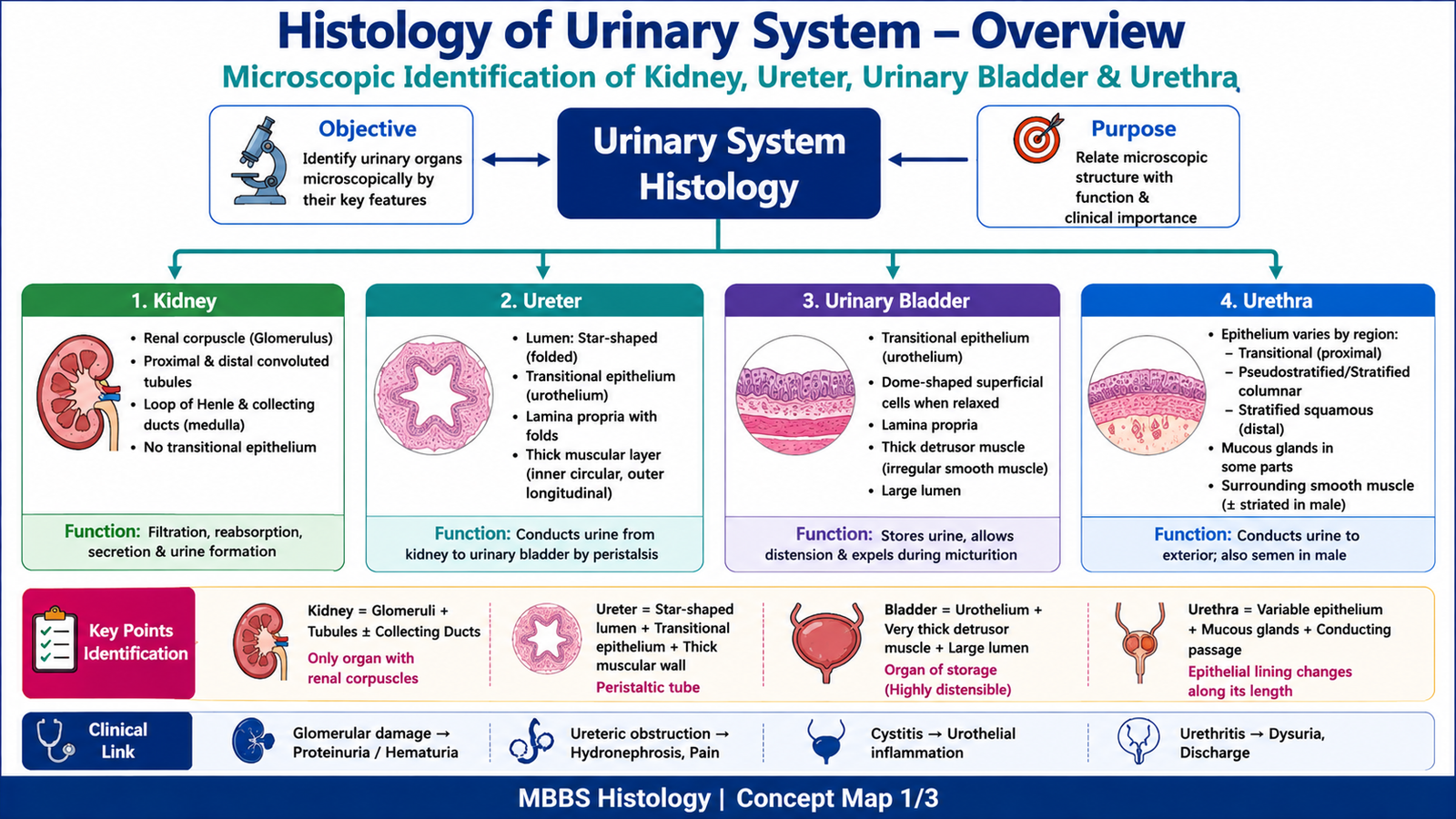
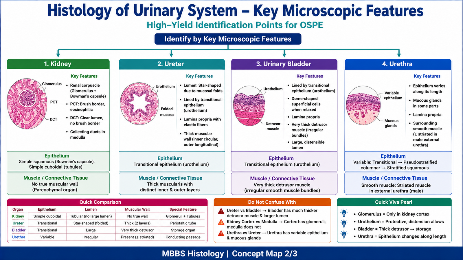
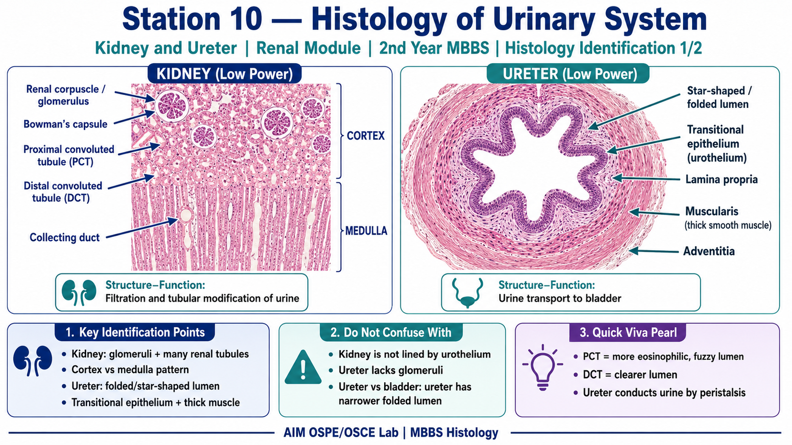
Kidney
- Presence of renal corpuscles / glomeruli in cortex
- Numerous proximal and distal convoluted tubules
- Medulla shows collecting ducts and loops of Henle
- No transitional epithelium
- Functionally related to filtration, reabsorption, secretion, and urine formation
Ureter
- Lumen is usually star-shaped or folded
- Lined by transitional epithelium / urothelium
- Thick muscular wall
- Mucosa forms folds
- Functionally conducts urine from kidney to urinary bladder
Urinary Bladder
- Lined by transitional epithelium / urothelium
- Large lumen
- Thick irregular smooth muscle layer called detrusor muscle
- Surface cells may appear dome-shaped when relaxed
- Functionally stores urine and allows distension
Urethra
- Lumen may be irregular
- Epithelium varies according to region:
- Transitional epithelium near bladder
- Pseudostratified / stratified columnar epithelium in middle part
- Stratified squamous epithelium near external opening
- May show mucous glands
- Functionally conducts urine to exterior
Result / Interpretation
| Slide / Organ | Key Microscopic Identification | Interpretation / Clinical Significance |
|---|---|---|
| Kidney | Glomeruli with renal tubules | Site of filtration and tubular modification of urine |
| Ureter | Folded mucosa with transitional epithelium and thick muscle | Conducts urine by peristalsis |
| Urinary bladder | Transitional epithelium with thick detrusor muscle | Stores urine and allows distension |
| Urethra | Variable epithelium with conducting lumen | Conducts urine to exterior; epithelial lining changes with region |
Principle:
Urinary system organs are identified microscopically by their epithelial lining, lumen shape, mucosal folds, glands, tubules, glomeruli, and muscle arrangement.
Clinical Significance:
Histological identification helps students understand renal filtration, urine transport, storage, and urinary tract pathology such as cystitis, ureteric obstruction, and glomerular disease.
Viva Questions
1. Which epithelium lines the ureter and urinary bladder?
Transitional epithelium, also called urothelium.
2. What is the main identifying feature of kidney cortex?
Presence of glomeruli and convoluted tubules.
3. Why does the urinary bladder have transitional epithelium?
It allows stretching and distension during urine storage.
4. What is the detrusor muscle?
It is the thick smooth muscle layer of the urinary bladder wall responsible for bladder contraction during micturition.
5. How can ureter be differentiated from urinary bladder histologically?
Ureter has a folded, often star-shaped lumen and a smaller thick muscular wall, while bladder has a larger lumen and very thick irregular detrusor muscle.
Common Student Mistakes
- Confusing ureter with urinary bladder because both have transitional epithelium.
- Identifying kidney medulla as cortex without looking for glomeruli.
- Writing only the organ name without mentioning histological features.
- Forgetting that urethral epithelium changes in different regions.
AIM Feedback
In histology OSPE, do not memorize the slide name alone. First look for the main diagnostic feature: glomeruli for kidney, folded urothelium for ureter, thick detrusor muscle for bladder, and variable epithelium for urethra. Always write organ identification with two microscopic features and one functional link. This improves both OSPE scoring and viva performance.
🖼️ Visual / Image Support
🧩 Concept Map / Interpretation Support