🩺 SStation 9 — Surface Anatomy of Perineum and Radiology
AIM OSPE/OSCE Lab — Practical Station | KMU Style | MBBS Practical + Viva
📋 Complete OSPE Station Content
OSPE Station Name
Station 9 — Surface Anatomy of Perineum and Radiology
Module: Renal
Year: 2nd Year MBBS
Subject / Integration: Anatomy / Clinical / Radiology Integrated
Learning Target
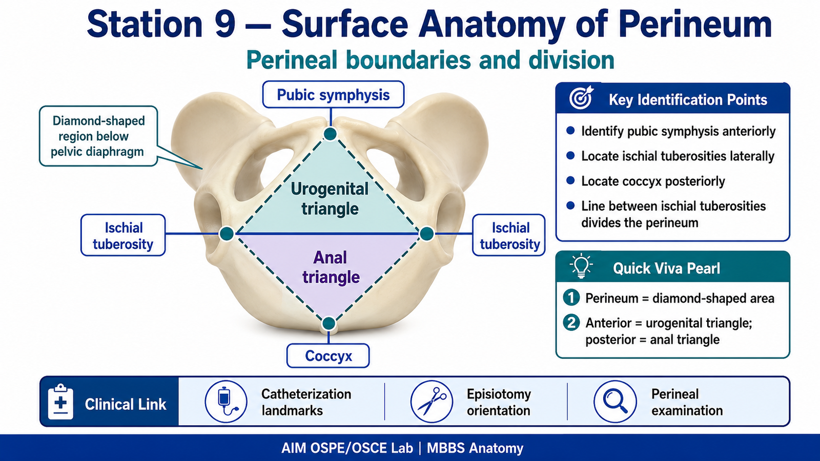
- Identify important surface anatomical structures and boundaries of the perineum on a model.
- Recognize key bony and radiological landmarks related to the perineum on pelvic radiographs.
Required Material
- Perineum model / pelvic model
- Labeled or unlabeled perineal surface anatomy diagram
- AP pelvis radiograph or pelvic outlet radiograph
- Pointer
- Station card / answer sheet
- Pen
Student Task / Procedure
- Observe the perineal model carefully.
- Identify the boundaries of the perineum.
- Identify the urogenital triangle and anal triangle.
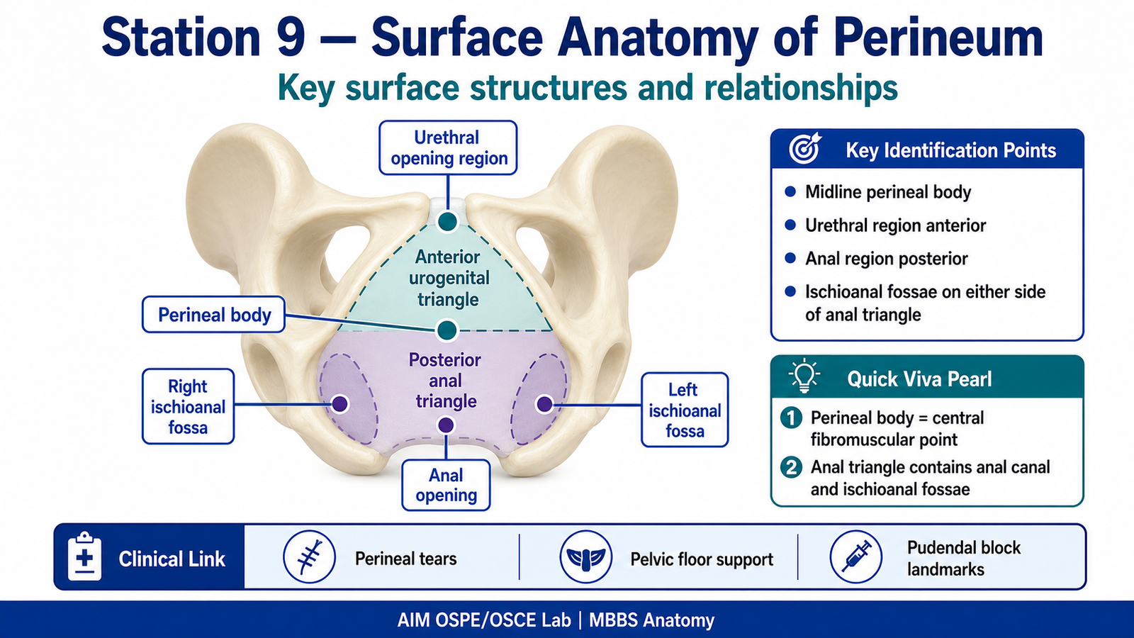
- Point out important surface structures on the model.
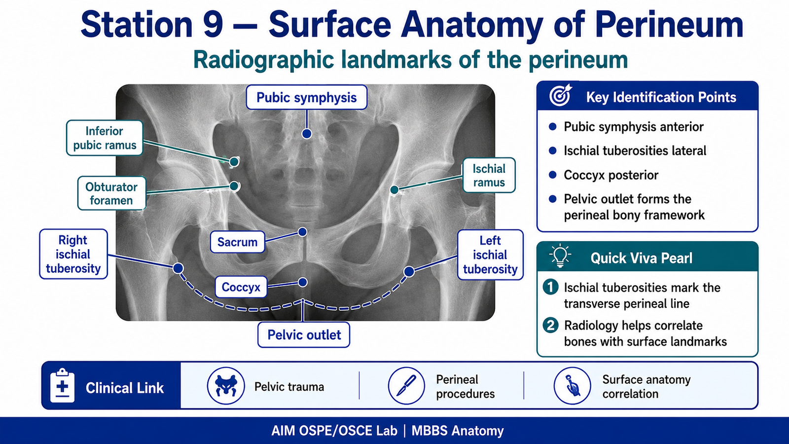
- Observe the pelvic radiograph provided.
- Identify the main bony landmarks related to the perineum.
- Write one clinical significance of correct perineal landmark identification.
- Answer the viva questions briefly.
Observation / Identification Points
The student should be able to identify:
Perineal Surface Anatomy
- Pubic symphysis
- Ischial tuberosities
- Coccyx
- Urogenital triangle
- Anal triangle
- Perineal body
- Anus
- External anal sphincter region
- Urethral opening region
- Ischioanal / ischiorectal fossa region
Radiological Landmarks
- Pubic symphysis
- Ischial tuberosity
- Inferior pubic ramus
- Ischial ramus
- Obturator foramen
- Sacrum
- Coccyx
- Pelvic outlet region
Result / Interpretation
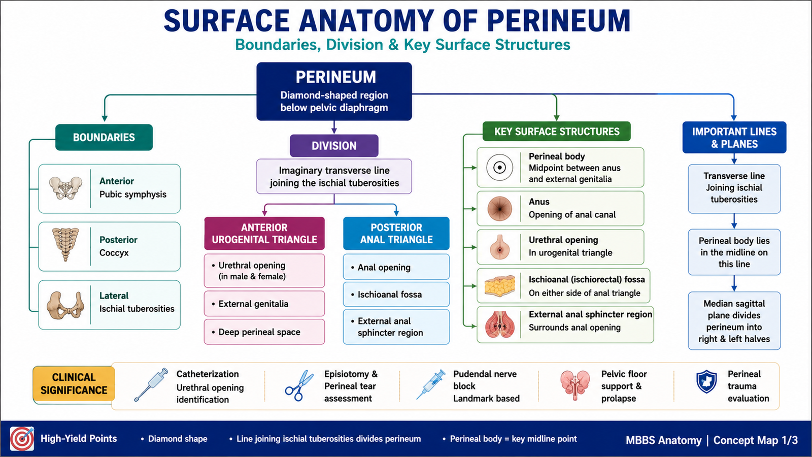
The perineum is a diamond-shaped region below the pelvic diaphragm. It is divided into the urogenital triangle anteriorly and the anal triangle posteriorly by an imaginary line joining the two ischial tuberosities.
Correct identification of perineal landmarks is clinically important for:
- Catheterization
- Perineal examination
- Episiotomy and perineal tear assessment
- Pudendal nerve block
- Understanding pelvic floor support
- Interpreting pelvic trauma on radiographs
Radiological identification of bony landmarks helps correlate surface anatomy with pelvic outlet anatomy and clinical procedures.
Viva Questions
1. What are the boundaries of the perineum?
Ideal answer: Anteriorly pubic symphysis, posteriorly coccyx, and laterally the right and left ischial tuberosities.
2. How is the perineum divided into two triangles?
Ideal answer: By a transverse line joining the two ischial tuberosities; anterior is urogenital triangle and posterior is anal triangle.
3. What is the clinical importance of the perineal body?
Ideal answer: It supports the pelvic floor; injury may lead to pelvic organ prolapse or weakness of perineal support.
4. Which radiological landmark helps identify the lateral boundary of the perineum?
Ideal answer: Ischial tuberosity.
5. Why is perineal anatomy important during catheterization?
Ideal answer: It helps identify the urethral opening correctly and prevents trauma or incorrect insertion.
Common Student Mistakes
- Confusing the urogenital triangle with the anal triangle.
- Forgetting the ischial tuberosities as key lateral landmarks.
- Identifying pelvic bones on radiograph without linking them to surface anatomy.
- Giving long theoretical answers instead of direct landmark identification.
AIM Feedback
In this station, focus on landmark-based anatomy. First identify the diamond shape of the perineum, then divide it into anterior urogenital and posterior anal triangles. On radiology, always search for the pubic symphysis, ischial tuberosities, obturator foramina, sacrum, and coccyx. Linking surface anatomy with radiological landmarks will help you answer both identification and viva questions confidently.
🖼️ Visual / Image Support
🧩 Concept Map / Interpretation Support