🩺Station 3 — KUB and IVU Identification
AIM OSPE/OSCE Lab — Practical Station | KMU Style | MBBS Practical + Viva
📋 Complete OSPE Station Content
OSPE Station Name
Station 3 — KUB and IVU Identification
Module: Renal
Year: 2nd Year MBBS
Integration: Anatomy + Clinical Radiology + Physiology
Learning Target
By the end of this station, the student should be able to:
- Identify major urinary tract parts on KUB and IVU radiographs.
- State the basic clinical significance of KUB and IVU in renal system assessment.
Required Material
- KUB radiograph / printed image
- IVU radiograph / printed image
- Pointer or marker
- Answer sheet
- Marking checklist
Student Task / Procedure
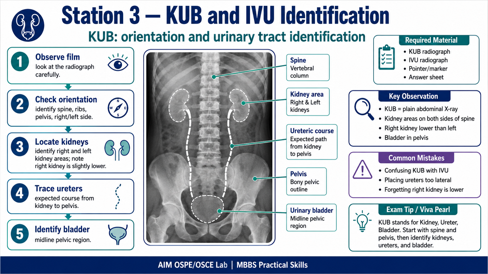
- Observe the given KUB and IVU radiographs.
- Identify the side and orientation of the image.
- On the KUB image, identify the expected areas of:
- Kidneys
- Ureters
- Urinary bladder
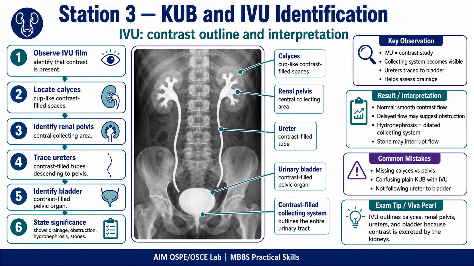
- On the IVU image, identify:
- Renal calyces
- Renal pelvis
- Ureters
- Urinary bladder
- State one clinical use of KUB.
- State one clinical use of IVU.
Observation / Identification Points
The student should identify or demonstrate:
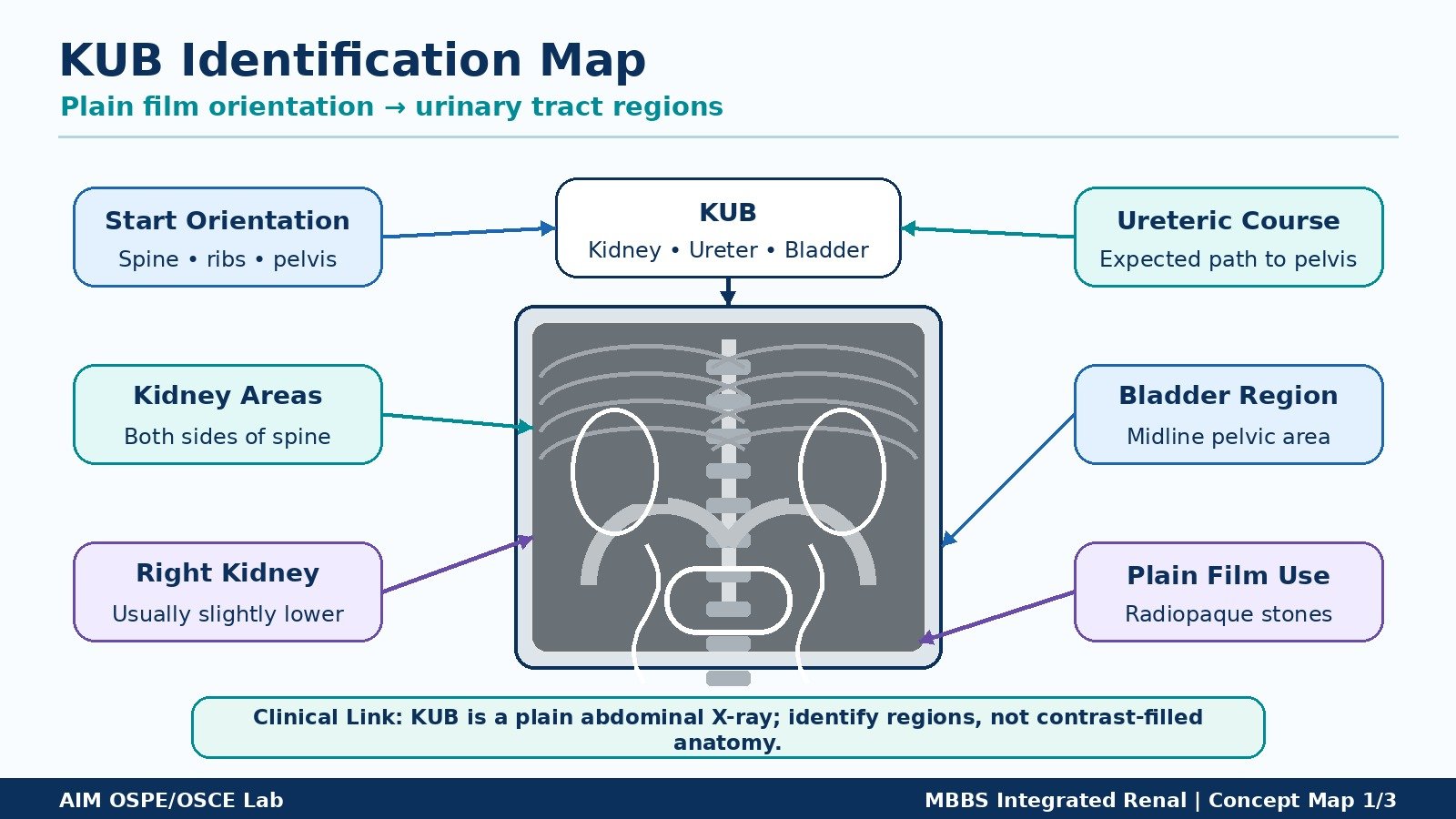
- KUB = Kidney, Ureter, Bladder plain radiograph.
- Kidney shadows lie approximately on either side of the vertebral column.
- Right kidney is usually slightly lower than the left kidney.
- Ureters descend from renal pelvis toward the urinary bladder.
- Urinary bladder is located in the pelvic region.
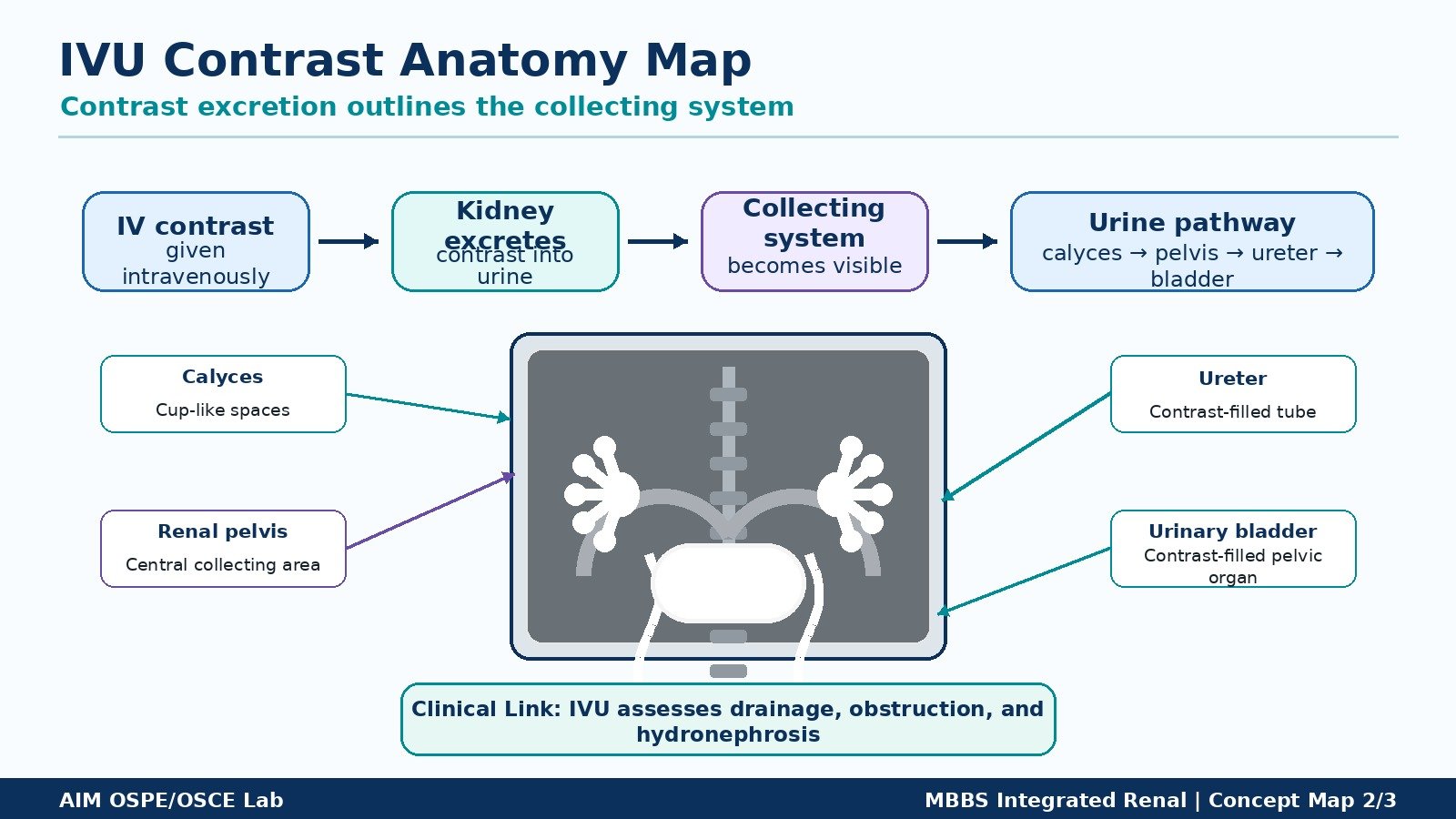
- In IVU, contrast outlines the:
- Calyces
- Renal pelvis
- Ureters
- Urinary bladder
- IVU helps assess urinary tract drainage and obstruction.
Result / Interpretation
KUB Radiograph:
A plain X-ray of the abdomen used to assess the urinary tract region. It may help identify radiopaque renal or ureteric stones, kidney area, and bladder region.
IVU Radiograph:
An intravenous contrast study in which contrast is excreted by the kidneys and outlines the collecting system, ureters, and bladder.
Clinical significance:
KUB and IVU help in identifying urinary tract anatomy and detecting abnormalities such as:
- Renal stone
- Ureteric obstruction
- Hydronephrosis
- Delayed renal excretion
- Abnormal urinary tract outline
Viva Questions
1. What does KUB stand for?
KUB stands for Kidney, Ureter, and Bladder.
2. What type of radiograph is KUB?
KUB is a plain abdominal X-ray used to assess the urinary tract region.
3. What structures are outlined in IVU?
IVU outlines the renal calyces, renal pelvis, ureters, and urinary bladder.
4. Why is contrast used in IVU?
Contrast is filtered and excreted by the kidneys, allowing visualization of the urinary collecting system.
5. Give one clinical use of IVU.
IVU is used to assess urinary tract obstruction, renal drainage, stones, or hydronephrosis.
Common Student Mistakes
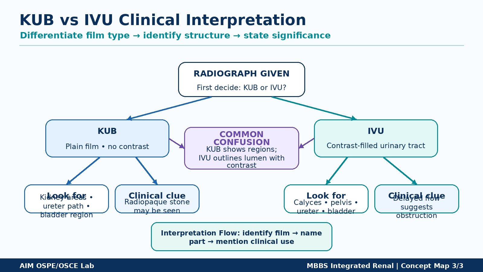
- Confusing KUB with IVU and forgetting that IVU uses contrast.
- Not identifying the renal pelvis and calyces separately on IVU.
- Marking ureters too laterally instead of following their course toward the bladder.
- Forgetting that the right kidney is usually slightly lower than the left.
- Giving long theoretical answers instead of focused radiological identification.
AIM Feedback
In this station, focus on recognition of urinary tract anatomy on radiographs. First orient yourself using the spine and pelvis, then locate the kidney areas, follow the ureters downward, and identify the bladder in the pelvis. Remember: KUB shows the urinary tract region on a plain film, while IVU outlines the collecting system using contrast.
🖼️ Visual / Image Support
🧩 Concept Map / Interpretation Support