
Study tip: This chapter follows the KMU learning outcomes in a logical sequence. First understand how pump failure causes congestion, then connect the pathology with investigations and drug actions. Use the final high-yield review only after reading the explanations.
📖 AIM Learning Material
Heart Failure, Cardiomyopathies, Pulmonary Edema and Heart-Failure Pharmacology
CVS Module — Pathology, Pharmacology, Medicine and PRIME Integration
Topic Introduction
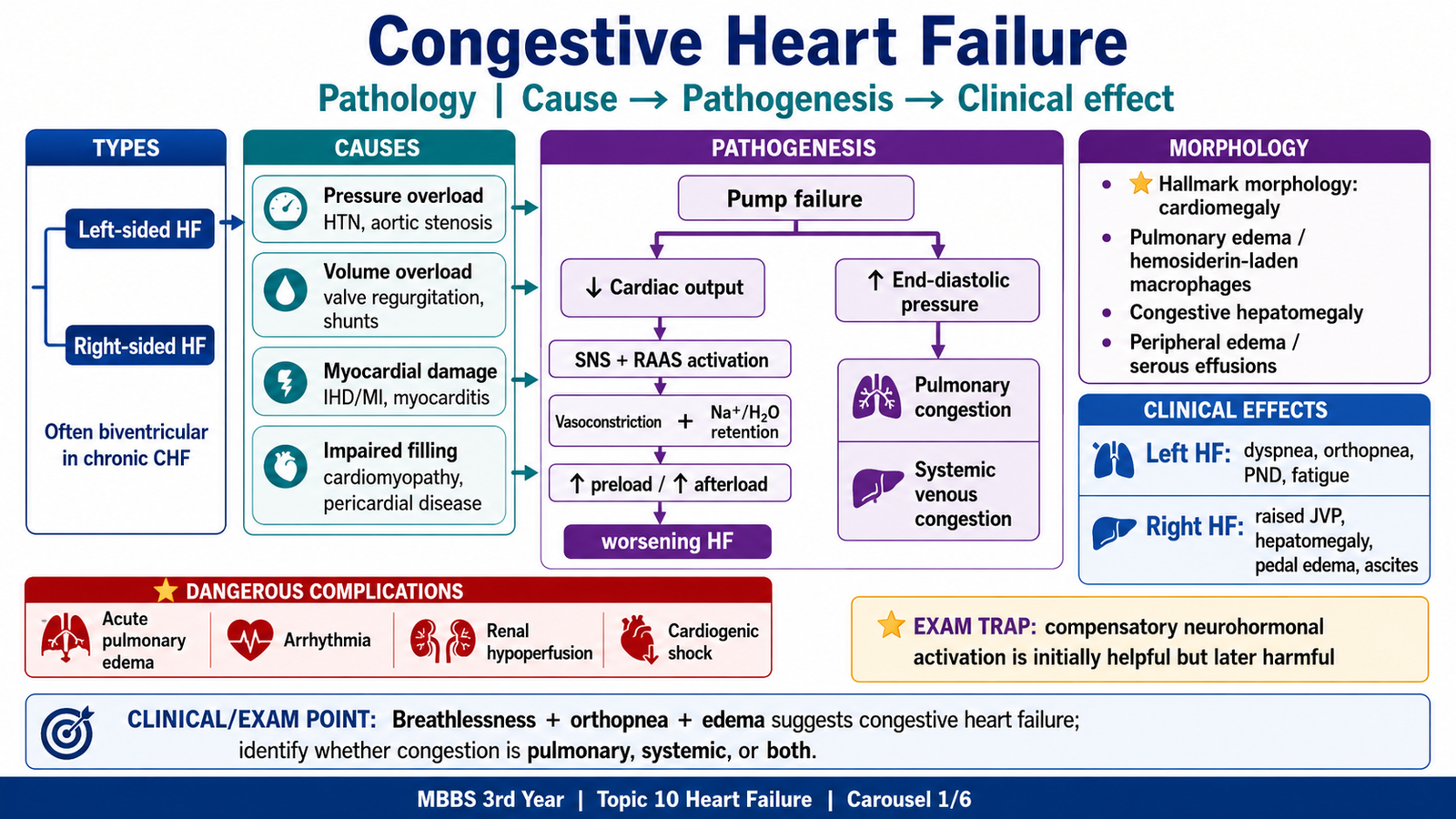
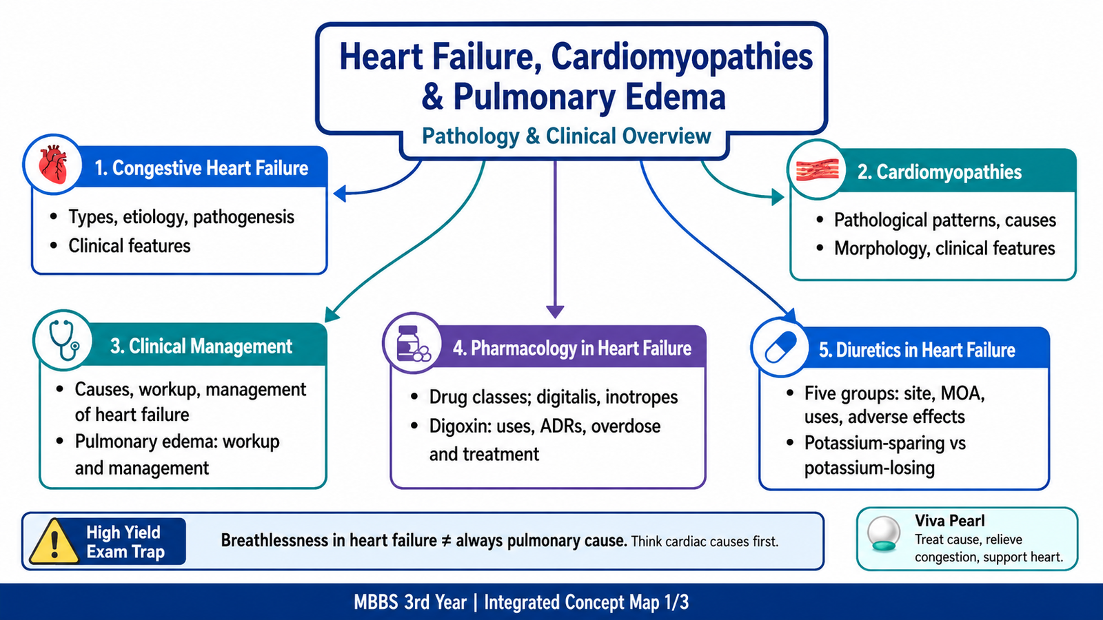
Heart failure is a syndrome in which the heart cannot pump enough blood for the body’s needs, or can do so only at abnormally high filling pressures. The problem may arise from weak contraction, poor relaxation, structural myocardial disease or excessive pressure or volume load. As cardiac output falls, compensatory mechanisms initially support circulation but later cause fluid retention, congestion and ventricular remodeling. This chapter explains the types and causes of heart failure, the pathological changes in the lungs and systemic organs, the major cardiomyopathies, the clinical workup, pulmonary edema, and the drugs used to relieve symptoms or improve cardiac function.
A. Heart Failure: Types, Causes and Pathogenesis
What is heart failure?
Heart failure is a clinical syndrome in which a structural or functional cardiac abnormality prevents the heart from providing adequate output to meet the body’s needs, or allows adequate output only at abnormally high filling pressures.
It is not synonymous with complete stoppage of the heart. The heart continues to pump, but its output is inadequate, its filling pressures are excessive, or both.
Major clinical types
Heart failure may be classified in several clinically useful ways.
According to the side predominantly affected
- Left-sided heart failure: pulmonary congestion and reduced systemic output predominate.
- Right-sided heart failure: systemic venous congestion predominates.
- Biventricular or congestive heart failure: features of both sides are present.
According to onset
- Acute heart failure: develops rapidly, as in acute myocardial infarction, severe hypertension, acute valvular failure or acute myocarditis.
- Chronic heart failure: develops progressively due to sustained pressure overload, volume overload or myocardial damage.
- Acute-on-chronic heart failure: sudden deterioration in a patient with established chronic heart failure.
According to ventricular function
- HFrEF: heart failure with reduced ejection fraction, generally EF ≤40%; predominantly systolic dysfunction.
- HFmrEF: heart failure with mildly reduced ejection fraction, generally EF 41–49%.
- HFpEF: heart failure with preserved ejection fraction, generally EF ≥50%; impaired relaxation and raised filling pressure predominate.
A preserved ejection fraction does not mean that the heart is normal. A stiff ventricle may eject a normal percentage of a reduced end-diastolic volume while still producing inadequate effective output.
According to cardiac output
- Low-output failure: the usual form, caused by ischemic heart disease, hypertension, valvular disease or cardiomyopathy.
- High-output failure: cardiac output is increased but remains insufficient for markedly raised tissue requirements.
Causes of high-output failure include:
- Severe anemia
- Thyrotoxicosis
- Large arteriovenous fistula
- Beriberi due to thiamine deficiency
- Extensive Paget disease of bone
Major causes of heart failure
Important causes include:
- Ischemic heart disease and previous myocardial infarction
- Chronic hypertension
- Valvular stenosis or regurgitation
- Dilated, hypertrophic or restrictive cardiomyopathy
- Myocarditis
- Persistent tachyarrhythmia or severe bradyarrhythmia
- Congenital heart disease
- Alcohol and cardiotoxic drugs such as anthracyclines
- Pericardial disease
- Severe pulmonary hypertension and chronic lung disease
- Metabolic or endocrine disorders such as thyroid disease
- Severe anemia or arteriovenous shunting
The compensatory response
A fall in cardiac output activates several mechanisms intended to preserve blood pressure and organ perfusion.
Frank–Starling mechanism
Increased end-diastolic volume stretches myocardial fibers and initially increases contraction strength. However, excessive ventricular dilation eventually increases wall stress and reduces mechanical efficiency.
Sympathetic nervous system activation
Reduced arterial pressure activates the sympathetic nervous system:
- Heart rate increases.
- Contractility increases.
- Peripheral vasoconstriction maintains blood pressure.
- Renal vasoconstriction contributes to sodium retention.
Prolonged sympathetic stimulation increases myocardial oxygen demand, promotes arrhythmias and contributes to myocyte injury.
Renin–angiotensin–aldosterone system
Reduced renal perfusion activates renin:
Renin → angiotensin I → angiotensin II → vasoconstriction and aldosterone secretion.
Aldosterone increases sodium and water retention. This raises preload and may initially support cardiac output, but persistent retention produces pulmonary and peripheral congestion.
Angiotensin II and aldosterone also promote hypertrophy, fibrosis and adverse ventricular remodeling.
Antidiuretic hormone
ADH promotes water retention, which may worsen congestion and contribute to dilutional hyponatremia.
Natriuretic peptides
Stretch of the ventricles stimulates BNP release, while atrial stretch promotes ANP release. These hormones attempt to increase natriuresis and vasodilation, but their effects are insufficient to overcome strong sympathetic and RAAS activation.
⭐ Basic mechanism: An initially helpful compensatory response becomes harmful when chronic vasoconstriction, fluid retention and remodeling further increase cardiac workload.
Ventricular remodeling
Myocardial infarction, pressure overload or volume overload causes:
Cause → myocyte injury or increased mechanical stress → myocyte hypertrophy and loss → ventricular dilation or wall thickening → interstitial fibrosis → impaired contraction or relaxation → progressive heart failure.
In the scenario, previous infarction caused loss of functioning myocardium, while hypertension maintained pressure overload. The remaining myocardium hypertrophied and the ventricle gradually dilated, producing systolic failure.
Diagnostic and management conclusion
The clinical syndrome must not be treated as fluid overload alone. Effective management must:
- Identify and treat the cause.
- Reduce congestion.
- interrupt harmful neurohormonal pathways.
- Prevent further remodeling.
- Recognize acute deterioration early.
Exam trap: Compensatory mechanisms are beneficial initially but become maladaptive during chronic heart failure.
B. Congestion in Heart Failure: Left-Sided and Right-Sided Changes
Left-sided heart failure
Pathogenesis
Left ventricular failure causes:
Reduced LV contraction or relaxation
→ increased LV end-diastolic pressure
→ increased left atrial pressure
→ increased pulmonary venous pressure
→ increased pulmonary capillary hydrostatic pressure
→ interstitial and alveolar pulmonary edema.
⭐ Normal mechanism whose failure explains disease: The pulmonary circulation normally operates at low pressure. A rise in left atrial and pulmonary venous pressure directly produces pulmonary congestion.
Causes
Common causes include:
- Ischemic heart disease
- Hypertension
- Aortic or mitral valve disease
- Dilated cardiomyopathy
- Hypertrophic or restrictive cardiomyopathy
- Myocarditis
- Congenital abnormalities
Clinical features explained by the pathology
Exertional dyspnea: pulmonary congestion reduces lung compliance and impairs oxygen diffusion.
Orthopnea: lying flat increases venous return from the lower limbs and redistributes fluid from dependent tissues into the central circulation.
Paroxysmal nocturnal dyspnea: the patient awakens after sleep with severe breathlessness because of increased central blood volume and reduced sympathetic support during sleep.
Cough and basal crackles: fluid accumulates within pulmonary interstitium and alveoli.
Pink, frothy sputum: air mixes with edema fluid containing small amounts of blood.
Fatigue and weakness: reduced forward cardiac output produces inadequate skeletal-muscle perfusion.
Cool peripheries and reduced urine output: severe low-output failure causes peripheral and renal hypoperfusion.
Pulmonary morphology
Gross appearance
The lungs become:
- Heavy
- Wet
- Boggy
- Congested
Frothy, blood-tinged fluid may be present in airways during acute pulmonary edema.
Microscopic appearance
Acute changes include:
- Engorged alveolar capillaries
- Interstitial edema
- Intra-alveolar transudate
- Small intra-alveolar hemorrhages
Chronic pulmonary congestion causes:
- Hemosiderin accumulation in alveolar macrophages
- Thickening and fibrosis of alveolar septa
- Brown discoloration and firmness of the lungs
⭐ Hallmark morphology: Hemosiderin-laden alveolar macrophages in chronic left-sided heart failure are called heart-failure cells.
These macrophages form when erythrocytes escape congested capillaries, are phagocytosed and their hemoglobin iron is converted into hemosiderin.
Right-sided heart failure
Pathogenesis
Right ventricular failure causes:
Reduced right ventricular output
→ increased right ventricular and right atrial pressure
→ systemic venous congestion
→ increased hydrostatic pressure in systemic veins and capillaries
→ edema, hepatic congestion, ascites and raised JVP.
Causes
The most common cause is left-sided heart failure.
Other causes include:
- Pulmonary hypertension
- Chronic obstructive pulmonary disease
- Recurrent pulmonary emboli
- Right ventricular myocardial infarction
- Pulmonary or tricuspid valve disease
- Congenital heart disease
- Restrictive cardiomyopathy
- Arrhythmogenic right ventricular cardiomyopathy
Right-sided failure caused by pulmonary disease or pulmonary hypertension is termed cor pulmonale.
Clinical features explained by pathology
- Raised JVP
- Dependent pitting edema
- Tender congested hepatomegaly
- Ascites
- Pleural effusions
- Weight gain
- Positive hepatojugular reflux
- Nocturia during earlier stages because recumbency improves renal perfusion
Systemic morphology
Liver
The liver becomes enlarged and congested.
Grossly, chronic congestion produces a mottled red-brown and yellow appearance called a nutmeg liver.
Microscopically:
- Centrilobular sinusoids are congested.
- Centrilobular hepatocytes undergo hypoxic injury or necrosis.
- Periportal hepatocytes may develop fatty change.
Prolonged severe congestion may result in cardiac fibrosis or, rarely, cardiac cirrhosis.
Other organs and tissues
- Peripheral subcutaneous edema
- Ascites
- Pleural and pericardial effusions
- Congestive splenomegaly
- Renal congestion and reduced filtration
- Cerebral hypoxia in advanced low-output failure
Biventricular failure
Most advanced cases eventually involve both ventricles. A patient may therefore have pulmonary crackles together with raised JVP, hepatomegaly and peripheral edema.
⭐ Most likely diagnosis clue: Orthopnea, PND and crackles point toward left-sided congestion; raised JVP, hepatomegaly and dependent edema point toward right-sided congestion.
Exam trap: Peripheral edema is not specific for heart failure. Renal, hepatic and venous disorders must also be considered.
C. Cardiomyopathies: Patterns, Causes and Morphology
Cardiomyopathies are primary disorders of the myocardium associated with mechanical or electrical dysfunction. They commonly produce heart failure because the ventricle becomes dilated, abnormally thick or excessively stiff.
Dilated cardiomyopathy
Core functional defect
Dilated cardiomyopathy produces:
Myocyte injury or genetic cytoskeletal abnormality
→ impaired contractility
→ increased end-systolic volume
→ progressive chamber dilation
→ reduced ejection fraction
→ systolic heart failure.
Causes
- Genetic abnormalities
- Previous viral or immune-mediated myocarditis
- Chronic alcohol consumption
- Anthracyclines such as doxorubicin
- Peripartum cardiomyopathy
- Hemochromatosis
- Nutritional deficiency, particularly thiamine deficiency
- Persistent tachycardia
- Some cases remain idiopathic
Morphology
Gross changes
- Enlarged, heavy and often flabby heart
- Dilation of all four chambers
- Ventricular walls may appear thinned, normal or mildly thickened
- Mural thrombi may form in poorly contracting chambers
- Functional mitral or tricuspid regurgitation may result from annular dilation
Microscopic changes
Changes are often nonspecific:
- Myocyte hypertrophy
- Variation in myocyte size
- Myocyte loss
- Interstitial and endocardial fibrosis
Clinical features and complications
- Progressive exertional dyspnea
- Orthopnea and PND
- Fatigue
- S3 gallop
- Peripheral edema
- Functional mitral regurgitation
- Atrial or ventricular arrhythmias
- Mural thrombosis and systemic embolism
⭐ Most likely diagnosis clue: A markedly dilated ventricle with global hypokinesia and reduced ejection fraction suggests dilated cardiomyopathy.
Hypertrophic cardiomyopathy
Core functional defect
Hypertrophic cardiomyopathy commonly results from inherited mutations in sarcomeric proteins.
Sarcomeric abnormality
→ abnormal myocyte growth
→ marked ventricular hypertrophy
→ reduced ventricular compliance
→ impaired diastolic filling
→ raised filling pressure and breathlessness.
Some patients also develop dynamic left ventricular outflow obstruction.
Morphology
Gross changes
- Marked left ventricular hypertrophy
- Disproportionate hypertrophy of the interventricular septum in many cases
- Small, narrow ventricular cavity
- Possible thickening of the anterior mitral valve leaflet due to repeated contact
Microscopic changes
⭐ Hallmark morphology: Disorganized, branching hypertrophied cardiac myocytes arranged in a chaotic pattern, known as myofiber disarray.
Additional changes include:
- Interstitial fibrosis
- Thickened small intramural coronary arteries
Clinical features
- Exertional dyspnea due to impaired ventricular filling
- Angina
- Palpitations
- Exertional dizziness or syncope
- Systolic murmur where outflow obstruction is present
- Signs of diastolic heart failure
The sudden-death and detailed risk-stratification aspects belong to the previously covered topic and are not repeated here.
Basic management logic
- Specialist assessment is required.
- Beta-blockers may improve filling by reducing heart rate.
- Diuretics must be used cautiously where significant outflow obstruction is present.
- Excessive reduction of preload may worsen dynamic obstruction.
⭐ Common clinical trap: Aggressive diuresis or vasodilation may worsen obstructive hypertrophic cardiomyopathy by reducing ventricular filling.
Restrictive cardiomyopathy
Core functional defect
Restrictive cardiomyopathy produces:
Infiltration or endomyocardial fibrosis
→ stiff ventricular walls
→ impaired diastolic filling
→ raised atrial and venous pressures
→ pulmonary and systemic congestion.
Systolic function may remain relatively preserved during early disease.
Causes
- Cardiac amyloidosis
- Endomyocardial fibrosis
- Loeffler endomyocarditis
- Hemochromatosis
- Sarcoidosis
- Previous mediastinal radiation
- Some inherited storage or infiltrative disorders
Morphology
- Ventricles are usually normal in size or only mildly enlarged.
- Ventricular walls are firm and poorly compliant.
- Both atria may become markedly dilated because they pump against stiff ventricles.
- Morphology depends on the underlying disease.
In amyloidosis, extracellular amyloid deposition separates myocardial fibers and may produce thickened, firm ventricular walls.
Clinical features
- Exertional dyspnea
- Fatigue
- Raised JVP
- Peripheral edema
- Ascites
- Signs resembling constrictive pericarditis
- Arrhythmias or conduction abnormalities
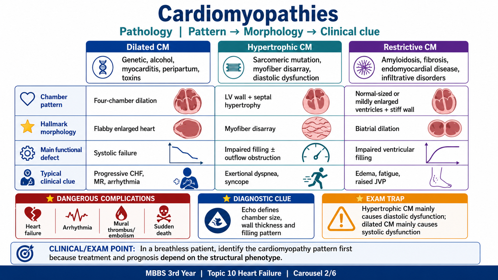
⭐ Differentiating clue: Restrictive cardiomyopathy usually has nondilated stiff ventricles with biatrial enlargement, whereas dilated cardiomyopathy has enlarged ventricles with reduced systolic function.
Arrhythmogenic right ventricular cardiomyopathy
This inherited disorder causes progressive loss of right ventricular myocytes with fibrofatty replacement.
Morphological consequences include:
- Right ventricular wall thinning
- Right ventricular dilation
- Fibrofatty replacement
- Ventricular arrhythmias
- Progressive right-sided failure in advanced disease
Because arrhythmia may occur before overt failure, unexplained ventricular arrhythmia in a young patient requires specialist evaluation.
Integrated conclusion
The three major mechanical patterns are:
- Dilated: large, weak ventricle → systolic failure.
- Hypertrophic: thick, poorly compliant ventricle → diastolic failure, sometimes obstruction.
- Restrictive: stiff, nondilated ventricle → impaired filling and venous congestion.
D. Clinical Workup, Management and Acute Pulmonary Edema
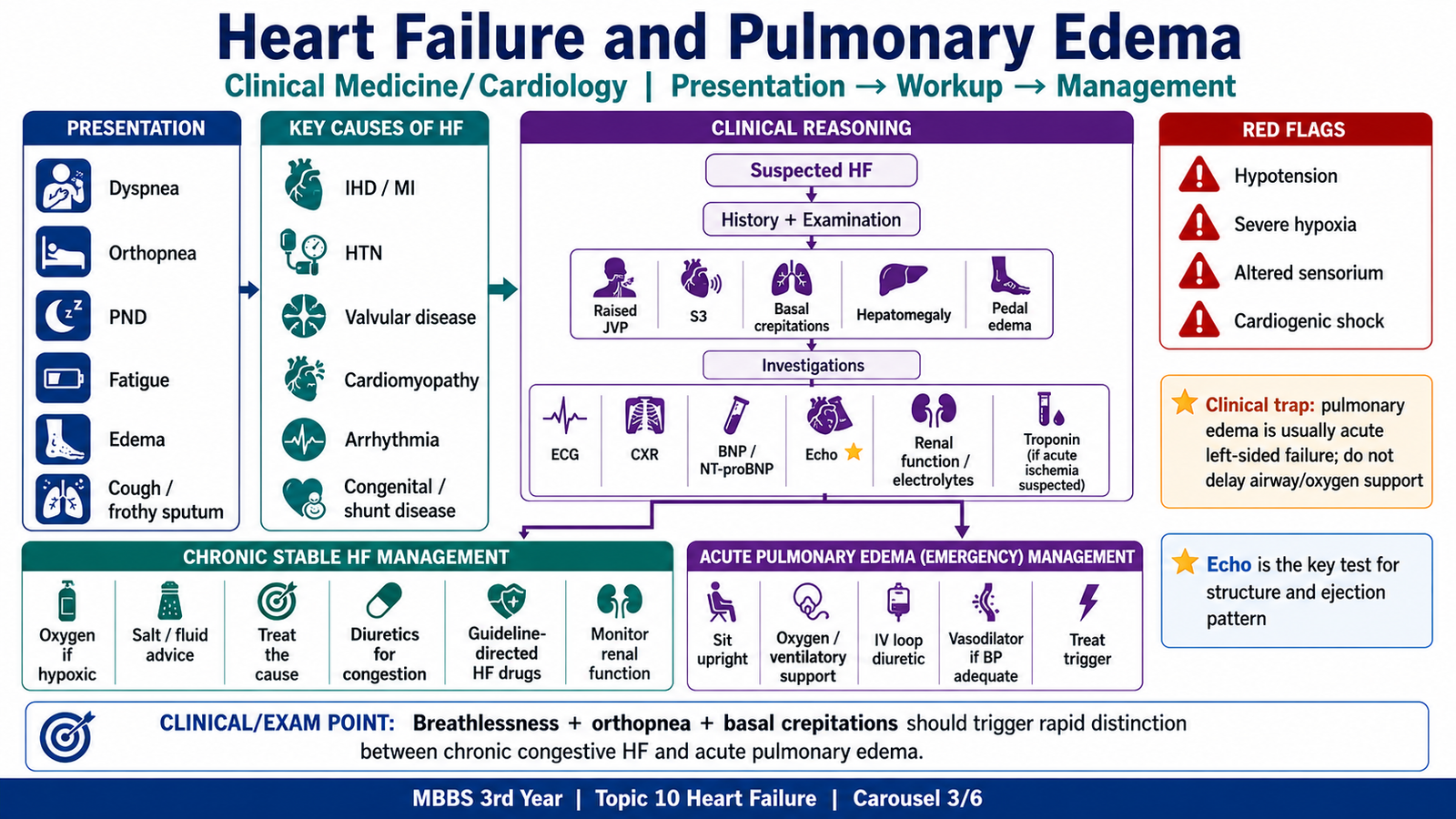
Presenting complaints
Common complaints include:
- Exertional breathlessness
- Orthopnea
- Paroxysmal nocturnal dyspnea
- Fatigue
- Reduced exercise tolerance
- Ankle swelling
- Abdominal distension
- Nocturnal cough
- Palpitations
- Rapid weight gain
Key examination findings
- Tachycardia or an irregular pulse
- Low blood pressure in severe low-output failure
- Raised JVP
- Displaced apex beat in ventricular dilation
- S3 gallop in volume-overloaded systolic failure
- S4 in a stiff, poorly compliant ventricle
- Cardiac murmurs suggesting valvular disease
- Pulmonary crackles
- Peripheral pitting edema
- Hepatomegaly or ascites
- Cool peripheries and reduced capillary refill in hypoperfusion
Initial diagnostic questions
The workup should answer four questions:
- Does the patient have heart failure?
- Is it acute or chronic?
- Is ventricular systolic function reduced or preserved?
- What caused or precipitated the failure?
Important history
Ask about:
- Previous myocardial infarction or angina
- Hypertension
- Valvular or congenital disease
- Diabetes and renal disease
- Alcohol use
- Viral illness
- Pregnancy or recent delivery
- Cardiotoxic chemotherapy
- Thyroid symptoms
- Palpitations or syncope
- Recent infection
- Drug adherence
- Excess salt intake
- Recent use of NSAIDs
- Drugs that cause fluid retention or myocardial depression
Common precipitating factors for acute decompensation include:
- Acute coronary syndrome
- Uncontrolled hypertension
- Arrhythmia
- Infection
- Anemia
- Renal deterioration
- Pulmonary embolism
- Nonadherence to treatment
- Excessive salt or fluid intake
- NSAID use
Basic investigations
Electrocardiogram
ECG may identify:
- Previous or acute myocardial infarction
- Left ventricular hypertrophy
- Atrial fibrillation
- Tachyarrhythmia or bradyarrhythmia
- Conduction block
- Clues to electrolyte disturbance or digoxin effect
A normal ECG makes major structural heart disease less likely but does not independently exclude heart failure.
Chest radiograph
Possible findings include:
- Cardiomegaly
- Upper-lobe venous diversion
- Kerley B lines due to interstitial edema
- Perihilar alveolar or “bat-wing” edema
- Pleural effusions
Natriuretic peptides
BNP or NT-proBNP rises when ventricular wall stress increases.
⭐ Biochemical marker: A low natriuretic peptide level helps make significant heart failure less likely, while an elevated level supports further cardiac assessment.
Interpretation must consider that levels may rise with age and renal impairment and may be lower than expected in obesity.
Echocardiography
Echocardiography is the key basic imaging investigation because it assesses:
- Ejection fraction
- Chamber size
- Ventricular wall thickness
- Regional or global wall-motion abnormalities
- Valvular disease
- Diastolic function
- Right ventricular function
- Pericardial disease
- Possible intracardiac thrombus
⭐ Most important diagnostic investigation: Echocardiography distinguishes reduced-EF failure from preserved-EF failure and may reveal the underlying structural cause.
Laboratory workup
Relevant tests include:
- Complete blood count for anemia or infection
- Serum electrolytes
- Urea and creatinine
- Liver-function tests
- Blood glucose or HbA1c
- Thyroid-function tests
- Lipid profile
- Troponin when acute ischemia is suspected
- Iron studies where iron deficiency or hemochromatosis is suspected
- Digoxin level where toxicity is suspected
Selected patients may require cardiac MRI, coronary assessment, genetic evaluation or endomyocardial biopsy under specialist care.
Clinical red flags
⭐ Urgent red flags include:
- Severe breathlessness at rest
- Oxygen desaturation
- Pink frothy sputum
- Hypotension
- Altered mental state
- Cold peripheries or reduced urine output
- Ongoing chest pain
- Sustained serious arrhythmia
- Syncope
- Rapidly worsening edema
- Evidence of cardiogenic shock
These findings require urgent hospital assessment rather than routine outpatient adjustment of medication.
Common clinical trap: An elevated BNP supports heart failure but does not identify its cause; echocardiography and clinical evaluation remain necessary.
The patient in the opening scenario has severe breathlessness, hypoxemia, diffuse crackles and pink frothy sputum. These findings indicate acute pulmonary edema.
How pulmonary edema develops
Acute left ventricular dysfunction causes a sudden rise in LV end-diastolic pressure. This pressure is transmitted backward to the left atrium, pulmonary veins and pulmonary capillaries.
When pulmonary capillary hydrostatic pressure exceeds the capacity of lymphatic drainage:
- Fluid enters the pulmonary interstitium.
- Interstitial edema reduces lung compliance.
- Fluid enters alveoli.
- Gas exchange becomes severely impaired.
- Hypoxemia and respiratory distress develop.
This is a hydrostatic, usually low-protein transudative process, although severe capillary stress may permit some erythrocyte leakage.
Causes and precipitants
- Acute myocardial infarction or ischemia
- Hypertensive emergency
- Acute mitral or aortic regurgitation
- Severe chronic heart failure with fluid overload
- Rapid atrial fibrillation or another arrhythmia
- Acute myocarditis
- Renal failure and fluid retention
- Medication nonadherence
- Excessive salt intake
- Infection
Basic diagnostic approach
Assessment should include:
- Airway and breathing
- Oxygen saturation
- Respiratory rate
- Blood pressure
- Perfusion and mental state
- ECG
- Chest radiograph or bedside lung imaging where available
- Cardiac biomarkers when ischemia is suspected
- Electrolytes and renal function
- Echocardiography once the patient is stabilized
Immediate management logic
Position and monitoring
- Sit the patient upright if blood pressure permits.
- Establish continuous monitoring.
- Obtain intravenous access.
- Monitor blood pressure, ECG, oxygen saturation and urine output.
Oxygen and ventilatory support
Oxygen is given when the patient is hypoxemic. Non-invasive positive-pressure ventilation, such as CPAP or BiPAP, may be required in marked respiratory distress or persistent hypoxemia.
Routine high-flow oxygen is not required in a patient with normal oxygen saturation.
Loop diuretic
Intravenous loop diuretic therapy is appropriate when pulmonary edema is accompanied by volume overload.
Furosemide:
- Produces powerful natriuresis.
- Reduces intravascular volume.
- Lowers ventricular filling pressure.
- Relieves pulmonary and systemic congestion.
Vasodilator therapy
Intravenous nitrates may be particularly useful when pulmonary edema occurs with severe hypertension and adequate blood pressure.
Venodilation reduces preload, while higher doses also reduce arterial resistance. This decreases pulmonary capillary pressure and ventricular workload.
Inotropes and vasopressors
Positive inotropes are not routine treatment for every patient with pulmonary edema. They are reserved for patients with hypotension, reduced cardiac output and evidence of organ hypoperfusion.
Treat the precipitating cause
- Acute coronary syndrome requires urgent cardiac management.
- Severe hypertension requires controlled blood-pressure reduction.
- Rapid arrhythmia requires rate or rhythm management.
- Infection requires appropriate treatment.
- Mechanical valve failure requires urgent specialist intervention.
⭐ Emergency point: Severe pulmonary edema with hypoxemia is a medical emergency requiring immediate stabilization and treatment of the underlying trigger.
Current guideline-based acute management emphasizes oxygen for hypoxemia, non-invasive ventilation for significant respiratory distress, IV loop diuretics for congestion and vasodilators in appropriately hypertensive patients. Routine morphine is not recommended.
Common clinical trap: Mannitol is not used to treat cardiogenic pulmonary edema. Its initial expansion of extracellular volume can worsen pulmonary congestion.
E. Long-Term Heart-Failure Pharmacology
Management begins with the cause and the clinical phenotype rather than with a single drug.
General management logic
Treat the underlying cause
Examples include:
- Revascularization or ischemic-heart-disease management
- Control of hypertension
- Management of valvular disease
- Correction of severe anemia or thyroid disease
- Treatment of infection
- Control of arrhythmia
- Stopping alcohol or cardiotoxic drugs where relevant
Patient education and monitoring
Patients should understand:
- The purpose of each medicine
- The importance of adherence
- The danger of self-adjusting medication
- Daily weight monitoring where feasible
- Moderation of salt intake
- Fluid restriction only when clinically indicated
- Recognition of worsening breathlessness or edema
- Avoidance of unnecessary NSAIDs
- The need for scheduled follow-up
A rapid increase in weight may indicate fluid accumulation before obvious edema appears.
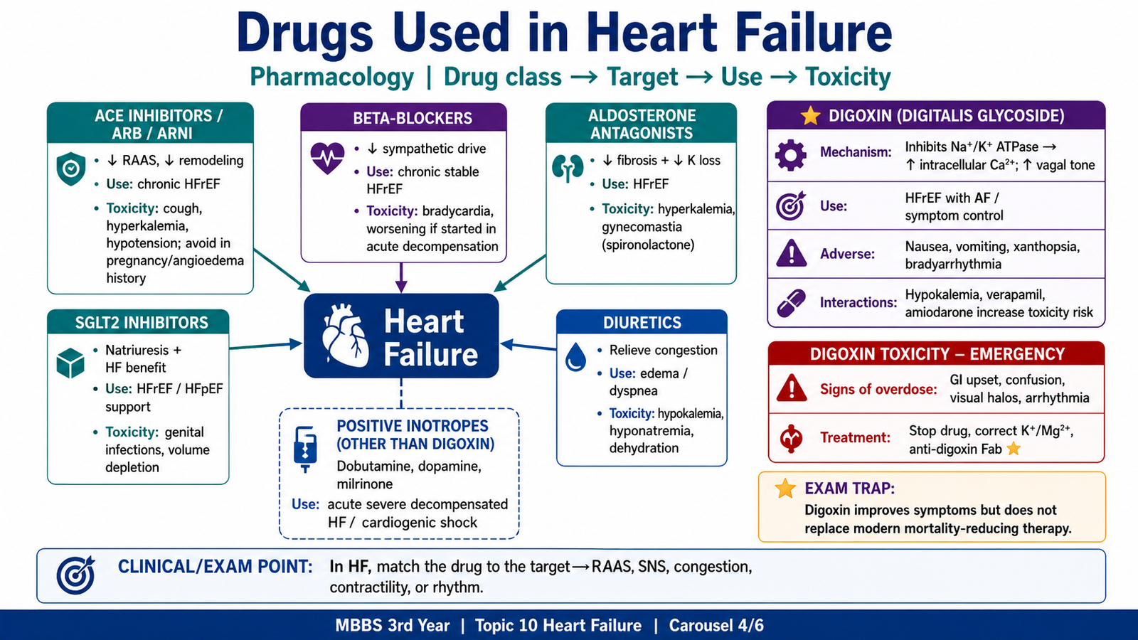
Drug classes used in heart failure
The major classes include:
Neurohormonal and remodeling-directed drugs
- ACE inhibitors
- Angiotensin-receptor blockers
- Angiotensin-receptor–neprilysin inhibitors
- Evidence-based beta-blockers
- Mineralocorticoid-receptor antagonists
- SGLT2 inhibitors
Drugs mainly used to control congestion
- Loop diuretics
- Thiazide or thiazide-like diuretics
- Selected potassium-sparing diuretics
Vasodilators and selected additional therapies
- Hydralazine plus nitrate
- Nitrates in selected acute situations
- Ivabradine in selected patients
- Digoxin in selected patients
- Intravenous inotropes in low-output states or shock
Foundational treatment of HFrEF
Modern guideline-directed therapy for symptomatic HFrEF is built around four major disease-modifying groups:
- ARNI, ACE inhibitor or ARB
- Evidence-based beta-blocker
- Mineralocorticoid-receptor antagonist
- SGLT2 inhibitor
These drugs target different harmful mechanisms and are used together when tolerated. Contemporary AHA/ACC/HFSA guidance identifies these four groups as foundational HFrEF therapy.
ARNI, ACE inhibitors and ARBs
These drugs reduce angiotensin-II-mediated vasoconstriction, aldosterone effects and remodeling.
Clinical benefits include:
- Reduced afterload
- Reduced sodium retention
- Slower adverse remodeling
- Reduced hospitalization and mortality
Important adverse effects of ACE inhibitors include:
- Cough
- Hyperkalemia
- Renal-function deterioration
- Angioedema
- Hypotension
ARNI therapy must not be given simultaneously with an ACE inhibitor because of increased angioedema risk.
Evidence-based beta-blockers
Carvedilol, bisoprolol and sustained-release metoprolol are established examples.
They reduce persistent sympathetic stimulation, lower heart rate and improve ventricular function over time.
They should be introduced in stable patients at low doses and gradually increased. They are not initiated or rapidly increased during cardiogenic shock or severe uncontrolled congestion.
Mineralocorticoid-receptor antagonists
Spironolactone and eplerenone:
- Block aldosterone.
- Reduce sodium retention.
- Reduce fibrosis and remodeling.
- Improve outcomes in suitable patients with HFrEF.
Renal function and serum potassium must be monitored.
SGLT2 inhibitors
Dapagliflozin and empagliflozin reduce heart-failure hospitalization and improve outcomes, including in patients without diabetes. Their benefits extend beyond glucose lowering.
Important adverse effects include:
- Genital fungal infection
- Volume depletion
- Rare euglycemic ketoacidosis
Current ESC guidance also supports SGLT2 inhibitors as important treatment in heart failure with mildly reduced or preserved ejection fraction.
Diuretics
Loop diuretics improve breathlessness and edema by reducing congestion. However, relief of fluid overload should not be confused with reversal of the underlying remodeling process.
⭐ Common KMU trap: Loop diuretics improve symptoms of congestion but are not the main mortality-reducing therapy in chronic HFrEF.
Basic approach to HFpEF
The main logic is to:
- Relieve congestion with diuretics.
- Control hypertension.
- Treat atrial fibrillation and ischemia.
- Manage diabetes, obesity, renal disease and sleep-disordered breathing.
- Consider an SGLT2 inhibitor.
- Avoid excessive reduction of filling pressure in a stiff, preload-dependent ventricle.
When referral is required
Referral to cardiology or urgent specialist care is appropriate for:
- Suspected cardiomyopathy
- Severe valve disease
- Recurrent admissions
- Persistent severe symptoms despite treatment
- Syncope or serious arrhythmia
- Suspected inherited heart disease
- Cardiogenic shock
- Consideration of device or advanced therapies
F. Digoxin and Other Positive Inotropes
Digitalis glycosides
Digitalis glycosides include digoxin and digitoxin. Digoxin is the clinically important prototype in most contemporary undergraduate teaching.
⭐ Prototype drug: Digoxin.
Mechanism of action
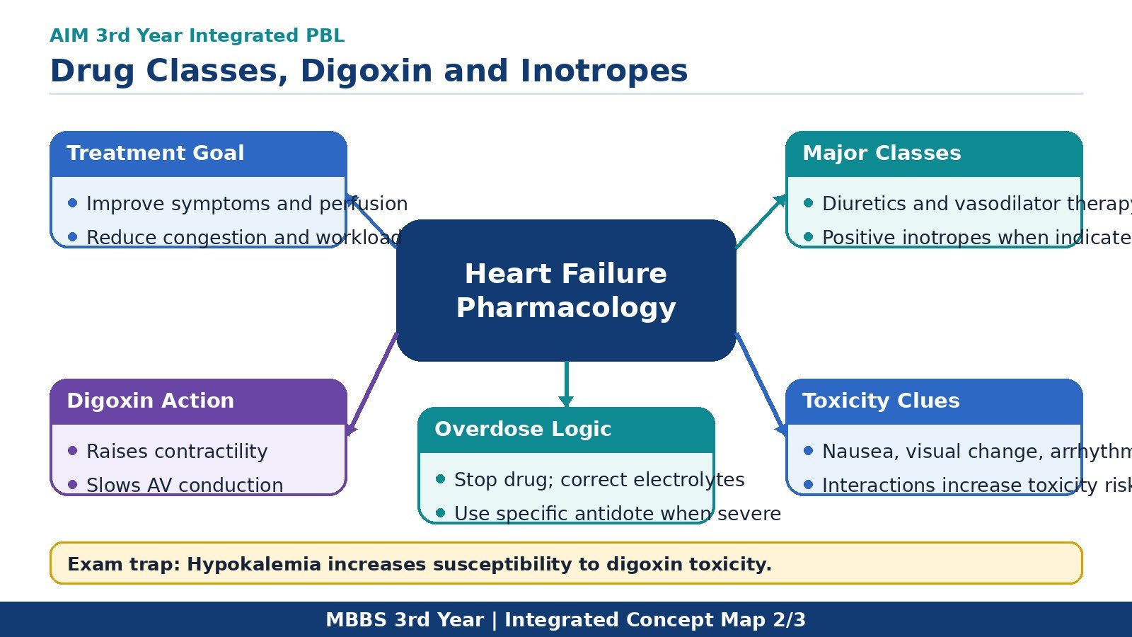
Digoxin inhibits the myocardial sodium–potassium ATPase pump.
Na⁺/K⁺ ATPase inhibition
→ increased intracellular sodium
→ reduced activity of the sodium–calcium exchanger
→ increased intracellular calcium
→ greater calcium storage in the sarcoplasmic reticulum
→ increased calcium release during contraction
→ positive inotropic effect.
Digoxin also increases vagal activity and reduces sympathetic activity.
Pharmacological effects
On myocardial contraction
- Increased force of contraction: positive inotropic effect
On the sinoatrial node
- Reduced heart rate: negative chronotropic effect
On the atrioventricular node
- Slower AV nodal conduction: negative dromotropic effect
- Increased AV nodal refractory period
These AV nodal effects explain its use in selected patients with atrial fibrillation.
⭐ Key mechanism: Digoxin increases intracellular calcium while slowing AV nodal conduction through enhanced vagal activity.
Pharmacokinetic points
- Usually administered orally for chronic use.
- Has a large volume of distribution.
- Is predominantly eliminated by the kidneys.
- Has a narrow therapeutic index.
- Its half-life becomes prolonged in renal impairment.
Dose reduction is therefore required in older patients and those with impaired renal function.
Clinical uses
Digoxin may be used for:
- Selected patients with symptomatic HFrEF despite appropriate foundational therapy
- Ventricular-rate control in selected patients with atrial fibrillation, particularly when heart failure is present
It may reduce symptoms or hospitalization in selected patients but does not provide the same mortality benefit as foundational HFrEF therapy.
Adverse effects
Gastrointestinal
- Anorexia
- Nausea
- Vomiting
- Abdominal discomfort
- Diarrhea
Neurological and visual
- Fatigue
- Weakness
- Confusion
- Delirium
- Blurred vision
- Yellow-green vision or xanthopsia
Cardiac
Almost any arrhythmia may occur, including:
- Sinus bradycardia
- AV block
- Premature ventricular complexes
- Atrial tachycardia with block
- Ventricular tachycardia
- Bidirectional ventricular tachycardia
⭐ Serious adverse effect: Digoxin-induced cardiac arrhythmia.
Factors increasing toxicity
- Renal impairment
- Older age
- Excessive dose
- Hypokalemia
- Hypomagnesemia
- Hypercalcemia
- Drug interactions
Why hypokalemia increases toxicity
Potassium normally competes with digoxin for binding to Na⁺/K⁺ ATPase. When serum potassium falls, more digoxin can bind to the pump, increasing its effects and toxicity.
This explains the opening scenario: excess furosemide caused potassium loss, which increased susceptibility to digoxin toxicity.
Important drug interactions
Drugs that may raise digoxin concentration include:
- Amiodarone
- Quinidine
- Verapamil
- Some macrolide antibiotics
- Certain azole antifungals
- Spironolactone in some circumstances
Loop and thiazide diuretics may indirectly increase toxicity by causing hypokalemia.
The official digoxin prescribing information particularly warns that potassium-depleting diuretics contribute to digitalis toxicity and that drugs such as quinidine, verapamil and amiodarone may increase digoxin exposure.
Contraindications and major cautions
⭐ Major contraindication: Ventricular fibrillation.
Digoxin must also be avoided or used with specialist caution in:
- Pre-excited atrial fibrillation associated with WPW syndrome
- Significant AV block without a pacemaker
- Marked bradycardia
- Renal impairment
- Severe electrolyte disturbance
- Obstructive hypertrophic cardiomyopathy in some circumstances
Recognition of digoxin overdose
Suspect digoxin toxicity when a patient taking digoxin develops a combination of:
- Anorexia, nausea or vomiting
- Confusion or weakness
- Visual disturbance
- Bradycardia or irregular pulse
- New AV block or ventricular arrhythmia
- Renal deterioration
Acute major overdose may produce hyperkalemia because severe Na⁺/K⁺ ATPase inhibition prevents potassium entry into cells. Chronic toxicity commonly occurs in patients with renal impairment or potassium depletion.
Treatment of digoxin toxicity
The basic approach is:
- Stop digoxin.
- Obtain ECG monitoring.
- Check serum digoxin, potassium, magnesium and renal function.
- Correct hypokalemia and hypomagnesemia carefully.
- Consider activated charcoal after a recent significant oral ingestion.
- Use atropine for clinically important bradycardia or AV block.
- Lidocaine or phenytoin may be used for selected ventricular arrhythmias.
- Give digoxin-specific antibody fragments when toxicity is severe.
⭐ Antidote: Digoxin-specific antibody fragments, commonly called digoxin immune Fab.
Indications include:
- Life-threatening ventricular arrhythmia
- Severe symptomatic bradyarrhythmia not responding to usual measures
- Major acute ingestion
- Significant hyperkalemia associated with acute toxicity
- Hemodynamic instability
Positive inotropic drugs other than digoxin
Dobutamine
A predominantly β₁-adrenergic agonist.
Effects:
- Increased contractility
- Increased cardiac output
- Some increase in heart rate
Use:
- Short-term support in severe low-output heart failure or cardiogenic shock with hypoperfusion
Adverse effects:
- Tachycardia
- Arrhythmias
- Increased myocardial oxygen demand
Dopamine
Dose-dependent adrenergic effects may increase contractility and blood pressure. It may be considered in selected hypotensive states but can provoke tachyarrhythmias.
Milrinone
A phosphodiesterase-3 inhibitor.
Mechanism:
PDE-3 inhibition
→ increased intracellular cAMP
→ increased calcium availability in myocardium
→ increased contractility.
In vascular smooth muscle, increased cAMP causes vasodilation. Milrinone is therefore an inodilator.
Adverse effects include:
- Hypotension
- Ventricular arrhythmias
Levosimendan
A calcium-sensitizing inodilator used in some healthcare settings. Availability and local practice vary.
⭐ Emergency pharmacology point: Intravenous positive inotropes are reserved for low-output failure with hypoperfusion; routine use in stable chronic heart failure may increase arrhythmias and mortality.
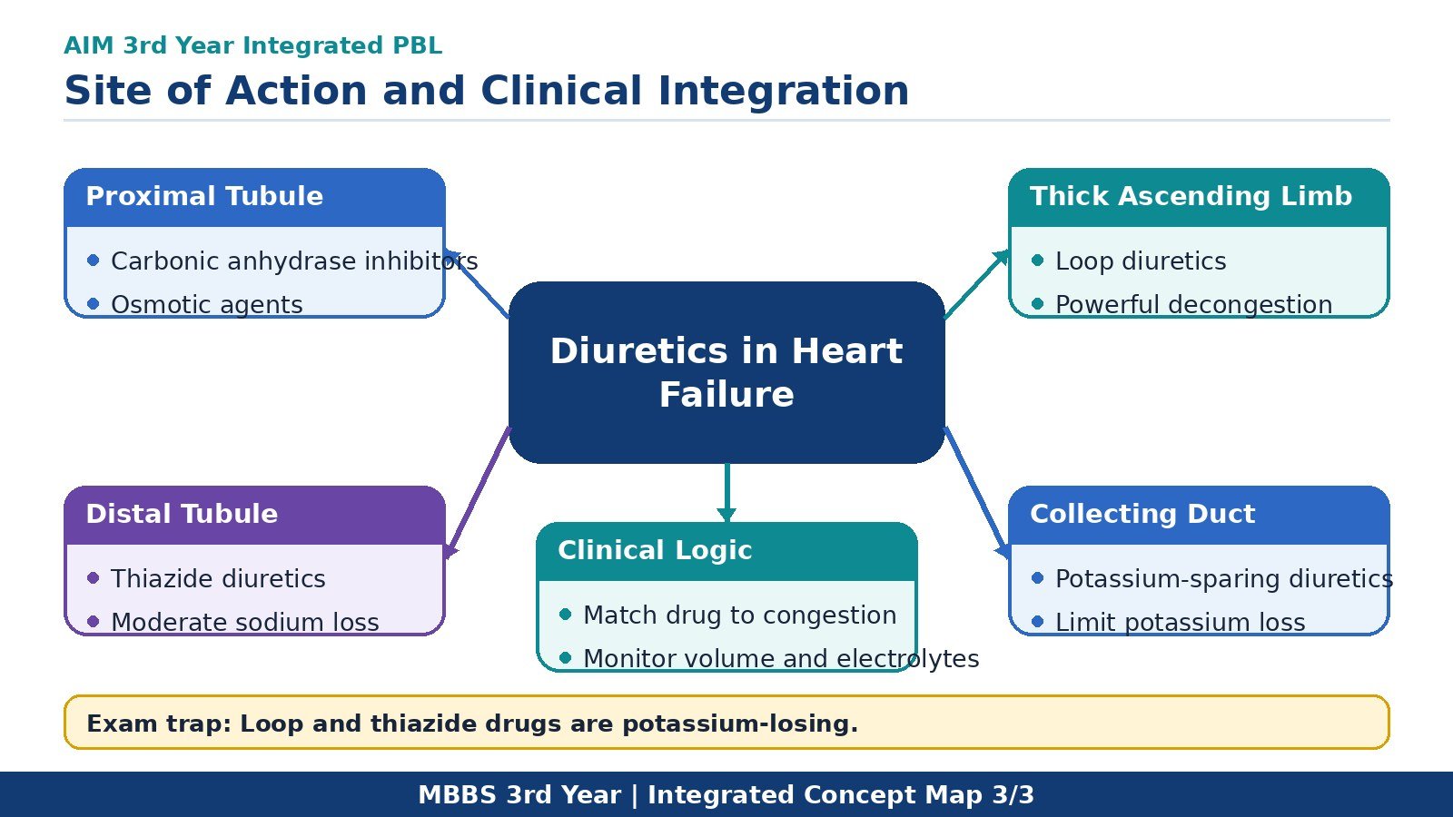
G. Diuretics: Classification, Nephron Sites and Clinical Use
Diuretics increase renal sodium and water excretion. Their actions depend on the nephron segment in which they inhibit reabsorption.
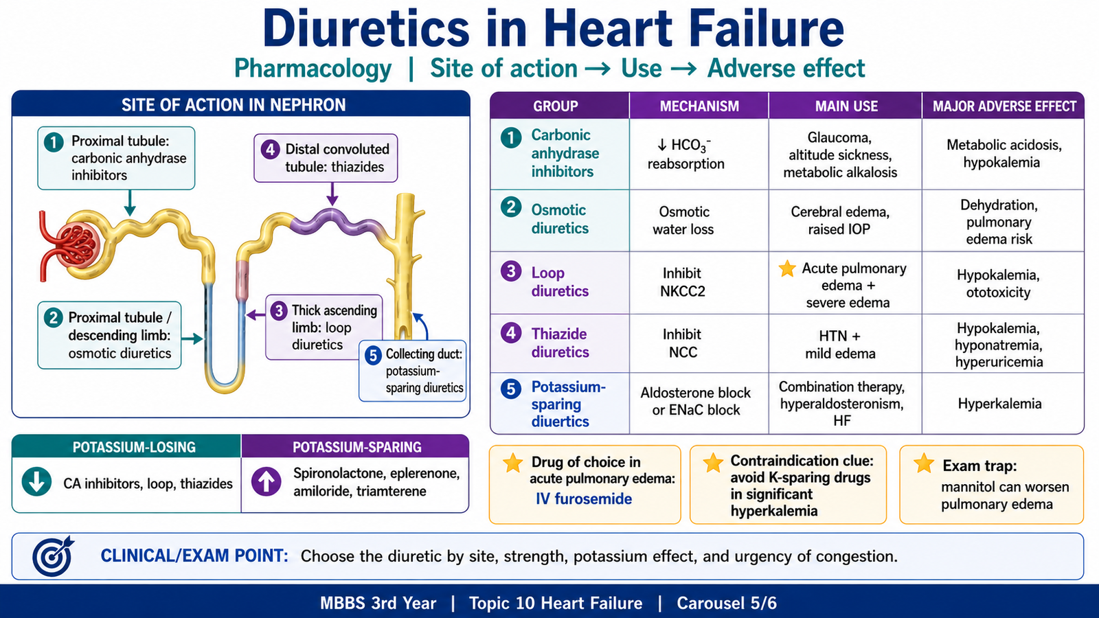
The five major groups and their sites
| Diuretic group | Prototype/examples | Main site of action |
|---|---|---|
| Carbonic anhydrase inhibitors | Acetazolamide | Proximal convoluted tubule |
| Osmotic diuretics | Mannitol | Proximal tubule and descending limb |
| Loop diuretics | Furosemide, bumetanide, torsemide | Thick ascending limb |
| Thiazide diuretics | Hydrochlorothiazide, chlorthalidone, metolazone | Early distal convoluted tubule |
| Potassium-sparing diuretics | Spironolactone, eplerenone, amiloride, triamterene | Late distal tubule and collecting duct |
Carbonic anhydrase inhibitors
⭐ Prototype: Acetazolamide.
Mechanism
Carbonic anhydrase inhibition in the proximal tubule reduces formation of hydrogen ions and bicarbonate reabsorption.
This causes:
- Increased bicarbonate excretion
- Increased sodium and water excretion
- Alkaline urine
- Hyperchloremic metabolic acidosis
Its diuretic effect becomes weaker after several days because systemic bicarbonate depletion reduces the filtered bicarbonate load.
Clinical applications
- Glaucoma
- Acute mountain sickness
- Metabolic alkalosis
- Idiopathic intracranial hypertension
- Selected adjunctive use in edematous states
It is not the principal routine diuretic for chronic heart failure.
Adverse effects
- Hyperchloremic metabolic acidosis
- Hypokalemia
- Renal calcium-phosphate stones
- Paresthesia
- Drowsiness
- Sulfonamide hypersensitivity
- Worsening of hepatic encephalopathy by reducing urinary ammonium trapping
Osmotic diuretics
⭐ Prototype: Mannitol.
Mechanism
Mannitol is filtered by the glomerulus but is not significantly reabsorbed.
It increases tubular-fluid osmolality and reduces water reabsorption, especially in nephron segments highly permeable to water.
Clinical applications
- Reduction of raised intracranial pressure
- Reduction of acute intraocular pressure
- Maintenance of urine flow in selected circumstances
Adverse effects
An initial movement of water into the extracellular compartment may cause:
- Expansion of extracellular volume
- Hyponatremia
- Pulmonary edema
Later water loss may cause:
- Dehydration
- Hypernatremia
- Headache
- Nausea
⭐ Major contraindication: Mannitol should not be given to a patient with pulmonary edema or significant heart failure because initial plasma-volume expansion can worsen congestion.
Loop diuretics
⭐ Prototype: Furosemide.
Other examples include bumetanide, torsemide and ethacrynic acid.
Mechanism
Loop diuretics inhibit the Na⁺–K⁺–2Cl⁻ cotransporter in the thick ascending limb.
Consequences include:
- Marked sodium, chloride and water excretion
- Loss of the medullary concentration gradient
- Increased calcium excretion
- Increased magnesium excretion
- Reduced intravascular volume and filling pressure
Because the thick ascending limb normally reabsorbs a large proportion of filtered sodium, loop diuretics are the most powerful conventional diuretics.
Clinical applications
- Acute pulmonary edema
- Severe peripheral edema due to heart failure
- Edema due to renal or hepatic disease
- Hypercalcemia
- Hyperkalemia as part of broader management when renal function permits
⭐ Most important clinical use: Rapid relief of significant congestion in acute or chronic heart failure.
Adverse effects
- Hypokalemia
- Metabolic alkalosis
- Hyponatremia
- Hypomagnesemia
- Hypocalcemia
- Volume depletion and hypotension
- Hyperuricemia and gout
- Ototoxicity
- Sulfonamide hypersensitivity
Ethacrynic acid is not a sulfonamide but has a comparatively high risk of ototoxicity.
Important interactions
- Hypokalemia increases digoxin toxicity.
- NSAIDs may reduce the diuretic response.
- Other ototoxic drugs may increase hearing damage.
- Concomitant antihypertensive drugs may worsen hypotension.
Thiazide and thiazide-like diuretics
⭐ Prototype: Hydrochlorothiazide.
Other important examples include chlorthalidone and metolazone.
Mechanism
Thiazides inhibit the Na⁺–Cl⁻ cotransporter in the early distal convoluted tubule.
They produce:
- Moderate natriuresis
- Increased potassium and hydrogen-ion loss
- Increased calcium reabsorption
Clinical applications
- Hypertension
- Mild edema
- Combination with a loop diuretic in resistant edema
- Prevention of recurrent calcium-containing renal stones in hypercalciuria
- Nephrogenic diabetes insipidus
Metolazone may remain effective at reduced renal function and may be combined with a loop diuretic for resistant congestion under close monitoring.
Adverse effects
- Hyponatremia
- Hypokalemic metabolic alkalosis
- Hyperuricemia
- Hyperglycemia
- Hyperlipidemia
- Hypercalcemia
- Sulfonamide hypersensitivity
- Sexual dysfunction
A useful memory pattern is that thiazides may produce several “hyper” effects: hyperGLUC—hyperglycemia, hyperlipidemia, hyperuricemia and hypercalcemia.
Potassium-sparing diuretics
These drugs act in the late distal tubule and collecting duct.
They are divided into two groups.
Aldosterone-receptor antagonists
- Spironolactone
- Eplerenone
Epithelial sodium-channel blockers
- Amiloride
- Triamterene
Mechanisms
Spironolactone and eplerenone block mineralocorticoid receptors, reducing aldosterone-induced synthesis of sodium channels and Na⁺/K⁺ ATPase.
Amiloride and triamterene directly block epithelial sodium channels.
Both actions reduce sodium reabsorption and decrease potassium and hydrogen-ion secretion.
Clinical applications
- Prevention or correction of diuretic-induced hypokalemia
- HFrEF, particularly with spironolactone or eplerenone
- Hyperaldosteronism
- Resistant hypertension
- Ascites due to cirrhosis
- Amiloride for lithium-induced nephrogenic diabetes insipidus
Why aldosterone antagonists are important in HFrEF
Spironolactone and eplerenone are not used merely for their weak diuretic effect. They also reduce aldosterone-mediated myocardial fibrosis and remodeling.
⭐ Important distinction: Mineralocorticoid-receptor antagonists can improve HFrEF outcomes, whereas amiloride and triamterene mainly preserve potassium.
Adverse effects
All potassium-sparing diuretics may cause:
- Hyperkalemia
- Metabolic acidosis
Spironolactone may additionally cause:
- Gynecomastia
- Reduced libido
- Erectile dysfunction
- Menstrual irregularity
Eplerenone produces fewer endocrine adverse effects because it is more selective for the mineralocorticoid receptor.
Important interactions
Risk of dangerous hyperkalemia increases with:
- ACE inhibitors
- ARBs
- ARNIs
- Potassium supplements
- Other potassium-sparing drugs
- Renal impairment
- Trimethoprim-containing medicines
⭐ Dangerous clinical point: Potassium-sparing diuretics require monitoring of serum potassium and renal function.
Potassium-sparing versus potassium-losing diuretics
Potassium-sparing
- Spironolactone
- Eplerenone
- Amiloride
- Triamterene
Potassium-losing
- Loop diuretics
- Thiazide diuretics
- Carbonic anhydrase inhibitors
Osmotic diuretics may increase urinary electrolyte losses, but they are not usually classified clinically in the same simple potassium-wasting group.
Integrated diuretic logic in the scenario
The patient doubled his furosemide dose:
Excessive loop-diuretic effect
→ increased distal sodium delivery
→ increased potassium secretion
→ hypokalemia
→ increased digoxin binding to Na⁺/K⁺ ATPase
→ digoxin toxicity.
This explains his nausea, visual disturbance, bradycardia and irregular pulse.
Common KMU trap: Spironolactone is potassium-sparing, but it is not automatically “safe”; severe hyperkalemia may develop when it is combined with RAAS blockers or used in renal failure.
H. PRIME Integration: SWOT Analysis for a Heart-Failure Task
The task
A clinical teaching unit plans to introduce a structured discharge and follow-up program for patients admitted with heart failure.
The program includes:
- Medication counselling
- Daily-weight education
- Recognition of red flags
- A written follow-up date
- Telephone contact after discharge
- Family involvement where appropriate
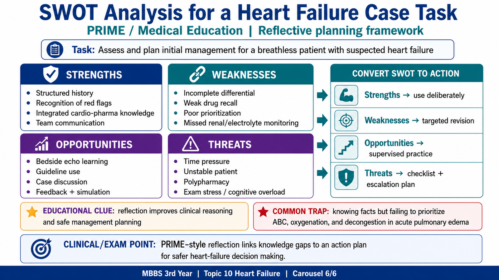
What is SWOT analysis?
SWOT is a structured planning tool used to assess:
- Strengths: internal favorable factors
- Weaknesses: internal unfavorable factors
- Opportunities: external favorable factors
- Threats: external unfavorable factors
SWOT analysis for the task
Strengths
- Standardizes patient education.
- Improves understanding of medicines.
- May identify fluid retention earlier.
- Encourages timely follow-up.
- Can involve doctors, nurses, pharmacists and family members.
- Uses relatively low-cost interventions.
Weaknesses
- Requires staff time and coordination.
- Discharge counselling may be rushed.
- Some patients may have limited health literacy.
- Not every patient has access to a weighing scale.
- Records may not be linked across departments.
- Telephone numbers may be inaccurate or change frequently.
Opportunities
- Use of SMS or WhatsApp reminders
- Collaboration with primary-care services
- Involvement of medical students in supervised education
- Development of local-language educational material
- Audit of readmission rates
- Use of telemedicine for selected patients
- Family-based support for elderly patients
Threats
- High medicine costs
- Poor access to transport
- Loss to follow-up
- Unavailability of medicines
- Misinformation from nonmedical sources
- Uncontrolled comorbidities
- Patients independently changing drug doses
- Limited emergency-care access
Converting SWOT into action
A useful SWOT analysis should lead to practical decisions:
- Use simple bilingual discharge instructions.
- Verify the patient’s contact details.
- Prioritize three to five essential red flags.
- Involve a family member when appropriate.
- Arrange a realistic follow-up pathway.
- Audit whether the plan reduces preventable readmissions.
⭐ Common PRIME trap: Strengths and weaknesses are usually internal to the organization or task, whereas opportunities and threats arise mainly from the external environment.
AIM High-Yield Review
- Heart failure means inadequate cardiac output, abnormally high filling pressure, or both; it does not mean complete cardiac arrest.
- Chronic sympathetic and RAAS activation becomes maladaptive by increasing vasoconstriction, sodium retention, myocardial oxygen demand and ventricular remodeling.
- Left-sided failure produces pulmonary congestion, dyspnea, orthopnea, paroxysmal nocturnal dyspnea and crackles.
- Chronic pulmonary congestion produces hemosiderin-laden macrophages called heart-failure cells and may cause brown induration of the lungs.
- Right-sided failure produces raised JVP, dependent edema, hepatomegaly, ascites and systemic venous congestion; chronic liver congestion produces a nutmeg appearance.
- Dilated cardiomyopathy mainly causes systolic dysfunction, hypertrophic cardiomyopathy impairs filling and may obstruct outflow, and restrictive cardiomyopathy produces a stiff, poorly compliant ventricle.
- Echocardiography is central for assessing ejection fraction, chamber size, wall thickness, valves and possible causes of heart failure.
- Acute cardiogenic pulmonary edema results from a rapid rise in pulmonary capillary hydrostatic pressure and requires urgent stabilization and reduction of congestion.
- Long-term treatment must address the cause, reduce congestion and limit harmful neurohormonal activation and remodeling.
- Digoxin inhibits Na⁺/K⁺-ATPase, raises intracellular calcium and increases contractility, but has a narrow therapeutic index.
- Nausea, vomiting, visual disturbance and arrhythmias suggest digoxin toxicity; hypokalemia increases the risk.
- Loop diuretics are powerful potassium-losing diuretics used for marked congestion and pulmonary edema.
- Potassium-sparing diuretics act in the collecting tubule and may cause hyperkalemia; carbonic anhydrase inhibitors, osmotic agents and thiazides have distinct nephron sites and uses.
- In SWOT analysis, strengths and weaknesses are internal factors, while opportunities and threats are external factors.
🎥 AIM Video Learning — Congestive Heart Failure
This video explains the causes, pathophysiology, clinical features, diagnosis and complications of congestive heart failure.
AIM Study Tip: First understand ventricular dysfunction, compensatory mechanisms and congestion. Then relate these mechanisms to pulmonary edema, clinical findings and heart-failure treatment.