
📌 AIM Study Tip
This chapter follows the KMU learning outcomes and explains congenital heart disease in a logical sequence. First understand how abnormal blood flow produces cyanosis, murmurs and cardiac overload, and then use the AIM High-Yield Review for revision.
KMU Curriculum
AIM Learning Cycle
📖 AIM Learning Material
Congenital Heart Disease: Cyanotic and Acyanotic Lesions
CVS Module — Topic 11
Understand how congenital structural defects alter cardiac blood flow, how cyanotic and acyanotic lesions differ, and how Tetralogy of Fallot, atrial septal defect, ventricular septal defect and pulmonary stenosis produce their characteristic clinical findings.
Topic Introduction
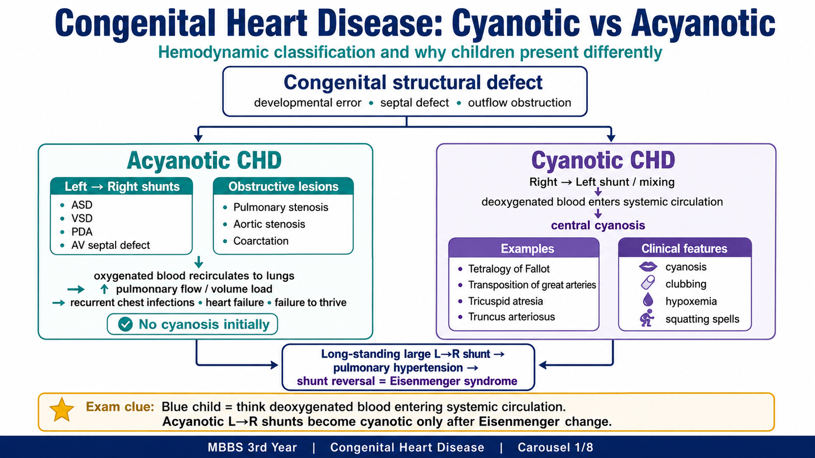
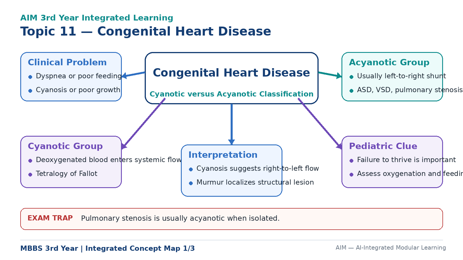
Congenital heart disease is a structural abnormality of the heart or great vessels that develops before birth. Some defects create an abnormal connection between cardiac chambers, while others obstruct the normal passage of blood. The direction of blood flow through the defect determines whether the child remains acyanotic or develops cyanosis. In acyanotic disease, oxygenated blood commonly moves from the left side of the heart to the right side, increasing pulmonary blood flow without initially lowering systemic oxygen levels. In cyanotic disease, deoxygenated blood reaches the systemic circulation. This chapter explains these principles and applies them to Tetralogy of Fallot, atrial septal defect, ventricular septal defect and pulmonary stenosis.
A. Basic Principles and Classification of Congenital Heart Disease
Congenital heart disease develops when formation of the fetal heart or great vessels is disturbed. A defect may remain clinically silent for some time or may produce symptoms soon after birth. Its effect depends not only on its anatomical appearance but also on the pressures on each side of the defect and the amount of blood passing through it.
The important factors that determine the clinical picture are:
- The type and site of the structural defect
- The size of an abnormal communication
- The direction and volume of the shunt
- The presence and severity of obstruction
- Pulmonary and systemic vascular resistance
- The resulting pressure or volume load on the cardiac chambers
Etiological basis
Most congenital cardiac defects are multifactorial, meaning that genetic susceptibility and environmental influences may act together. In many children, no single cause can be identified.
Important associations include:
- Abnormal cardiac embryogenesis
- Genetic or chromosomal abnormalities
- Family history of congenital heart disease
- Maternal diabetes mellitus
- Maternal infection, particularly congenital rubella
- Maternal exposure to teratogenic drugs or alcohol
- Certain inherited syndromes
The general sequence is:
Acyanotic congenital heart disease
In an acyanotic lesion, a significant amount of deoxygenated venous blood does not initially enter the systemic arterial circulation. These lesions are broadly divided into left-to-right shunts and obstructive defects.
Common left-to-right shunts:
- Atrial septal defect
- Ventricular septal defect
- Patent ductus arteriosus
- Atrioventricular septal defect
Common obstructive lesions:
- Pulmonary stenosis
- Aortic stenosis
- Coarctation of the aorta
In a left-to-right shunt, oxygenated blood moves from the higher-pressure left side to the lower-pressure right side. The blood is sent through the lungs again, so the child is initially not cyanosed. However, the excessive pulmonary flow may cause recurrent respiratory symptoms, chamber dilation, heart failure and pulmonary hypertension.
Cyanotic congenital heart disease
Cyanosis develops when deoxygenated blood enters the systemic arterial circulation through a right-to-left shunt or when oxygenated and deoxygenated blood mix abnormally.
Important cyanotic defects include:
- Tetralogy of Fallot
- Transposition of the great arteries
- Tricuspid atresia
- Total anomalous pulmonary venous connection
- Truncus arteriosus
- Pulmonary atresia
- Ebstein anomaly
- Single-ventricle defects
How an acyanotic defect can become cyanotic
A persistent left-to-right shunt exposes the pulmonary circulation to excessive blood flow and, in some defects, high pressure. The pulmonary arteries gradually develop medial hypertrophy and intimal thickening. These changes increase pulmonary vascular resistance.
When pulmonary vascular resistance becomes greater than systemic vascular resistance, the direction of the shunt reverses. Deoxygenated blood then passes from the right side to the left side and enters the systemic circulation. This late change is called Eisenmenger syndrome.
The child may then develop:
- Central cyanosis
- Clubbing
- Exercise intolerance
- Secondary polycythemia
- Right-sided cardiac strain
B. Clinical Recognition of Cyanotic and Acyanotic Disease
Congenital heart disease can present at different ages because the physiological effect of a defect changes with its size, the fall in pulmonary vascular resistance after birth and the development of cardiac compensation. The clinical pattern should therefore be interpreted together with the underlying blood flow.
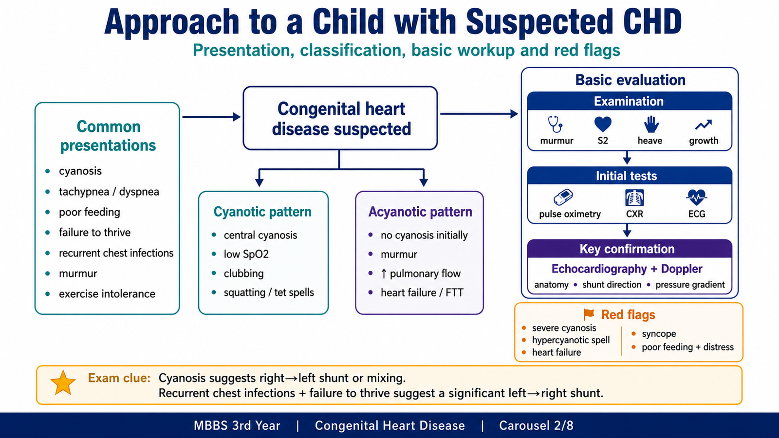
Common presentations
- Cyanosis
- Rapid breathing or shortness of breath
- Poor feeding
- Sweating during feeding
- Failure to thrive
- Recurrent respiratory infections
- Easy fatigability or exercise intolerance
- Squatting after exertion
- Syncope
- A cardiac murmur
Feeding requires considerable effort in an infant. A child with cardiac failure may therefore feed slowly, stop frequently, become sweaty or breathless and fail to gain weight.
Features suggesting cyanotic disease
- Blue discoloration of the tongue and oral mucosa
- Reduced oxygen saturation
- Clubbing in long-standing disease
- Hypercyanotic spells
- Squatting after exertion
- Secondary polycythemia
- Evidence of right-to-left shunting
Features suggesting acyanotic disease
- No central cyanosis initially
- Tachypnea
- Recurrent respiratory infections
- Poor weight gain
- Features of heart failure
- Signs of increased pulmonary blood flow
- A left-to-right shunt or an isolated obstructive lesion
Basic diagnostic approach
Clinical examination identifies cyanosis, abnormal pulses, chamber enlargement and characteristic murmurs. Investigations then define the anatomy and physiological effect of the lesion.
- Pulse oximetry: Detects reduced arterial oxygen saturation.
- Chest radiography: May demonstrate cardiac enlargement, altered pulmonary vascular markings or a characteristic cardiac silhouette.
- Electrocardiography: May show chamber hypertrophy, conduction changes or arrhythmias.
- Echocardiography with Doppler: Demonstrates the defect, shunt direction, chamber effects and pressure gradient across an obstruction.
Features requiring urgent assessment
- Severe central cyanosis
- Recurrent hypercyanotic spells
- Poor feeding with respiratory distress
- Syncope or seizures
- Signs of shock
- Severe failure to thrive
- Features of heart failure
- Suspected duct-dependent circulation
C. Tetralogy of Fallot
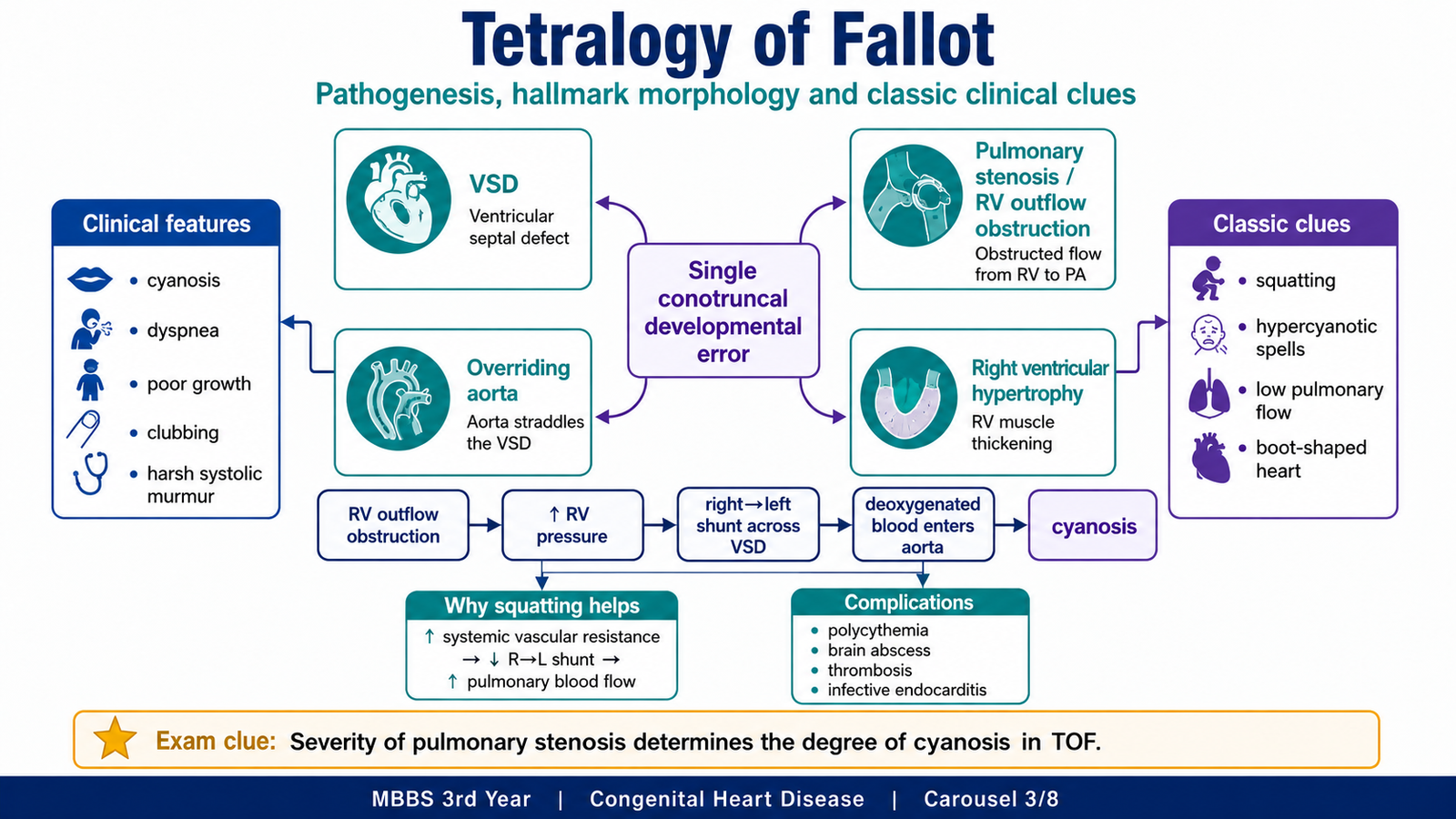
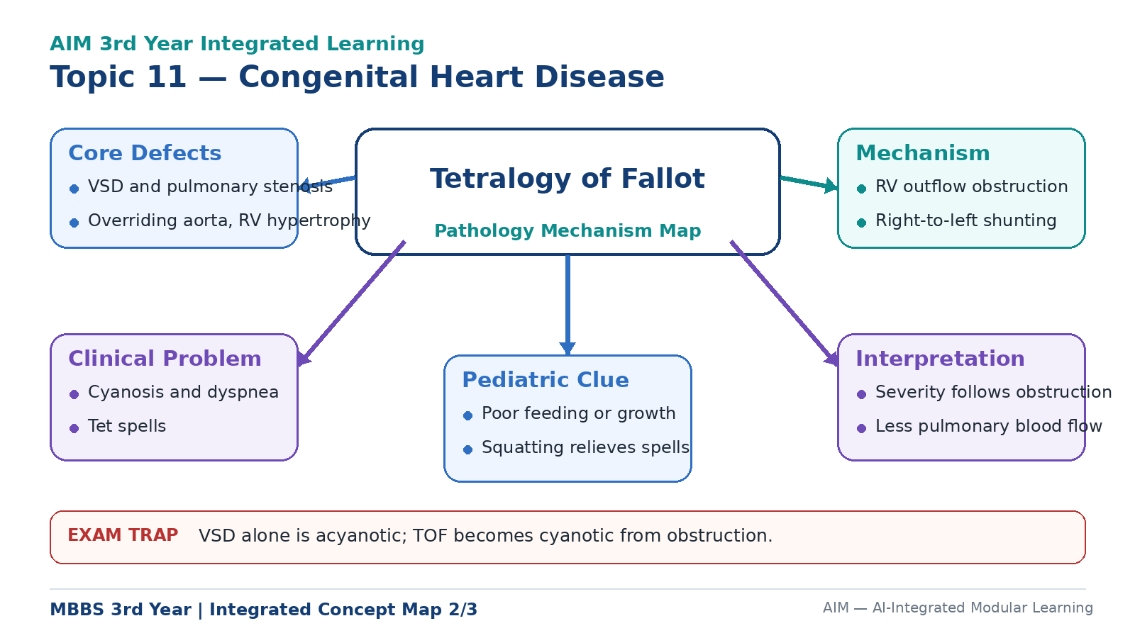
Tetralogy of Fallot is a cyanotic congenital heart disease in which obstruction of the right ventricular outflow tract causes deoxygenated blood to pass through a ventricular septal defect and enter the aorta. It is a classical cause of cyanosis beyond the neonatal period.
The four components
- Ventricular septal defect
- Right ventricular outflow tract obstruction, usually pulmonary stenosis
- Overriding of the aorta over the ventricular septal defect
- Right ventricular hypertrophy
These abnormalities are connected consequences of one developmental disturbance rather than four unrelated defects.
Etiology and developmental basis
Tetralogy of Fallot results from abnormal development of the conotruncal region. Anterosuperior displacement of the infundibular septum divides the outflow tract unequally. The pulmonary outflow tract becomes narrow, while the ventricular septum remains open and the aorta lies above the defect. The right ventricle then hypertrophies because it must pump against the obstruction.
It may occur as an isolated lesion or in association with genetic abnormalities, particularly 22q11-related syndromes.
Pathogenesis
The large VSD allows communication between both ventricles. Right ventricular outflow obstruction raises right ventricular pressure. Blood then passes from the right ventricle through the VSD into the overriding aorta, allowing deoxygenated blood to enter the systemic circulation.
The VSD is usually large, so pressures in the two ventricles may become nearly equal. The severity of cyanosis therefore depends mainly on the severity of pulmonary outflow obstruction.
- Mild obstruction: Limited right-to-left shunting; cyanosis may be absent initially.
- Severe obstruction: Marked right-to-left shunting, reduced pulmonary blood flow and early cyanosis.
Morphology
The heart shows a large VSD, an overriding aorta, narrowing of the pulmonary outflow tract and right ventricular hypertrophy. Right ventricular hypertrophy raises the cardiac apex, while the pulmonary segment is concave because the pulmonary trunk is small. Together, these changes produce the classical boot-shaped cardiac silhouette on chest radiography.
Microscopic changes mainly reflect hypertrophy of right ventricular myocardial fibres. Long-standing hypoxaemia may also cause secondary changes in systemic tissues and blood vessels.
Clinical features
- Central cyanosis
- Dyspnoea on exertion
- Poor growth
- Clubbing in long-standing disease
- Harsh systolic ejection murmur caused by pulmonary outflow obstruction
- Hypercyanotic or “tet” spells
- Squatting after exertion
The murmur is mainly caused by turbulent flow through the narrowed right ventricular outflow tract rather than by blood crossing the large VSD.
Hypercyanotic spells
A hypercyanotic spell is a sudden worsening of cyanosis caused by an acute increase in right-to-left shunting. Infundibular spasm, a fall in systemic vascular resistance or both may reduce pulmonary blood flow further.
The child may develop rapid breathing, irritability, deep cyanosis, weakness, syncope or seizures. Severe hypoxaemia makes the episode a medical emergency.
Why squatting helps
Squatting compresses the arteries of the lower limbs and raises systemic vascular resistance. This reduces the pressure difference that promotes right-to-left shunting and directs more blood towards the pulmonary circulation.
Complications
- Secondary polycythaemia and hyperviscosity
- Cerebral thrombosis or stroke
- Brain abscess
- Infective endocarditis
- Growth retardation
- Arrhythmias
- Sudden death in severe disease
D. Atrial Septal Defect
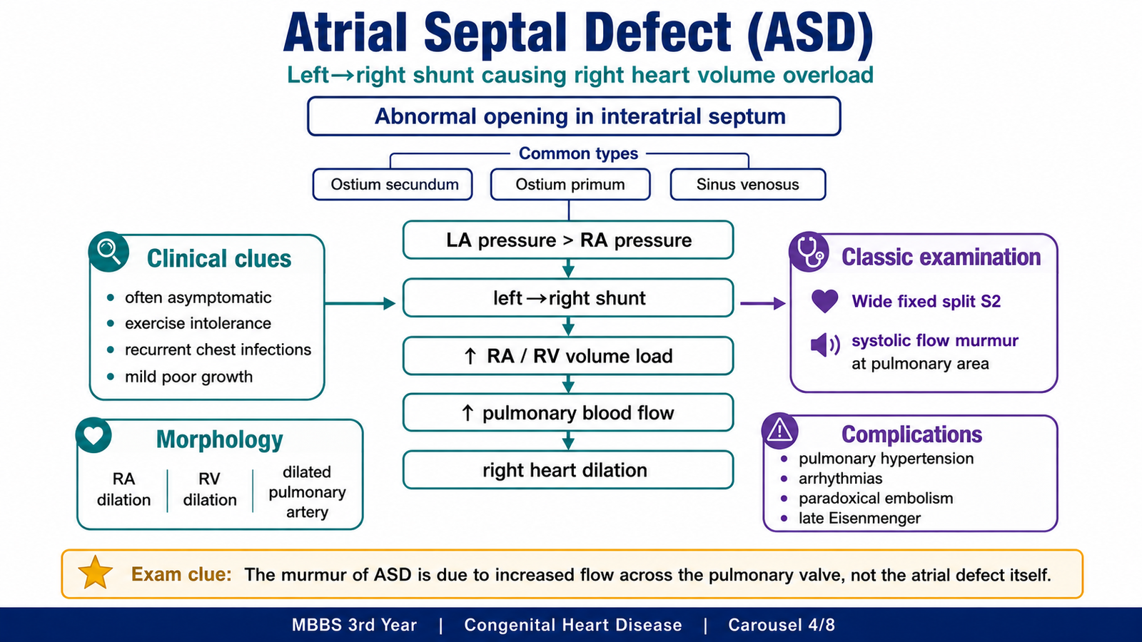
An atrial septal defect is an abnormal opening between the right and left atria. Because left atrial pressure is normally slightly higher than right atrial pressure, blood passes from the left atrium to the right atrium. This produces a left-to-right shunt and increases the volume handled by the right side of the heart.
Types
- Ostium secundum defect: Located near the fossa ovalis and is the most common type.
- Ostium primum defect: Located close to the atrioventricular valves and associated with endocardial cushion abnormalities.
- Sinus venosus defect: Located near the entry of the superior or inferior vena cava.
- Coronary sinus defect: A rare communication involving the wall between the coronary sinus and left atrium.
A patent foramen ovale is not a true deficiency of atrial septal tissue. It results from failure of the septum primum and septum secundum to fuse after birth.
Etiology and pathogenesis
ASD results from abnormal formation of the interatrial septum. The left-to-right shunt increases right atrial filling, right ventricular filling and pulmonary blood flow. Over time, the right atrium and right ventricle dilate because of chronic volume overload.
The size of the shunt depends on the size of the defect, ventricular compliance and pulmonary and systemic vascular resistance.
Morphology
A significant ASD produces right atrial dilation, right ventricular dilation and hypertrophy, dilation of the pulmonary artery and increased pulmonary vascular markings. The left ventricle is usually not enlarged because some left atrial blood is diverted into the right atrium.
Long-standing disease may produce pulmonary vascular changes and pulmonary hypertension.
Clinical features
Many children remain asymptomatic because the pressure difference between the atria is small and the shunt develops gradually. Larger defects may produce:
- Mild exercise intolerance
- Easy fatigability
- Recurrent respiratory infections
- Poor growth
- Right-sided volume overload
- Palpitations or atrial arrhythmias later in life
The characteristic findings are a systolic ejection murmur in the pulmonary area and wide, fixed splitting of the second heart sound. The murmur is caused by increased flow across the pulmonary valve rather than by flow through the atrial defect.
The splitting of S₂ remains relatively fixed during respiration because right ventricular stroke volume is chronically increased.
Complications and diagnosis
- Pulmonary hypertension
- Right-sided heart failure
- Atrial arrhythmias
- Paradoxical embolism
- Late Eisenmenger syndrome
Echocardiography demonstrates the site and size of the defect, the direction of flow, right atrial and right ventricular enlargement and associated abnormalities.
E. Ventricular Septal Defect
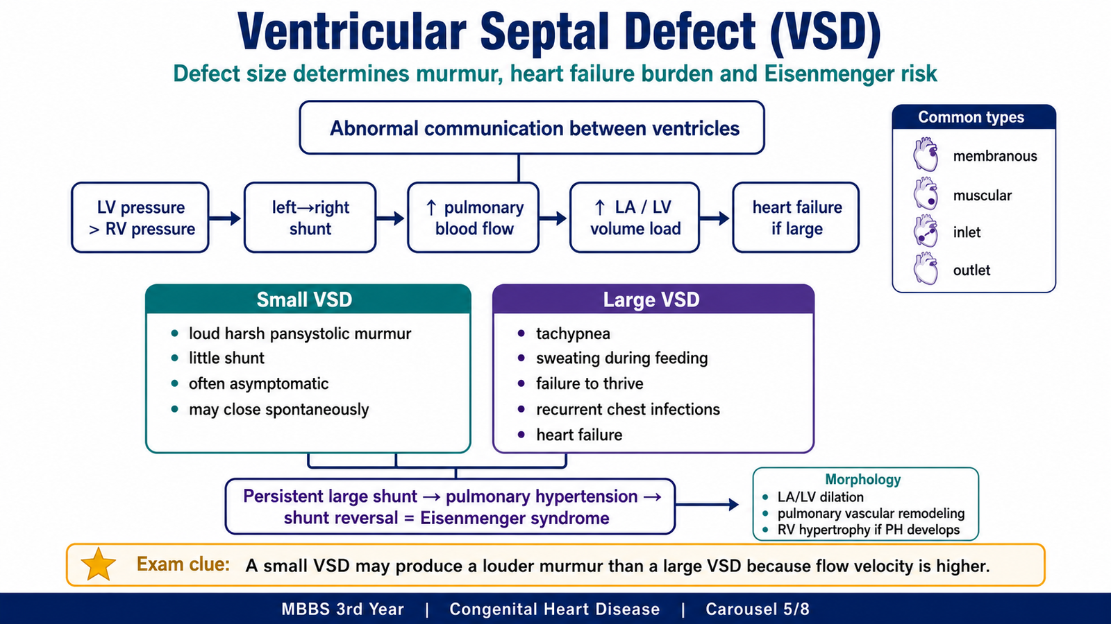
A ventricular septal defect is an abnormal communication between the right and left ventricles. After birth, left ventricular pressure is higher than right ventricular pressure, so blood usually moves from left to right. The clinical burden depends mainly on the size of the defect and the resistance of the pulmonary circulation.
Anatomical types
- Membranous or perimembranous VSD
- Muscular VSD
- Inlet VSD
- Outlet or supracristal VSD
The membranous or perimembranous type is the most common.
Etiology and pathogenesis
VSD results from incomplete formation or fusion of components of the interventricular septum. It may occur alone or as part of another congenital abnormality, including Tetralogy of Fallot and atrioventricular septal defects.
Effect of defect size
A small defect offers high resistance to flow. Only a limited volume of blood crosses it, but the blood travels at high velocity and produces a loud murmur. The child usually has no heart failure, and a small muscular defect may close spontaneously.
A large defect offers little resistance. A large volume of blood moves into the pulmonary circulation, returns to the left atrium and ventricle and produces left-sided volume overload. The infant may develop heart failure, recurrent respiratory infections and failure to thrive.
Morphology
A haemodynamically significant VSD produces dilation of the left atrium and left ventricle because increased pulmonary blood flow returns to the left side of the heart. The pulmonary artery may also dilate. If pulmonary hypertension develops, the right ventricle hypertrophies.
Microscopic pulmonary vascular changes include medial hypertrophy, intimal proliferation and progressive narrowing of pulmonary arteries.
Clinical features
A small VSD may be detected during routine examination because of a harsh pansystolic murmur at the lower left sternal border. A palpable thrill may be present, while cyanosis is absent.
A large VSD may present during infancy with:
- Tachypnoea
- Feeding difficulty
- Sweating during feeding
- Poor weight gain
- Recurrent respiratory infections
- Features of congestive heart failure
Eisenmenger transformation
Persistent pulmonary overcirculation causes progressive pulmonary vascular remodelling. Pulmonary vascular resistance eventually becomes high enough to reverse the shunt from right-to-left. The child then develops late cyanosis and clubbing.
A large untreated VSD may progress to Eisenmenger syndrome earlier than an ASD because the pulmonary circulation is exposed to higher pressure.
Complications
- Congestive heart failure
- Pulmonary hypertension
- Eisenmenger syndrome
- Infective endocarditis
- Aortic valve prolapse or regurgitation in some outlet defects
- Growth failure
F. Pulmonary Stenosis
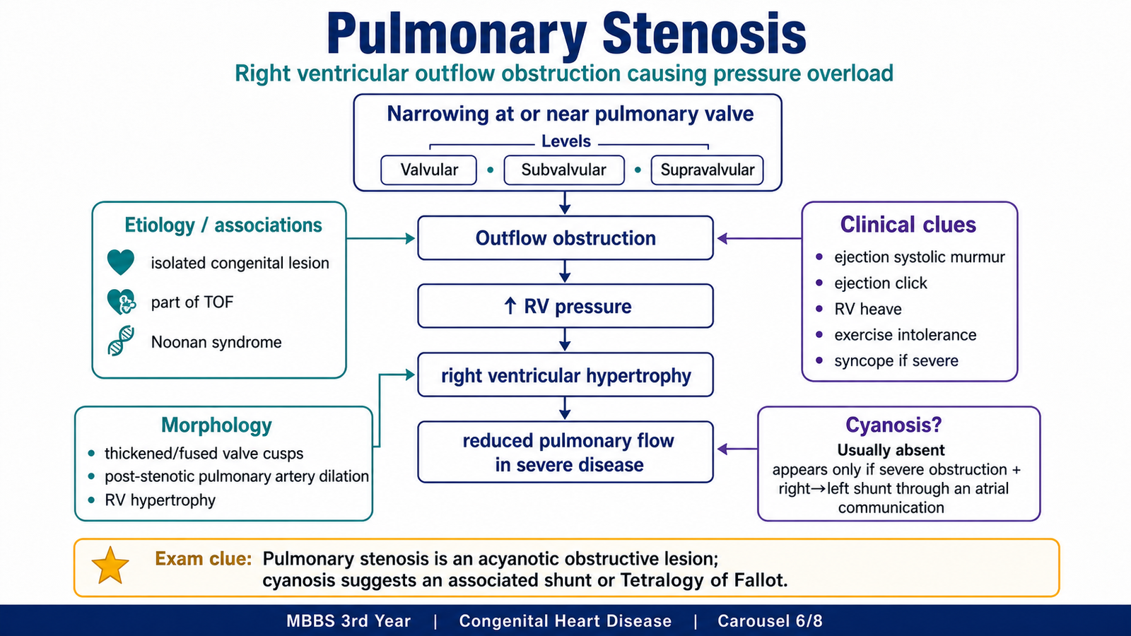
Pulmonary stenosis is narrowing at or near the pulmonary valve that obstructs blood flow from the right ventricle into the pulmonary artery. Unlike ASD and VSD, isolated pulmonary stenosis is primarily an obstructive lesion rather than a shunt lesion.
Types and etiology
- Valvular pulmonary stenosis: Narrowing at the pulmonary valve; the most common isolated form.
- Subvalvular or infundibular stenosis: Narrowing below the valve.
- Supravalvular stenosis: Narrowing above the valve.
The lesion results from abnormal development of the pulmonary valve or right ventricular outflow tract. It may occur alone, as part of Tetralogy of Fallot, with Noonan syndrome or with other right-sided cardiac abnormalities.
Pathogenesis
The narrowed valve or outflow tract resists right ventricular ejection. The right ventricle must generate greater pressure to move blood through the obstruction. This pressure load causes concentric right ventricular hypertrophy. Severe obstruction reduces pulmonary blood flow and may eventually cause right ventricular failure.
Morphology
- Thickened or fused pulmonary valve cusps
- Dome-shaped pulmonary valve during systole
- Post-stenotic dilation of the pulmonary artery
- Concentric right ventricular hypertrophy
- Right atrial dilation in severe disease
Clinical features
Mild pulmonary stenosis may remain asymptomatic. Moderate or severe obstruction may produce exercise intolerance, dyspnoea, fatigue, chest discomfort, syncope or right-sided heart failure.
Examination may show:
- Systolic ejection murmur at the upper left sternal border
- An ejection click
- Reduced intensity of the pulmonary component of S₂
- A right ventricular heave
Cyanosis is not usually present in isolated pulmonary stenosis because there is no obligatory pathway for deoxygenated blood to enter the systemic circulation. Cyanosis may occur if severe obstruction causes right-to-left shunting through an associated atrial communication.
Diagnostic distinction from Tetralogy of Fallot
Echocardiography with Doppler identifies the level of obstruction, valve morphology, pressure gradient, right ventricular hypertrophy and associated septal defects.
Isolated pulmonary stenosis produces obstruction without a necessary VSD or overriding aorta. Tetralogy of Fallot contains pulmonary obstruction together with a large VSD and overriding aorta, allowing right-to-left shunting and marked cyanosis.
G. Linking the Defect with Blood Flow, Murmur and Clinical Effect
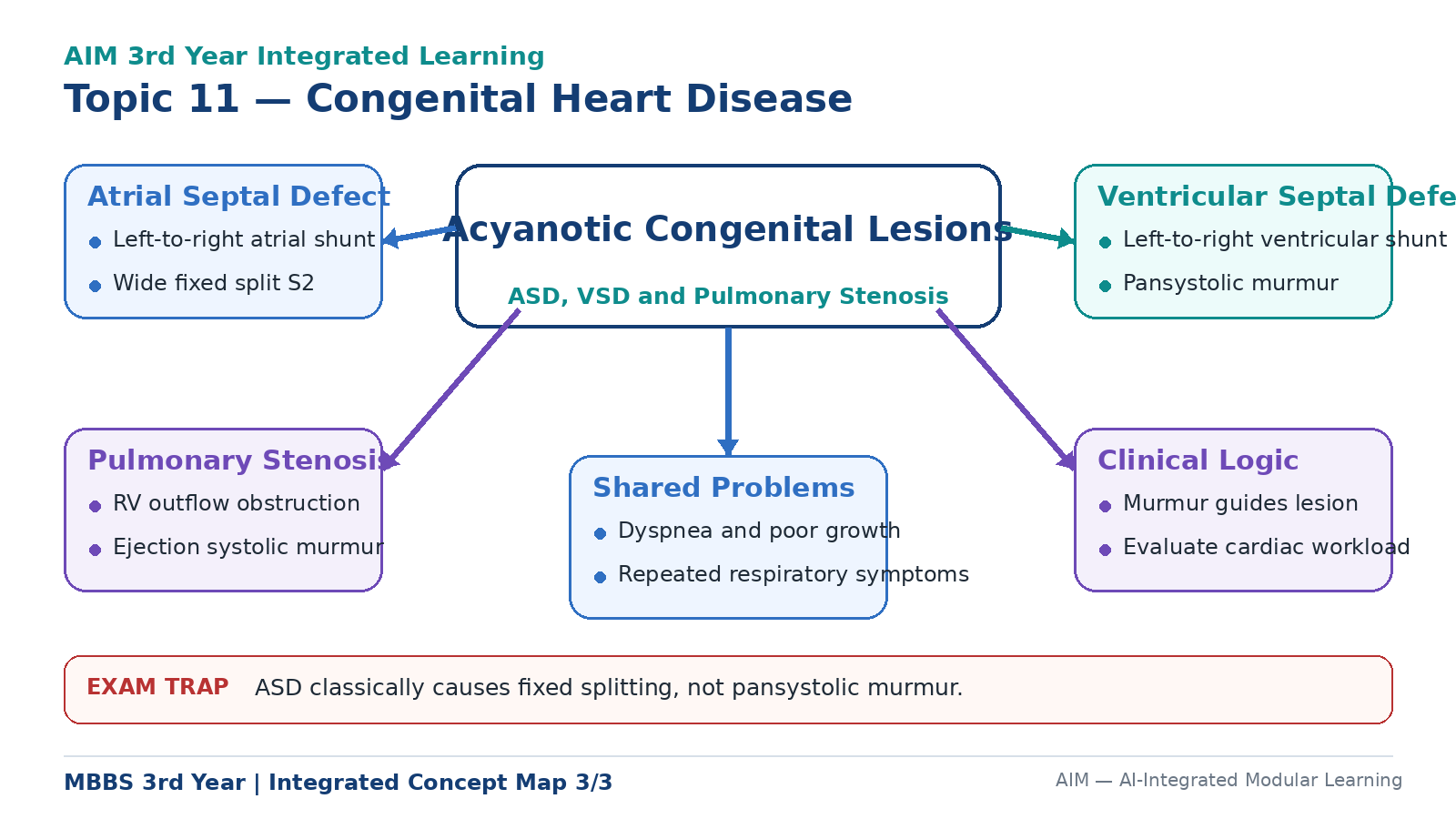
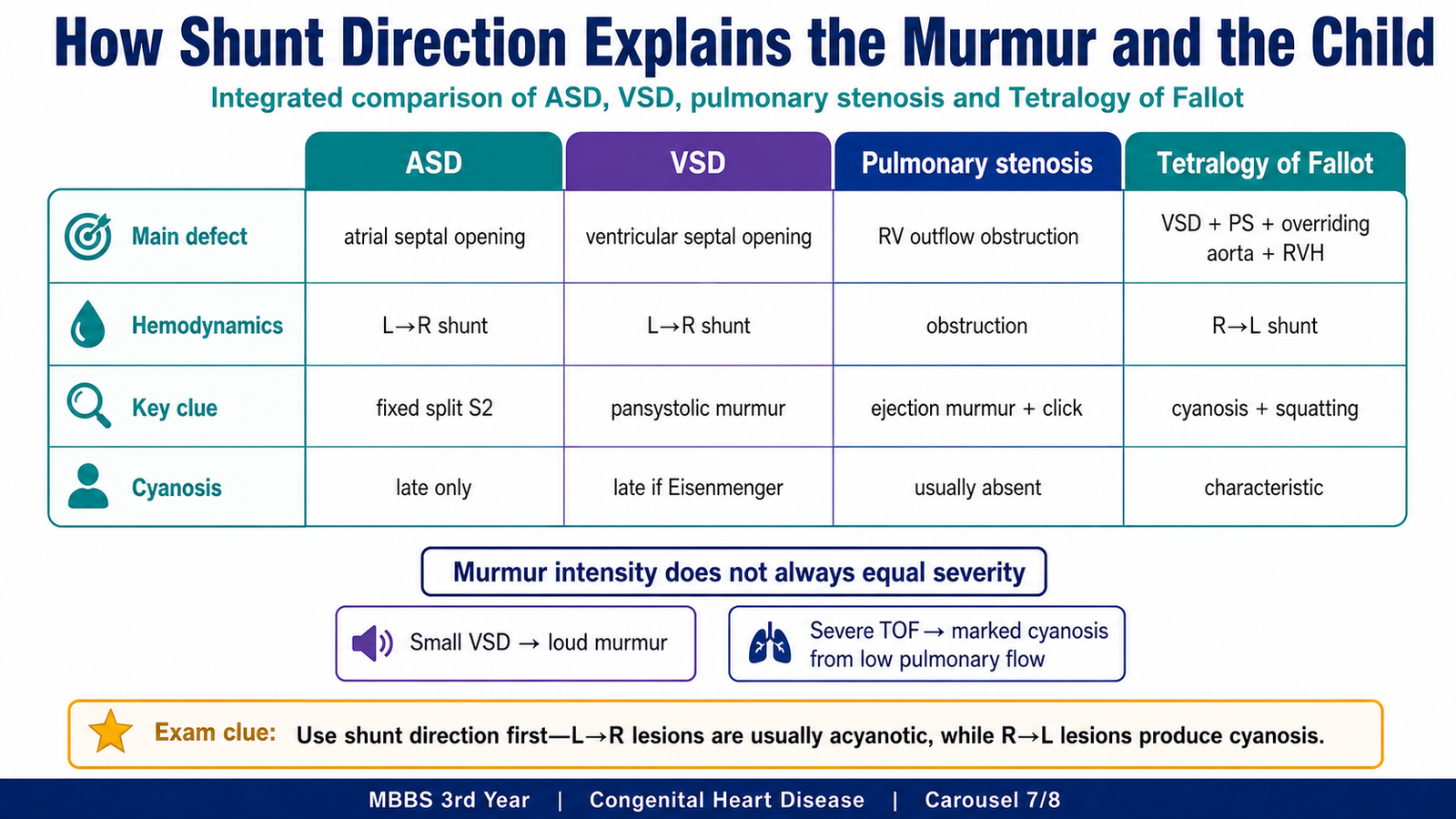
The same symptom or murmur can arise through different haemodynamic mechanisms. A useful approach is to identify whether the lesion produces a volume load, a pressure load or systemic delivery of deoxygenated blood.
| Lesion | Main abnormality | Cardiac burden | Typical clue | Cyanosis |
|---|---|---|---|---|
| ASD | Left-to-right atrial shunt | Right atrial and right ventricular volume overload | Wide fixed splitting of S₂ | Absent unless late shunt reversal occurs |
| VSD | Left-to-right ventricular shunt | Pulmonary overcirculation and left-sided volume overload | Pansystolic murmur at lower left sternal border | Absent initially; may develop with Eisenmenger syndrome |
| Pulmonary stenosis | Right ventricular outflow obstruction | Right ventricular pressure overload | Systolic ejection murmur at upper left sternal border | Usually absent |
| Tetralogy of Fallot | Pulmonary obstruction with right-to-left ventricular shunting | Reduced pulmonary flow and systemic hypoxaemia | Cyanosis, squatting and hypercyanotic spells | Characteristic |
Why murmur intensity does not always indicate severity
A murmur is produced by turbulent blood flow. Its loudness depends on the velocity and pressure difference across the abnormal opening, not simply on the physical size of the defect.
- A small VSD may produce a loud murmur because flow through it has high velocity.
- A very large VSD may produce a softer murmur when ventricular pressures become similar.
- Severe Tetralogy of Fallot may produce a shorter or softer outflow murmur when pulmonary blood flow is extremely reduced.
Important consequences
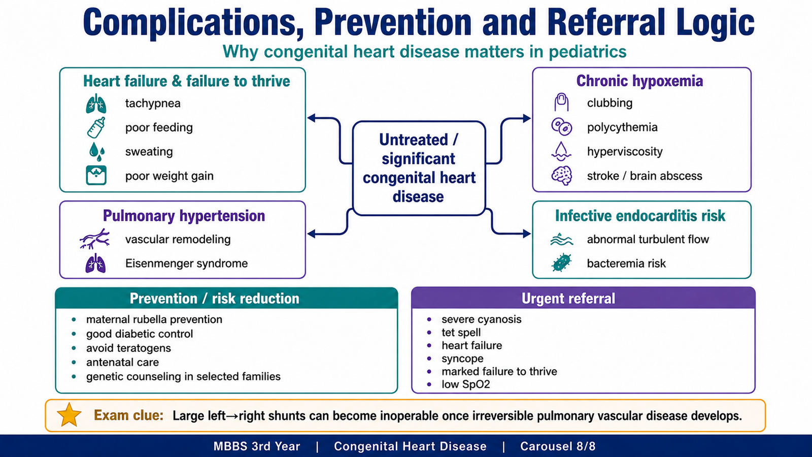
Large left-to-right shunts increase pulmonary blood flow and cardiac workload. This may cause tachypnoea, feeding difficulty, sweating, hepatomegaly, recurrent respiratory infections and failure to thrive. Growth is affected because feeding becomes difficult, caloric intake falls and energy expenditure rises.
Chronic hypoxaemia stimulates erythropoietin release and increases red-cell production. This secondary polycythaemia initially improves oxygen-carrying capacity, but excessive red-cell mass increases blood viscosity and raises the risk of headache, dizziness, thrombosis and stroke.
Abnormal high-velocity blood flow may damage endocardium and predispose to infective endocarditis during bacteraemia. Persistent pulmonary overcirculation may cause irreversible pulmonary vascular disease and Eisenmenger syndrome.
Integrated Mechanism Flow
→ septal communication, outflow obstruction or abnormal great-vessel arrangement
→ left-to-right shunt, right-to-left shunt or pressure obstruction
→ pulmonary overcirculation, reduced pulmonary flow or chamber overload
→ murmur, cyanosis, respiratory symptoms or heart failure
→ pulmonary hypertension, chronic hypoxaemia or cardiac dysfunction
→ Eisenmenger syndrome, hyperviscosity, infective endocarditis or growth failure
⭐ AIM High-Yield Review
- Congenital heart disease causes symptoms through abnormal shunting, obstruction, altered pulmonary blood flow or mixing of oxygenated and deoxygenated blood.
- Acyanotic disease includes left-to-right shunts such as ASD and VSD and obstructive lesions such as pulmonary stenosis.
- Cyanosis occurs when deoxygenated blood enters the systemic circulation; Tetralogy of Fallot is the classical example.
- Tetralogy of Fallot consists of pulmonary outflow obstruction, VSD, overriding aorta and right ventricular hypertrophy.
- The severity of cyanosis in Tetralogy of Fallot depends mainly on the degree of pulmonary outflow obstruction.
- Squatting improves Tetralogy of Fallot by increasing systemic vascular resistance and reducing right-to-left shunting.
- Wide fixed splitting of S₂ is the classical examination finding in ASD.
- The murmur of ASD is caused mainly by increased flow across the pulmonary valve, not by flow through the atrial defect.
- A VSD classically produces a harsh pansystolic murmur at the lower left sternal border.
- A small VSD may produce a louder murmur than a large VSD because flow velocity is higher.
- Large VSDs can cause early heart failure, pulmonary hypertension and Eisenmenger syndrome.
- Pulmonary stenosis produces right ventricular pressure overload, right ventricular hypertrophy and a systolic ejection murmur.
- Isolated pulmonary stenosis is usually acyanotic because it does not provide an obligatory route for right-to-left shunting.
- A long-standing left-to-right shunt may become cyanotic when pulmonary vascular disease reverses the shunt.
- Echocardiography with Doppler defines the cardiac anatomy, shunt direction, chamber effects and severity of obstruction.
Concept Not Clear? Ask AIM AI
Use the AIM Learning Assistant to clarify difficult concepts from this topic before proceeding to assessment.
You may ask AIM AI to:
- Explain a difficult concept in simpler words.
- Describe a mechanism step by step.
- Compare two confusing diseases, drugs or findings.
- Provide a clinically relevant example.
- Highlight a high-yield KMU examination point.
- Explain a common MCQ trap related to the topic.
Best use: Ask one focused question at a time. AIM AI supports your understanding but does not replace the complete learning material.
🎥 Video Explanation
Watch this video to reinforce the classification and haemodynamic basis of congenital heart disease, including cyanotic and acyanotic lesions, atrial septal defect, ventricular septal defect and Tetralogy of Fallot.
Focus while watching: Compare left-to-right and right-to-left shunts, and relate each structural defect to pulmonary blood flow, cyanosis, chamber overload and characteristic clinical findings.
If the embedded player does not load, open the video on YouTube.